. 2024 Dec:509:153971.
doi: 10.1016/j.tox.2024.153971. Epub 2024 Oct 11.

Cadmium-induced lung injury disrupts immune cell homeostasis in the secondary lymphoid organs in mice

Affiliations

Cadmium-induced lung injury disrupts immune cell homeostasis in the secondary lymphoid organs in mice

Chandrashekhar Prasad et al. Toxicology. 2024 Dec.

Abstract

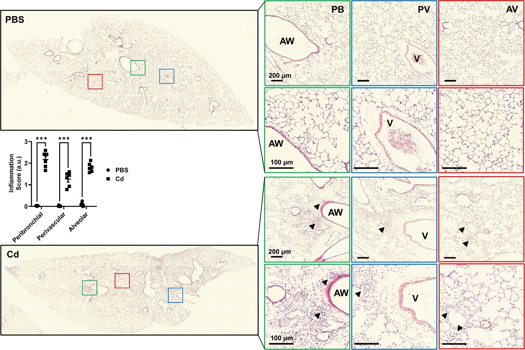
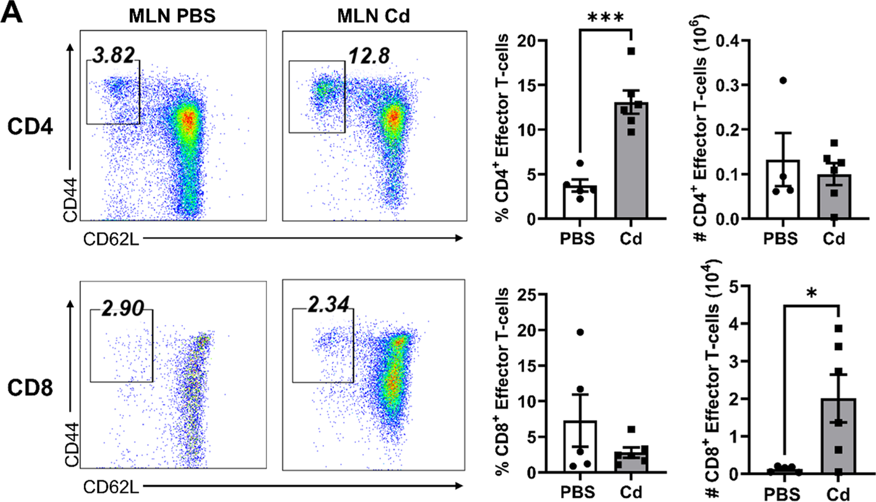
Cadmium (Cd) is a well-known toxic heavy metal that poses significant health risks, particularly through inhalation, smoking, and the consumption of contaminated food. Exposure to cadmium is linked to the development and exacerbation of chronic lung diseases such as pulmonary fibrosis and chronic obstructive pulmonary disease (COPD). This study investigated the systemic effects of intratracheal cadmium chloride (0.5 mg/kg) instillation in C57BL/6 mice. All parameters, including inflammation assessment, lung function evaluation (using Flexi-vent), and immunophenotyping of T-cells in secondary lymphoid organs (mediastinal lymph nodes and spleen), were analyzed 14 days after cadmium exposure. The results demonstrated that cadmium exposure led to significant immune cell infiltration in bronchoalveolar lavage (BAL) fluid, altered pro-inflammatory cytokine levels, and was associated with impaired lung function, characterized by increased lung resistance and Newtonian resistance. Analysis of T-cell populations revealed no significant changes in total T-cells in mediastinal lymph nodes and spleen, but a decrease in CD4+ T-cells and an increase in CD8+ T-cells were observed. These findings suggest that cadmium disrupts T-cell homeostasis in secondary lymphoid organs. Further research is crucial to elucidate the mechanisms underlying cadmium-induced lung injury and immune dysregulation, essential for developing effective therapeutic interventions against chronic lung diseases caused by cadmium exposure.

Keywords: Cadmium exposure; Lung function; Lung inflammation; Lymphoid organs; T-cells; mouse model.


Conflict of interest statement

Declaration of Competing Interest The authors declare that they have no known competing financial interests or personal relationships that could have appeared to influence the work reported in this paper.

Figures

Fig. 1.
Fig. 1.. Cadmium exposure causes an increased lung inflammatory response in mice.
C57BL/6 (WT) mice, ~2–3 months old males, were exposed to PBS or CdCl2 (0.5 mg/kg at ZT6; 12:00 pm). (A) Differential cell counts in BAL fluid were analyzed using Diff-Quik staining after 14 days. (B) Inflammatory mediators such as IL-6, TNFα, IP-10/CXCL10, and KC in BAL fluid were analyzed using a custom-designed LegendPlex assay in PBS and CdCl2-exposed groups. Total protein in the BAL fluid of PBS- and Cd-exposed mice was determined using a BCA assay. The mice were given food ad libitum (ad lib) (fed throughout the 12:12 L:D cycle) and had continuous access to water. Data were shown as mean ± SEM (n =5–6/group). * P < 0.05, ** P < 0.01, compared to the respective PBS control.
Fig. 2.
Fig. 2.. Histological evaluation of Cd-exposed mouse lung showing increased inflammation.
Representative hematoxylin and eosin (H&E)-stained lung sections showed a difference in the degree of lung inflammation in the peribronchial (PB), perivascular (PV), and alveolar (AV) regions at day 14 in PBS- and Cd-exposed mice. BAL fluid was isolated before processing the tissues for fixation and sectioning. The graph shows the average lung inflammation scores using the scoring criteria described in the materials and methods section in a blinded manner. The arrow indicates the representative area showing inflammation around the airways (AW), blood vessels (V), and alveolar regions. Data are shown as mean ± SEM (n=5–6/group). ***P < 0.001, compared to their respective PBS controls.
Fig. 3.
Fig. 3.. Cadmium exposure altered lung mechanics in mice.
C57BL/6 (WT) mice, ~2–3 months old males, were exposed to PBS or CdCl2 (0.5 mg/kg at ZT6; 12:00 pm). The forced oscillation technique (FOT) was used to measure lung mechanics such as dynamic compliance (Crs), elastance (Ers), resistance (Rrs), and Newtonian resistance (Rn) in the lungs after 14 days. Additional parameters measured include the coefficient of elasticity (K) by the Salazar-Knowles equation from the PV curve. The representative pressure-volume (PV) curve illustrates the changes in lung mechanics after Cd exposure in mice. Data were shown as mean ± SEM (n =5–8/group). * P < 0.05, ** P < 0.01, compared to the respective PBS control.
Fig. 4.
Fig. 4.. Cadmium exposure altered the T-cell percentages and counts in the mediastinal lymph nodes and spleen in mice.
Representative gating for the percentage of CD4+ and CD8+ T-cells from the mediastinal lymph node (MLN) and spleen. Cells from the MLN and spleen were isolated, counted, and stained with fluorophore-conjugated antibodies for TCRβ, CD4, and CD8. (A) Percentages and counts for total T-cells (TCRβ+ population), CD4+, and CD8+ T-cells in the MLN. (B) Percentages and counts for total T-cells (TCRβ+ population), CD4+, and CD8+ T-cells in the spleen. Data were shown as mean ± SEM (n =5–8/group). * P < 0.05, ** P < 0.01, ***P < 0.001 compared to the respective PBS control.
Fig. 5.
Fig. 5.. Cadmium exposure altered activation of CD4+ and CD8+ T-cells in the spleen and mediastinal lymph nodes in mice.
(A-B) Representative gating for the percentage of activated CD4+ and CD8+ T-cells from the mediastinal lymph nodes (MLN) and spleen. Cells from the MLN and spleen were isolated, counted, and stained with fluorophore-conjugated antibodies for TCRβ, CD4, CD8, and CD25 to delineate activated CD4 (TCRβ+CD4+CD25+) and CD8 (TCRβ+CD4+CD25+) T-cells. (A) Activated CD4+ and CD8+ T-cell percentages and counts in the MLN. (B) Activated CD4+ and CD8+ T-cell percentages and counts in the spleen. Data were shown as mean ± SEM (n =5–8/group). ** P < 0.01, *** P < 0.001 compared to the respective PBS control.
Fig. 6.
Fig. 6.. Cadmium exposure changed CD4+ and CD8+ effector T-cells in the mediastinal lymph nodes and spleen in mice.
(A-B) Representative gating for the percentage of effector CD4+ and CD8+ T-cells from the mediastinal lymph nodes (MLN) and spleen. Cells from the MLN and spleen were isolated, counted, and stained with fluorophore-conjugated antibodies for TCRβ, CD8, CD44, and CD62L to delineate effector CD4+ (TCRβ+CD4+CD44+CD62L) and CD8+ (TCRβ+CD8+CD44+CD62L) T-cells. (A) Effector CD4+ and CD8+ T-cell percentages and counts in the MLN. (B) Effector CD4+ and CD8+ T-cell percentages and counts in the spleen. Data were shown as mean ± SEM (n =5–8/group). * P < 0.05, ** P < 0.01, ***P < 0.001 compared to the respective PBS control.

References

    1. Badawi K, El Sharazly BM, Negm O, Khan R, & Carter WG (2024). Is Cadmium Genotoxicity Due to the Induction of Redox Stress and Inflammation? A Systematic Review. Antioxidants (Basel), 13(8). 10.3390/antiox13080932 - DOI - PMC - PubMed
    1. Carlier FM, de Fays C, & Pilette C (2021). Epithelial Barrier Dysfunction in Chronic Respiratory Diseases. Front Physiol, 12, 691227. 10.3389/fphys.2021.691227 - DOI - PMC - PubMed
    1. Cetintepe SP, Iritas SB, Gunduzoz M, Alaguney ME, Wilson D, Bal C, Yilmaz OH, Tutkun L, & Park E-K (2019). Relation Between Lung Dysfunction and Blood Cadmium and Lead Levels Among Welders. Exposure and Health, 11(1), 13–19. 10.1007/s12403-017-0262-x - DOI
    1. Devos FC, Maaske A, Robichaud A, Pollaris L, Seys S, Lopez CA, Verbeken E, Tenbusch M, Lories R, Nemery B, Hoet PH, & Vanoirbeek JA (2017). Forced expiration measurements in mouse models of obstructive and restrictive lung diseases. Respir Res, 18(1), 123. 10.1186/s12931-017-0610-1 - DOI - PMC - PubMed
    1. Dong S, Shen HM, & Ong CN (2001). Cadmium-induced apoptosis and phenotypic changes in mouse thymocytes. Mol Cell Biochem, 222(1–2), 11–20. https://www.ncbi.nlm.nih.gov/pubmed/11678592 - PubMed

Publication types

MeSH terms