. 2024 Oct 14;10(1):145.
doi: 10.1038/s41378-024-00778-2.

Neuronal activity in the ventral tegmental area during goal-directed navigation recorded by low-curvature microelectrode arrays

Affiliations

Neuronal activity in the ventral tegmental area during goal-directed navigation recorded by low-curvature microelectrode arrays

Wei Xu et al. Microsyst Nanoeng. .

Abstract

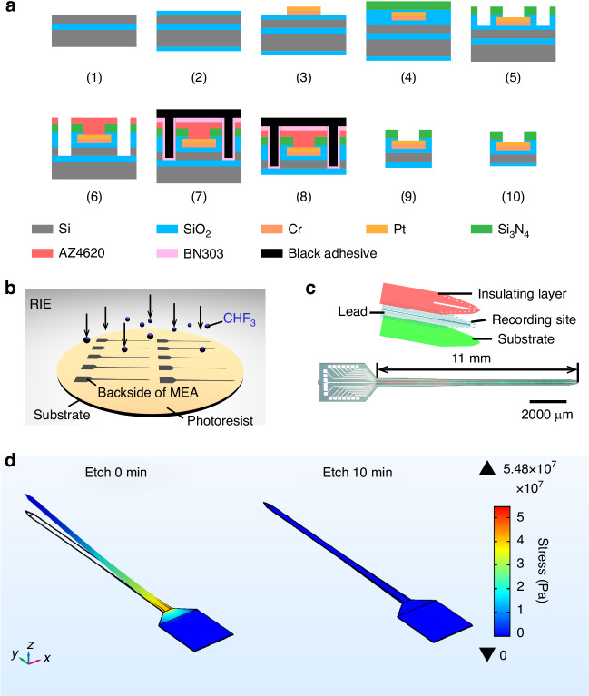
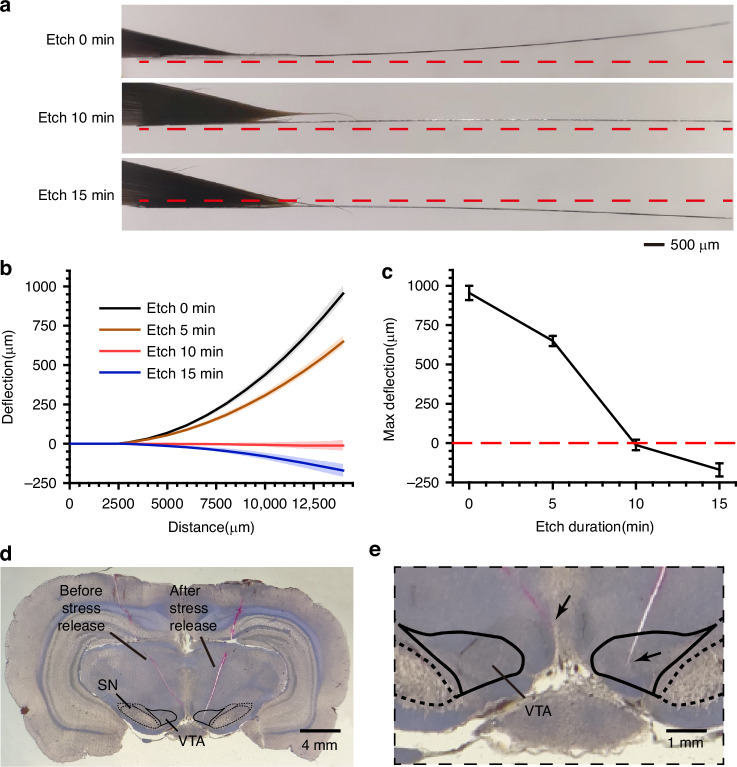
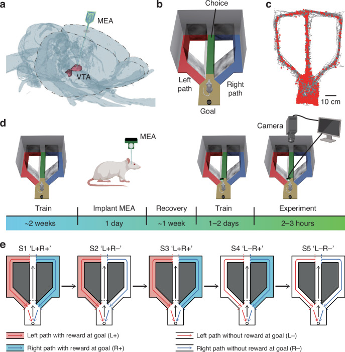
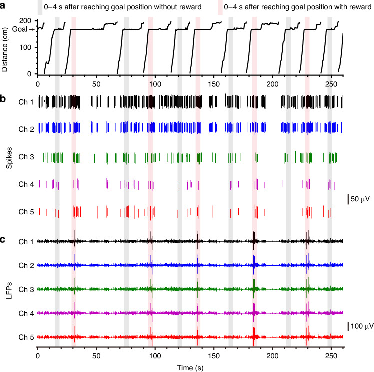
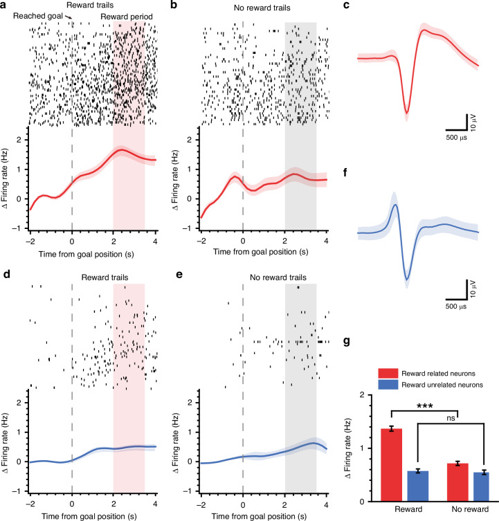
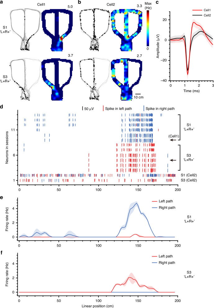
Navigating toward destinations with rewards is a common behavior among animals. The ventral tegmental area (VTA) has been shown to be responsible for reward coding and reward cue learning, and its response to other variables, such as kinematics, has also been increasingly studied. These findings suggest a potential relationship between animal navigation behavior and VTA activity. However, the deep location and small volume of the VTA pose significant challenges to the precision of electrode implantation, increasing the uncertainty of measurement results during animal navigation and thus limiting research on the role of the VTA in goal-directed navigation. To address this gap, we innovatively designed and fabricated low-curvature microelectrode arrays (MEAs) via a novel backside dry etching technique to release residual stress. Histological verification confirmed that low-curvature MEAs indeed improved electrode implantation precision. These low-curvature MEAs were subsequently implanted into the VTA of the rats to observe their electrophysiological activity in a freely chosen modified T-maze. The results of the behavioral experiments revealed that the rats could quickly learn the reward probability corresponding to the left and right paths and that VTA neurons were deeply involved in goal-directed navigation. Compared with those in no-reward trials, VTA neurons in reward trials presented a significantly greater firing rate and larger local field potential (LFP) amplitude during the reward-consuming period. Notably, we discovered place fields mapped by VTA neurons, which disappeared or were reconstructed with changes in the path-outcome relationship. These results provide new insights into the VTA and its role in goal-directed navigation. Our designed and fabricated low-curvature microelectrode arrays can serve as a new device for precise deep brain implantation in the future.


Conflict of interest statement

Xinxia Cai is an editor for the journal, no other author has reported any competing interest.

Figures

Fig. 1
Fig. 1. Fabrication and residual stress relief of the MEA.
a Manufacturing process of the MEA. (1) Clean the SOI (silicon on insulator). (2) Thermally oxidize the substrate to form the insulating layer. (3) Sputter the metal layer. (4) Deposit SiO2/Si3N4 insulating layers via plasma-enhanced chemical vapor deposition (PECVD). (5) Etch the insulating layer to expose the electrode sites. (6) Perform deep silicon etching to define the electrode morphology. (7) Coat the front side of the SOI with black adhesive. (8) Wet etch the backside Si layer of the SOI. (9) Dissolve the black adhesive to obtain individual electrodes. (10) Perform dry etching on the backside of the electrodes to release residual stress. b Release of residual stress by etching the backside of the MEAs. c Electrode structure, tip morphology (top), and actual MEA image (bottom). d Stress simulation results of electrodes with unrelieved residual stress (left) and with relieved residual stress (right)
Fig. 2
Fig. 2. Characterization of residual stress-relieved MEAs and histological verification.
a Photograph of the electrode taken from the side. The red dotted lines indicate the horizontal direction. b, c Electrode deflection and maximum deflection (deflection at the end of the electrode) for different etching durations. A positive deflection indicates that the electrode was bent toward the front. When etched for 10 minutes, the residual stress was relieved (max deflection = −11.8 ± 32.9 μm, mean ± SD, n = 5). d, e Histological verification. Micrograph of the coronal plane of the brain with 2 electrodes implanted simultaneously. The types of MEAs corresponding to the trajectories have been annotated. e Enlarged view of a portion of (d). The arrows in (e) indicate the electrode implantation sites
Fig. 3
Fig. 3. Electrophysiological recording and behavioral tasks.
a A microelectrode array was implanted in the VTA of the left hemisphere. b Schematic diagram of the modified T-maze. The maze is fully enclosed; some of the sidewalls are not drawn, and all the sidewalls and the ground are black and opaque. The black well at the bottom is the goal position where the food reward can be dropped from a feeder above. The end of the middle arm (green) is the choice position, where the rat chooses to enter the left path (red) or the right path (blue). c Rat trajectories (gray lines) and spike locations (red dots) are plotted. d Schematic representation of the behavior, surgery, and electrophysiological activity recordings. e Diagram of the experiment, which consists of 5 sessions. Whether the food reward is dropped at the goal depends on the chosen path, and the color-filled arm indicates the correct path. L: left path; R: right path; ‘+’: reward; ‘−’: no reward
Fig. 4
Fig. 4. Behavioral performance and electrophysiological recordings.
a Rat movements (distance from the start line, see Fig. S3) during several trials. b Recordings of spikes of a single unit in parts of the channels during 260 s of the ‘L + R−’ session (marked in a with the gray shaded window). Periods within 4 s after reaching the goal position were denoted by shaded windows (red, reward; gray, no reward). c Traces of LFPs corresponding to b
Fig. 5
Fig. 5. Neural activity related to reward.
ac An example neuron related to reward. df An example neuron unrelated to reward. a, d Neural activities when the rat received food reward at the goal position. The moment when the rat reaches the goal position is marked as time zero (dotted line), and the food reward is dropped at the goal approximately 2 seconds later. The spike raster across trials (each row indicates a trial) is plotted in the top panel. Evolution in time of the responses (i.e., Δ firing rate, baseline subtracted) to reward is plotted in the bottom panel. b, e Neural activities when the rat did not receive food reward at the goal position. c, f Spike waveform of an example neuron, mean ± SD. g Under the circumstances of receiving a reward and no reward at the goal, the response (i.e., the average firing rate during the shaded window in (ae)) of different population neurons is plotted (red, reward-related neurons, n = 33; blue, reward-unrelated neurons, n = 64). One-way ANOVA test
Fig. 6
Fig. 6. Remapping induced by reward.
a An example neuron (Cell1) with place fields. Left panel, gray line, the rat’s trace; black dot, spike. Right panel, corresponding 2D firing rate map (the maximum firing rate is marked at the upper right corner). Top panel, session 1 (S1, ‘L+R+’); bottom panel, session 3 (S3, ‘L+R+’). b An example neuron (Cell2) without a place field. c Comparison of spike waveforms (mean ± SD) between Cell1 and Cell2. d Spikes of neurons with or without place fields in session 1 (S1, ‘L+R+’) and session 3 (S3, ‘L+R+’) are displayed in the spatial domain. e Evolution in the linear position of the firing rate (red line, left path; blue line, right path; mean ± SD) for neurons with a place field during S1 (‘L+R+’). f Same as (e) but for S3 (‘L+R+’)

Similar articles

Cited by

References

    1. Nyberg, N., Duvelle, E., Barry, C. & Spiers, H. J. Spatial goal coding in the hippocampal formation. Neuron110, 394–422 (2022). - PubMed
    1. Sosa, M. & Giocomo, L. M. Navigating for reward. Nat. Rev. Neurosci.22, 472–487 (2021). - PMC - PubMed
    1. Redgrave, P. et al. Goal-directed and habitual control in the basal ganglia: implications for Parkinson’s disease. Nat. Rev. Neurosci.11, 760–772 (2010). - PMC - PubMed
    1. Watabe-Uchida, M., Eshel, N. & Uchida, N. Neural circuitry of reward prediction error. Annu. Rev. Neurosci.40, 373 (2016). - PMC - PubMed
    1. Ungless, M. A. & Grace, A. A. Are you or aren’t you? Challenges associated with physiologically identifying dopamine neurons. Trends Neurosci.35, 422–430 (2012). - PMC - PubMed

LinkOut - more resources