Review
. 2024 Oct 14;9(1):271.
doi: 10.1038/s41392-024-01969-z.

Iron homeostasis and ferroptosis in human diseases: mechanisms and therapeutic prospects

Affiliations
Review

Iron homeostasis and ferroptosis in human diseases: mechanisms and therapeutic prospects

Qin Ru et al. Signal Transduct Target Ther. .

Abstract

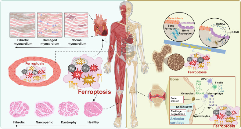
Iron, an essential mineral in the body, is involved in numerous physiological processes, making the maintenance of iron homeostasis crucial for overall health. Both iron overload and deficiency can cause various disorders and human diseases. Ferroptosis, a form of cell death dependent on iron, is characterized by the extensive peroxidation of lipids. Unlike other kinds of classical unprogrammed cell death, ferroptosis is primarily linked to disruptions in iron metabolism, lipid peroxidation, and antioxidant system imbalance. Ferroptosis is regulated through transcription, translation, and post-translational modifications, which affect cellular sensitivity to ferroptosis. Over the past decade or so, numerous diseases have been linked to ferroptosis as part of their etiology, including cancers, metabolic disorders, autoimmune diseases, central nervous system diseases, cardiovascular diseases, and musculoskeletal diseases. Ferroptosis-related proteins have become attractive targets for many major human diseases that are currently incurable, and some ferroptosis regulators have shown therapeutic effects in clinical trials although further validation of their clinical potential is needed. Therefore, in-depth analysis of ferroptosis and its potential molecular mechanisms in human diseases may offer additional strategies for clinical prevention and treatment. In this review, we discuss the physiological significance of iron homeostasis in the body, the potential contribution of ferroptosis to the etiology and development of human diseases, along with the evidence supporting targeting ferroptosis as a therapeutic approach. Importantly, we evaluate recent potential therapeutic targets and promising interventions, providing guidance for future targeted treatment therapies against human diseases.


Conflict of interest statement

The authors declare no competing interests.

Figures

Fig. 1
Fig. 1
The history and development of ferroptosis. In the figure, we outline the major events in the field of ferroptosis from 1955 to 2024, including the identification of the important role of cysteine in cell survival, the formal definition of ferroptosis, the discovery of system Xc, oxytosis, PUFA-phospholipids (PUFA-PLs), and the proposal of antioxidant systems GPX4 and FSP1. This figure was created with BioRender (https://biorender.com/)
Fig. 2
Fig. 2
The crosstalk of ferroptosis with apoptosis, autophagy and pyroptosis. In the figure, we illustrate that ferritin produced in the ferroptosis can be digested and degraded by the autophagic mechanism, releasing excessive ferric ions that participate in the Fenton reaction, thereby inducing a vicious cycle of ferroptosis. Moreover, inflammasome NACHT, LRR and PYD domains-containing protein 3 (NLRP3), besides inducing pyroptosis, can promote ferroptosis by inhibiting GPX4. Excessive oxygen free radicals and lipid peroxidation products in the apoptosis signal pathway also induced ferroptosis. This figure was created with BioRender (https://biorender.com/)
Fig. 3
Fig. 3
The role of ferroptosis in human diseases. The figure illustrates that ferroptosis plays a significant role in many human diseases. In neurodegenerative conditions, it is involved in the pathological process of Alzheimer’s disease (AD), Parkinson’s disease (PD), and Huntington’s disease (HD), as well as in cerebral ischemia-reperfusion (I/R) injury, brain and spinal cord injury, and pain. In the circulatory system, ferroptosis contributes to heart failure, coronary heart disease, and myocardial I/R injury. In the musculoskeletal system, it leads to functional decline in skeletal muscle, bones, and joints. In chronic metabolic diseases, ferroptosis can trigger the occurrence and development of diabetes mellitus, hyperlipidemia, fatty liver disease, and obesity. In cancer, ferroptosis mainly affects tumor growth, metastasis, invasion, and chemoresistance. In autoimmune diseases, it is mainly involved in the pathogenesis of rheumatoid arthritis, systemic lupus erythematosus, and myasthenia gravis. In genetic diseases, ferroptosis predominantly affects thalassemia and progressive muscular dystrophy. This figure was created with BioRender (https://biorender.com/)
Fig. 4
Fig. 4
Ferroptosis in cardiovascular diseases. In the circulatory system, iron overload, lipid peroxidation and oxidative stress associated with ferroptosis can gradually induce coronary atherosclerotic heart disease and myocardial infarction (a), dilated cardiomyopathy (b), hypertrophic cardiomyopathy (c), and inflammatory infiltration of myocardium (d). This occurs through the damage of vascular endothelial cells and myocardial cells, leading to inflammatory responses. This figure was created with BioRender (https://biorender.com/)
Fig. 5
Fig. 5
Ferroptosis in neurodegenerative diseases. In the nervous system, ferroptosis primarily induces and promotes neuronal damage and axonal degeneration. It also facilitates the aggregation of amyloid-beta (Aβ) in the brain and contributes to the formation of neurofibrillary tangles. Additionally, ferroptosis activates glial cells, triggering an inflammatory response. These complex pathological changes in the brain interact with each other, leading to neurodegenerative diseases such as Alzheimer’s disease (AD), Parkinson’s disease (PD), and Huntington’s disease (HD), as well as ischemia-reperfusion injury and inflammatory responses. This figure was created with BioRender (https://biorender.com/)
Fig. 6
Fig. 6
Ferroptosis in musculoskeletal diseases. Ferroptosis in the musculoskeletal system contributes to the occurrence and development of arthritis primarily through lipid peroxidation and oxidative stress, which to damage the joint synovial membrane, matrix, and hyperactivated immune cells. Additionally, ferroptosis disrupts the delicate balance of osteoblasts and osteoclasts in the bone tissue, leading to the development of osteoporosis. Ferroptosis also plays a crucial role in the decline of quality and function in skeletal muscle, cardiac muscle and smooth muscle, there by inducing the onset of sarcopenia. This figure was created with BioRender (https://biorender.com/)

References

    1. Stockwell, B. R. Ferroptosis turns 10: emerging mechanisms, physiological functions, and therapeutic applications. Cell185, 2401–2421 (2022). - PMC - PubMed
    1. Dixon, S. J. & Olzmann, J. A. The cell biology of ferroptosis. Nat. Rev. Mol. Cell Biol.25, 424–442 (2024). - PubMed
    1. Jiang, X., Stockwell, B. R. & Conrad, M. Ferroptosis: mechanisms, biology and role in disease. Nat. Rev. Mol. Cell Biol.22, 266–282 (2021). - PMC - PubMed
    1. Li, J. et al. Ferroptosis: past, present and future. Cell Death Dis.11, 88 (2020). - PMC - PubMed
    1. Berndt, C. et al. Ferroptosis in health and disease. Redox Biol.75, 103211 (2024). - PMC - PubMed

Publication types

MeSH terms