Case Reports
. 2024 Oct 14;15(1):8849.
doi: 10.1038/s41467-024-53072-0.

HEV ORF2 protein-antibody complex deposits are associated with glomerulonephritis in hepatitis E with reduced immune status

Affiliations
Case Reports

HEV ORF2 protein-antibody complex deposits are associated with glomerulonephritis in hepatitis E with reduced immune status

Anne-Laure Leblond et al. Nat Commun. .

Abstract

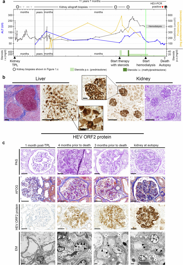
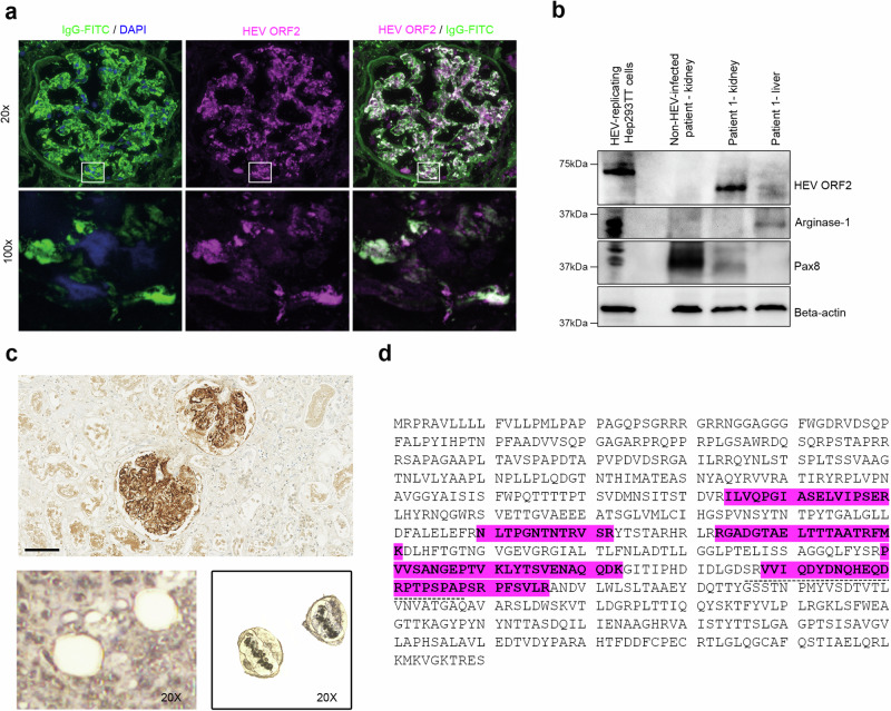
Hepatitis E virus (HEV) infection, one of the most common forms of hepatitis worldwide, is often associated with extrahepatic, particularly renal, manifestations. However, the underlying mechanisms are incompletely understood. Here, we report the development of a de novo immune complex-mediated glomerulonephritis (GN) in a kidney transplant recipient with chronic hepatitis E. Applying immunostaining, electron microscopy, and mass spectrometry after laser-capture microdissection, we show that GN develops in parallel with increasing glomerular deposition of a non-infectious, genome-free and non-glycosylated HEV open reading frame 2 (ORF2) capsid protein. No productive HEV infection of kidney cells is detected. Patients with acute hepatitis E display similar but less pronounced deposits. Our results establish a link between the production of HEV ORF2 protein and the development of hepatitis E-associated GN in the immunocompromised state. The formation of glomerular IgG-HEV ORF2 immune complexes discovered here provides a potential mechanistic explanation of how the hepatotropic HEV can cause variable renal manifestations. These findings directly provide a tool for etiology-based diagnosis of hepatitis E-associated GN as a distinct entity and suggest therapeutic implications.


Conflict of interest statement

The authors declare no competing interest.

Figures

Fig. 1
Fig. 1. Clinical course, autopsy findings and gradual development of immune complex glomerulonephritis with membranoproliferative pattern in a kidney transplant recipient with hepatitis E.
a Course of alanine transaminase (ALT, blue), proteinuria (yellow) and creatinine (grey), time points of therapeutic and diagnostic interventions as well as death / autopsy. b Histology of autopsy liver showing cirrhosis (Masson trichrome stain) and hepatitis with immune reactivity for HEV ORF2 protein in hepatocytes (left). Histology of transplant kidney at autopsy showing glomerulonephritis (periodic acid Schiff [PAS] stain) and (extracellular) immune reactivity for HEV ORF2 protein in glomeruli (right). Scale bars in overviews, 200 μm; scale bars in detail, 25 μm. Stainings performed more than 5 times on different tissues blocks. c Kidney histology. One month post transplantation: inconspicuous glomeruli on light microscopy (PAS and acid fuchsin-Orange G [AFOG] stains), no HEV ORF2 protein deposits, no electron dense deposits on electron microscopy (EM). Four months prior to death (biopsy 4): glomerulus with mild mesangial and endocapillary hypercellularity, segmental sclerosis and prominent podocytes (PAS stain). Mostly mesangial and few glomerular basement membrane protein deposits (AFOG stain). Moderate mesangial and glomerular basement membrane positivity for HEV ORF2 protein. Mesangial (white arrowheads), subendothelial (black arrowheads) and subepithelial (black and white arrowheads) on EM. Three months prior to death (biopsy 5): glomerulus with mild mesangial and endocapillary hypercellularity (PAS stain). Mostly mesangial and few glomerular basement membrane protein deposits (AFOG stain). Moderate to strong mesangial and glomerular basement membrane positivity for HEV ORF2 protein. Mesangial (white arrowheads), subendothelial (black arrowheads) and subepithelial (black and white arrowheads) on EM. Kidney at autopsy: glomerulus with mild mesangial and endocapillary hypercellularity (PAS stain). Mostly mesangial and few glomerular basement membrane protein deposits (AFOG stain). Strong mesangial and glomerular basement membrane positivity for HEV ORF2 protein. Mesangial (white arrowheads), subendothelial (black arrowheads) and subepithelial (black and white arrowheads) on EM. Scale bars in PAS, AFOG, and HEV ORF2 protein images: 50 μm; scale bars in EM images: 2 μm. Source data are provided as a Source Data file.
Fig. 2
Fig. 2. HEV replication detected in the liver but not in the kidney.
a Viral load using RT-PCR and corresponding semi-quantitative IHC results using 1E6 antibody on FFPE sections. b RNA in situ hybridization using Dap-B negative control (upper panel), PPIB positive control (middle panel) and HEV-specific (lower panel) probes, on non-HEV-replicating Hep293TT cells and HEV-replicating Hep293TT cells as well as autopsy liver and kidney transplant tissue (Patient 1). Scale bars: 2 mm; liver: 100 µm; kidney: 100 µm (n = 3 experiments, two sections stained per condition and per experiment). Source data are provided as a Source Data file.
Fig. 3
Fig. 3. Glomerular deposition of a 60 kDa HEV ORF2 protein in a kidney transplant recipient with hepatitis E.
a Visualization by immunofluorescence staining of a glomerulus from the autopsy transplant kidney (Patient 1) with IgG (left: green, FITC stain; DAPI counter-stain, blue) highlighting the co-localization with HEV ORF2 protein (middle: magenta, Alexa546 stain; right: white indicating co-localization). Overview at low magnification (top row, scale bar: 50 µm, 20x) and high resolution images (bottom row, scale bar: 5 µm, 100x) corresponding to the areas indicated by the white boxes. Highly significant IgG/HEV ORF2 protein co-localization was found on the scale of entire glomeruli (20x, Pearson’s correlation coefficient PPC = 0.838 ± 0.039; mean ± s.d., n = 25 glomeruli; p < 10E-10) as well as for small imaging fields (45–85 µm side length) acquired at high resolution (100x, Zeiss ApoTome; Pearson’s coefficient 0.668 ± 0.138, n = 16; p < 10E-10). For further glomeruli, please see Supplementary Fig. 2b. b Western blot analysis for HEV ORF2 protein, the liver marker Arginase-1, and kidney marker Pax8 in HEV-replicating Hep293TT cells, kidney tissue from a non-HEV-infected patient and kidney transplant of Patient 1 (Patient 1 - kidney), as well as liver tissue from Patient 1 (Patient 1 - liver)(n = 1 blot). c Laser-capturing of glomeruli positively stained for HEV ORF2 protein by IHC using 1E6 antibody. Scale bar: 100 µm. After excision (lower panel, 20x). Laser-captured glomeruli in the LCM cap (20x) (50 glomeruli per sample, n = 2 samples sourced from 1 stained section). d Mass spectrometry analysis of the HEV ORF2 sequence derived from laser-captured glomeruli from the transplant kidney tissue of Patient 1. Glomerular fragments of HEV ORF2 protein highlighted in magenta. The dashed line depicts the epitope of the 1E6 antibody. Source data are provided as a Source Data file.
Fig. 4
Fig. 4. Characterisation of the glomerular HEV ORF2 protein.
a Western Blot with 1E6 antibody combined with PNGase F treatment showing the presence of glycosylated HEV ORF2 protein (HEV ORF2g, 90 kDa) in supernatant of HEV-replicating Hep293TT cells (SN) and in the serum of Patient 1. No evidence of glycosylated protein in the kidney protein extract of Patient 1 (60 kDa bands), as observed in cell lysate of HEV-replicating Hep293TT cells containing intracellular non-glycosylated HEV ORF2intra (72 kDa bands). Detection of bands with higher molecular weight in kidney protein extract of Patient 1 likely corresponding to the immune complexes of HEV ORF2 proteins and immunoglobulins G (n = 2 blots). b Immunohistochemistry using 1E6 and P3H2 antibodies recognizing all the isoforms of HEV as well as P1H1, P2H1 and P2H2 mAbs recognizing only infectious HEV ORF2i. HEV-replicating Hep293TT cells, as well as liver (Patient 1 - liver) and kidney transplant tissue (Patient 1 - kidney). Scale bars: 50 µm (n = 3 experiments per tissue type, each condition in duplicate). Source data are provided as a Source Data file.

References

    1. Nimgaonkar, I., Ding, Q., Schwartz, R. E. & Ploss, A. Hepatitis E virus: advances and challenges. Nat. Rev. Gastroenterol. Hepatol.15, 96–110 (2018). - PMC - PubMed
    1. European Association for the Study of the Liver. Electronic address eee, European association for the study of the L. EASL clinical practice guidelines on hepatitis E virus infection. J. Hepatol.68, 1256–1271 (2018). - PubMed
    1. Kamar, N. et al. Hepatitis E virus infection. Nat. Rev. Dis. Prim.3, 17086 (2017). - PubMed
    1. Dalton, H. R. & Izopet, J. Transmission and epidemiology of hepatitis E virus genotype 3 and 4 infections. Cold Spring Harb. Perspect. Med8, a032144 (2018). - PMC - PubMed
    1. Dalton, H. R., Webb, G. W., Norton, B. C. & Woolson, K. L. Hepatitis E virus: time to change the textbooks. Dig. Dis.34, 308–316 (2016). - PubMed

Publication types

MeSH terms

LinkOut - more resources