. 2024 Oct 13;24(1):1272.
doi: 10.1186/s12885-024-13042-7.

A quinoline-2-thione derivative as a novel chemotherapy drug candidate displays anti-tumor activity in vitro and in vivo

Affiliations

A quinoline-2-thione derivative as a novel chemotherapy drug candidate displays anti-tumor activity in vitro and in vivo

Jin-Jin Zhao et al. BMC Cancer. .

Abstract

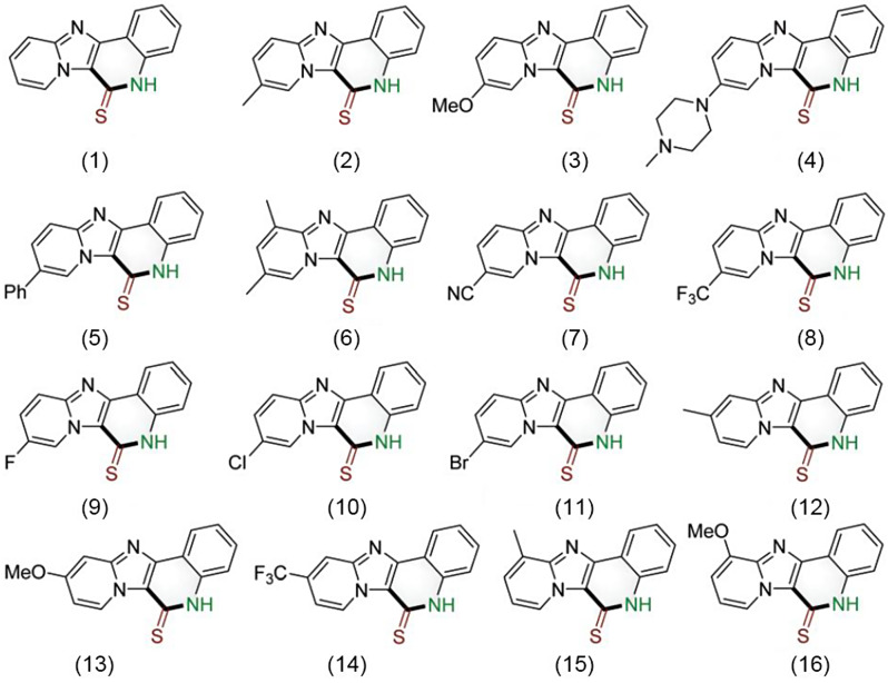
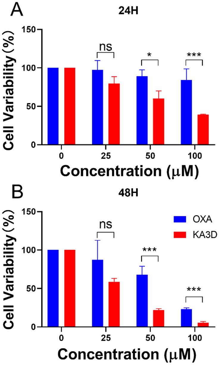
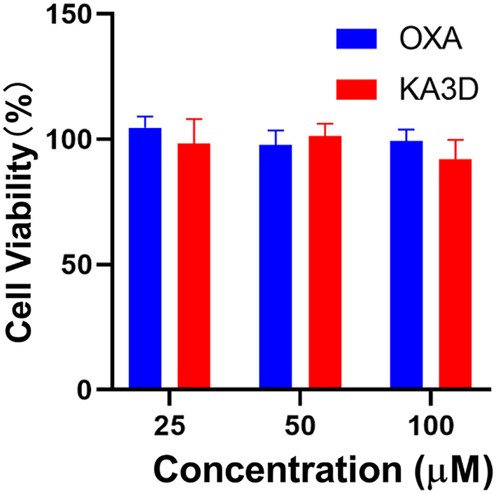
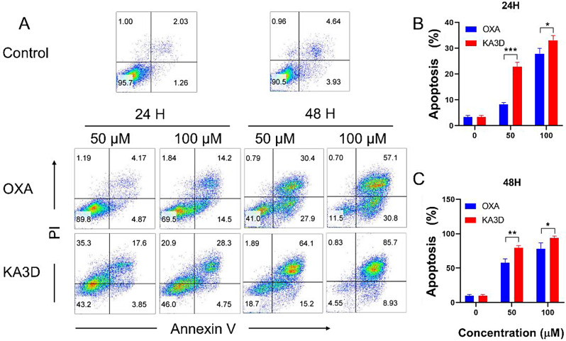
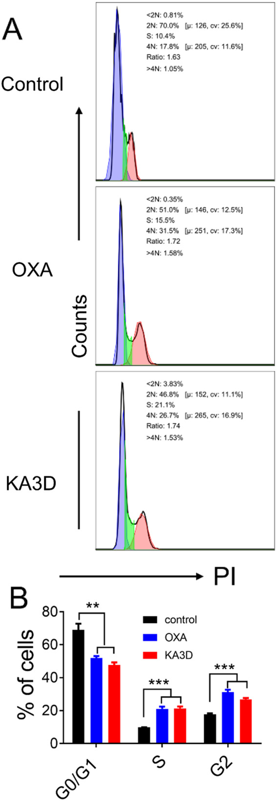
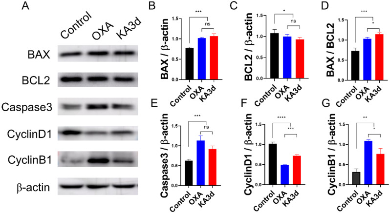
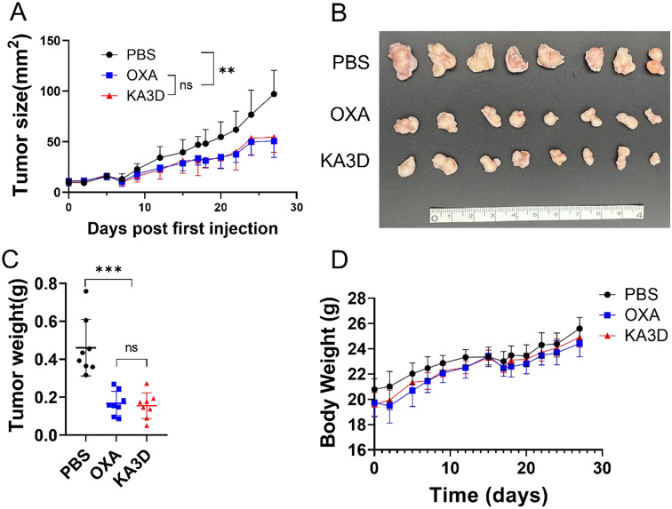
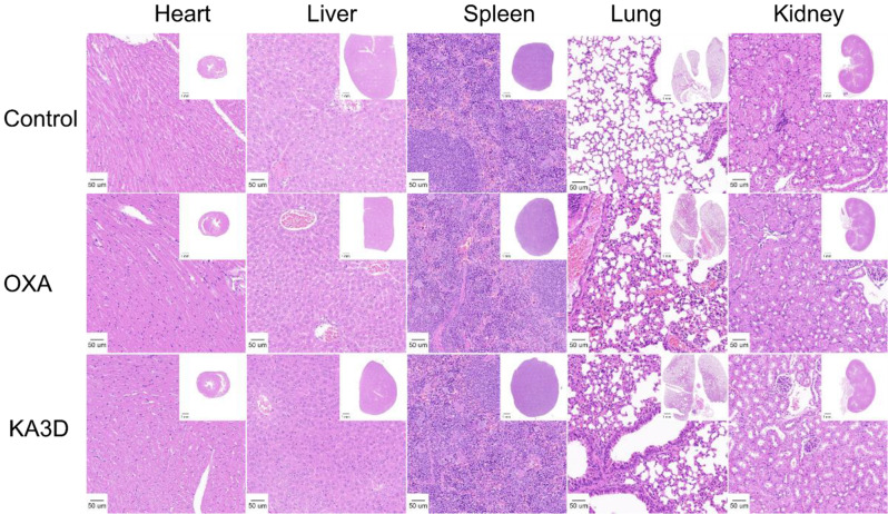
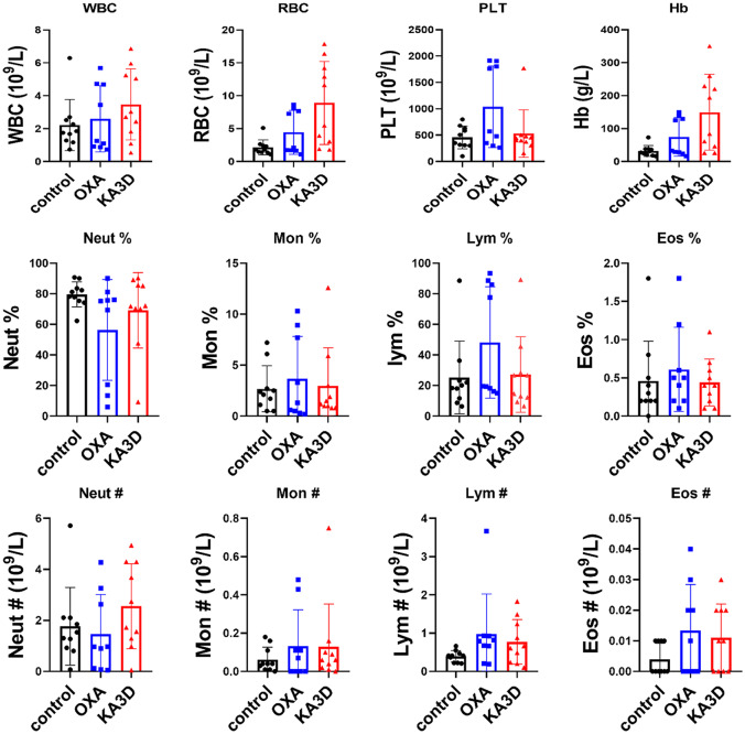
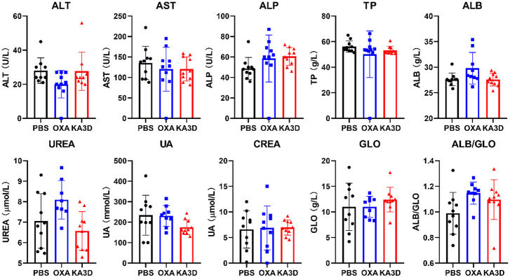
Ovarian cancer is the fifth most prevalent cancer in women. Chemotherapy is a major treatment option for patients with advanced ovarian cancer (OC). Quinoline-2-thione and its derivatives are potential candidates for tumor therapy. In this study, we investigated the anticancer activity of the quinoline-2-thione derivative KA3D against ovarian cancer. The effect of KA3D on the viability of ovarian cancer cells was evaluated using MTT assay, and its effects on apoptosis and the cell cycle were detected using flow cytometry. Western blotting was performed to identify apoptosis-and cell cycle-related proteins altered by KA3D treatment. A xenograft model was used to verify the inhibitory effect of KA3D in vivo. H&E staining, biochemical indicator detection, and blood cell counts were used to observe the toxicity and side effects of KA3D. KA3D treatment impeded cell viability, induced apoptosis, and impeded the G2 phase of the cell cycle in ovarian cancer cells. Mechanistically, we found that KA3D enhanced the expression of proapoptotic molecules such as BAX and Caspase 3, while antiapoptotic proteins such as BCL2 were inhibited. The G0/G1 phase-related protein cyclin D1 was reduced and the G2 phase-related protein cyclin B1 was upregulated. In vivo, KA3D displayed potent anticancer activity, with no apparent toxicity in BABLC/c nude mice bearing SKOV3 cells. KA3D demonstrated remarkable chemotherapeutic drug efficacy in terms of significant cancer suppression in vitro and in vivo with low toxicity.

Keywords: Anticancer activity; Apoptosis; Cell viability; KA3D; SKOV3.


Conflict of interest statement

The authors declare no competing interests.

Figures

Fig. 1
Fig. 1
Synthesis of the 16 small-molecule drugs used in this study
Fig. 2
Fig. 2
Among the 16 synthesized small-molecule drugs, only KA3D (4) inhibited SKOV3 cell proliferation
Fig. 3
Fig. 3
Cell viability was inhibited by KA3D and OXA. A–B Cell viability measured by MTT after KA3D or OXA treatment for 24 h (A) or 48 h (B) at various concentrations (25, 50 and 100 µM). The error bars represent the mean ± SD (n = 3). * P < 0.05, ** P < 0.01, ***P < 0.005, ****P < 0.001
Fig. 4
Fig. 4
KA3D had no effect on KGN ovarian granulosa cells
Fig. 5
Fig. 5
KA3D induced SKOV3 cell apoptosis. A Effects of different concentrations (50 and 100 µM) of KA3D and OXA on the apoptosis of SKOV3 cells at 24 h and 48 h, in the control group OXA or KA3D was replaced by PBS. B Statistical analysis of the results of the apoptosis assay. The error bars represent the mean ± SD (n = 3). * P < 0.05, ** P < 0.01, ***P < 0.005, ****P < 0.001
Fig. 6
Fig. 6
Effects of KA3D on apoptosis and cell cycle related proteins. (A) Expression of apoptosis- and cell cycle-related proteins after KA3D or OXA treatment for 24 h. B Statistical analysis of the protein expression of BAX (B), BCL2 (C), BAX/BCL2 (D), caspase3 (E), cyclinD1 (F), cyclinB1 (G). The error bars represent the mean ± SD (n = 3). * P < 0.05, ** P < 0.01, ***P < 0.005, ****P < 0.001
Fig. 7
Fig. 7
KA3D inhibited the cell cycle of SKOV3 cells. A Cell cycle assays using PI staining. B Statistical analysis of the results of the cell cycle analysis. The error bars represent the mean ± SD (n = 3). * P < 0.05, ** P < 0.01, ***P < 0.005, ****P < 0.001
Fig. 8
Fig. 8
KA3D inhibited tumor progression in the SKOV3 xenograft model in vivo. A size of subcutaneous tumors from the control, OXA and KA3D groups. B Dissected tumors were obtained from the mice in the control, OXA and KA3D groups. C Weights of subcutaneous tumors from the control, OXA and KA3D groups. D Body weights of nude mice in the control, OXA and KA3D groups. * P < 0.05, ** P < 0.01, ***P < 0.005, ****P < 0.001
Fig. 9
Fig. 9
KA3D and OXA do not elicit side toxicity in tumor-bearing mice. Representative HE images of hearts, livers, spleens, lungs, and kidneys from the control, OXA and KA3D groups are shown
Fig. 10
Fig. 10
KA3D and OXA do not elicit side toxicity in tumor-bearing mice. Representative results of blood cells from the control, OXA and KA3D groups
Fig. 11
Fig. 11
KA3D and OXA do not elicit side toxicity in tumor-bearing mice. Representative results of liver and kidney function in the control, OXA and KA3D groups

References

    1. Finch L, Chi DS. An overview of the current debate between using minimally invasive surgery versus laparotomy for interval cytoreductive surgery in epithelial ovarian cancer. J GYNECOL ONCOL 2023;34. - PMC - PubMed
    1. Mathew AT, Jain B, Patel TA, Hammond A, Dee EC, Chino F. Trends in Location of death for individuals with ovarian Cancer in the United States. OBSTET GYNECOL; 2023. - PMC - PubMed
    1. Morton M, Yao M, Chalif J, Lampert EJ, Chau D, Rose PG, et al. Association of Clinical Trial Participation with Improved overall survival for recurrent, platinum-resistant ovarian Cancer. OBSTET GYNECOL. 2023;142:459–66. - PubMed
    1. Han GYQ, Alexander M, Gattozzi J, Day M, Kirsch E, Tafreshi N, et al. Ecological and evolutionary dynamics to design and improve ovarian cancer treatment. CLIN TRANSL MED. 2024;14:e70012. - PMC - PubMed
    1. Giménez-Bonafé P, Tortosa A, Pérez-Tomás R. Overcoming drug resistance by enhancing apoptosis of tumor cells. CURR CANCER DRUG TAR. 2009;9:320–40. - PubMed

MeSH terms