. 2024 Oct 14;15(1):8848.
doi: 10.1038/s41467-024-53108-5.

Robotic microinjection enables large-scale transgenic studies of Caenorhabditis elegans

Affiliations

Robotic microinjection enables large-scale transgenic studies of Caenorhabditis elegans

Peng Pan et al. Nat Commun. .

Abstract

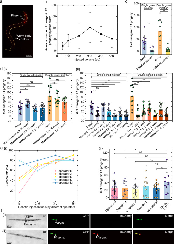
The nematode Caenorhabditis elegans is widely employed as a model organism to study basic biological mechanisms. However, transgenic C. elegans are generated by manual injection, which remains low-throughput and labor-intensive, limiting the scope of approaches benefitting from large-scale transgenesis. Here, we report a robotic microinjection system, integrating a microfluidic device capable of reliable worm immobilization, transfer, and rotation, for high-speed injection of C. elegans. The robotic system provides an injection speed 2-3 times faster than that of experts with 7-22 years of experience while maintaining comparable injection quality and only limited trials needed by users to become proficient. We further employ our system in a large-scale reverse genetic screen using multiplexed alternative splicing reporters, and find that the TDP-1 RNA-binding protein regulates alternative splicing of zoo-1 mRNA, which encodes variants of the zonula occludens tight junction proteins. With its high speed, high accuracy, and high efficiency in worm injection, this robotic system shows great potential for high-throughput transgenic studies of C. elegans.


Conflict of interest statement

The authors declare no competing interests.

Figures

Fig. 1
Fig. 1. Overview of robotic system for high-speed microinjection of C. elegans.
a Three-dimensional model of the robotic system for microinjection of C. elegans. b An enlarged view showing the detailed components of the robotic system. c Schematic of the microfluidic device. d Microscopic image of leftmost portion of microfluidic device mounted on the right micromanipulator under a 4× objective. Image is representative of at least 10 microfluidic devices. e Schematics of the microinjection procedure: (i) pick-up, (ii) transfer, rotation, and immobilization, (iii) injection, and (iv) release of the target worm. f Microscopic images showing the main operation steps during microinjection, including: (i)-(ii) rotation, (iii) immobilization, (iv) objectives switching from 4× to 10×, (v)-(vi) worm body penetration, (vii) plasmid DNA solution deposition, and (viii) pipette withdrawal. Images are representative of at least 20 microinjections. g Schematics of worm model and transverse sections of anterior/posterior part of worm at different orientations showing the relative position between intestine (purple area), distal gonad (blue area with yellow circles), and proximal gonad (blue area without yellow circles): (i) transverse sections of anterior worm body at different orientations, (ii) transverse sections of posterior worm body at different orientations. It should be noted that distal/proximal gonads are transparent, and intestine is opaque. h Mechanical model of worm rotation under the action of hydrodynamic forces and frictional force. i Continuous worm rotation at different orientations. Images are representative of at least 20 worm rotations.
Fig. 2
Fig. 2. Generation of extrachromosomal array-based transgenic lines.
a Generation of transgenic worms by the robotic injection. Images are representative of at least 150 transgenic worms from microinjections. b Calibration curve of the average transgenic F1 progeny vs. volume of injected plasmid DNA solution (number of injected worms: N = 10 for the injected volume of 50 pL, 100 pL, 300 pL, 400 pL; N = 11 for the injected volume of 200 pL, 500 pL; mean ± s.d.). c Number of transgenic F1 progeny obtained from robotic injection with/without the rotation procedure (number of independent injections: N = 10, 12, 11, 12 for single gonad injections w/wo rotation and double gonad injections w/wo rotation, respectively; mean ± s.d.). Two-sided t-test: single gonad arm injection pw/wo rotation = 0.00189; double gonad arm injection pw/wo rotation = 0.00019. **p < 0.005. d Number of transgenic F1 progeny obtained from robotic injection and manual injections: (i) comparison between robotic injection and manual injections of three experts with ~7–22 years’ experience (experts A, B, and C) (number of independent injections: N = 10, 11, 11, 12, 11, 11, 12 for single gonad injection by robotic system, experts A, B, C, and double gonad injections by robotic system, experts A, C, respectively; mean ± s.d.), (ii) comparison between robotic injection and manual injections of six proficient operators (operators D, E, F, G, H, and I) (number of independent injections: N = 10, 12, 12, 12, 12, 12, 14, 11, 12, 12 12, 12, 14, 14 for single gonad injection and double gonad injections by robotic system and six proficient operators, respectively; mean ± s.d.). Two-sided t-test: *p < 0.05, **p < 0.005, and ns (not significant): p > 0.05. e Robotic single gonad injections by five operators (operators E, J, K, L, M): (i) success rate of robotic injections when operators performed robotic injections for four trials, (ii) comparison between the control group and robotic injection results obtained from the fourth trial of five operators (number of independent robotic injections: N = 12, 11, 10, 10, 10, 10 for five operators and control group, respectively; mean ± s.d.). Two-sided t-test: ns (not significant): p > 0.05. f Robotic injection result of mutant strain (VC4040) which has small body diameter: (i) images of young adult worm from mutant strain-VC4040 showing it has a small diameter (~38 μm). Images are representative of VC4040 from which 100% of animals express GFP signals in the pharynx, (ii) images of transgenic progeny obtained from the successful robotic injection of plasmid pCFJ90 into the distal gonad of young adult worms from mutant strain-VC4040. Images are representative of at least 20 transgenic progeny obtained from the successful robotic injection of plasmid pCFJ90 into VC4040. Source data are provided as a Source Data file.
Fig. 3
Fig. 3. Generation of recombinant animals by CRISPR/Cas9 mediated genome editing.
a Dual-marker repair template for CRISPR/Cas9 plasmid DNA-mediated genome editing. Purple boxes, green box, yellow box, and red triangles represent appropriate homology arms for HDR, a pharyngeal GFP marker transgene, a neoR transgene, and loxP sites, respectively. b Visual strategy for identifying the recombinants obtained from the injection of CRISPR/Cas9 plasmid DNA solution into wild-type worm: (i) captured image of the recombinant under the brightfield mode, (ii) uniform GFP expression in the pharynx of recombinants, (iii) loss of mCherry expression in the recombinant, (iv) only uniform GFP expression in the recombinant demonstrating integration of the repair template and loss of the extrachromosomal array. Images are representative of recombinant worm generated via CRISPR/Cas9 plasmid DNA-mediated genome editing from three independent batch of injections. c Genotyping results at the klp-12 gene locus after CRISPR/Cas9 genome editing experiments using robotic microinjection platform. Images are representative of genotyping results of 6 lines from three independent batches of injections. d Visual strategy for identifying the recombinants obtained from the RNP complex injection: (i) mutant strain-JAC644 with GFP signal expressed in pharynx was chosen for the RNP complex injection, Image is representative of JAC644 from which 100% of animals express GFP signals in the pharynx, which was selected for the injection. (ii) recombinant worm which survived the G418 selection and possessed a uniform mCherry signal in the pharynx was successfully obtained by injecting the RNP solution into mutant (JAC644). Image is representative of recombinant worm generated via RNP-mediated CRISPR genome editing from one batch of injections. Source data are provided as a Source Data file.
Fig. 4
Fig. 4. Large-scale reverse genetic screen for alternative splicing regulation in neurons and intestine cells.
a Schematic of the two-color reporter system. Upon inclusion of the alternative exon (yellow box), the reading frame encoding mCherry is utilized, while the reading frame encoding GFP is not, indicated by the color change of the reading frame encoding GFP from deep green to light green. The exon skipping leads to a frameshift (+1) and translation of the GFP protein. Translated fluorescent proteins are uncoupled from upstream sequence through 2A peptides, and localized to the nucleus through two nuclear localization signals. Reporter-specific RT-PCR priming sites are also indicated in diagram. b Cocktail of four different splicing minigene reporters injected into worms to generate stable extrachromosomal array lines for the RNA collection and RT-PCR assays. c Stable extrachromosomal array lines generated from the injected mutants (RB1766): (i) bright field and fluorescent images of original mutant (RB1766). Images are representative of RB1766 from which 100% of animals show no fluorescent signals, which was selected for the injection. (ii) both GFP and mCherry signals observed in the neurons and intestine cells showing the transgenic progeny from injected mutants (RB1766) are successfully generated. Images are representative of at least 10 transgenic progeny obtained from the successful robotic injection of a cocktail of four plasmids into RB1766. d RT-PCR assays of the zoo-1 exon 9 reporter for selected six mutant strains. Images are representative of 5 RT-PCR assays of the zoo-1 exon 9 reporter for selected six mutant strains. Source data are provided as a Source Data file.

References

    1. Hirschhorn, J. N. & Daly, M. J. Genome-wide association studies for common diseases and complex traits. Nat. Rev. Genet.6, 95–108 (2005). - PubMed
    1. Botstein, D. & Risch, N. Discovering genotypes underlying human phenotypes: past successes for Mendelian disease, future approaches for complex disease. Nat. Genet.33, 228–237 (2003). - PubMed
    1. Shaye, D. D. & Greenwald, I. OrthoList: a compendium of C. elegans genes with human orthologs. PLoS One6, e20085 (2011). - PMC - PubMed
    1. Brenner, S. The genetics of Caenorhabditis elegans. Genetics77, 71–94 (1974). - PMC - PubMed
    1. White, J. G., Southgate, E., Thomson, J. N. & Brenner, S. The structure of the nervous system of the nematode Caenorhabditis elegans. Philos. Trans. R. Soc. L. B Biol. Sci.314, 1–340 (1986). - PubMed

Publication types

MeSH terms

LinkOut - more resources