. 2024 Oct 14;7(1):1318.
doi: 10.1038/s42003-024-07030-7.

ARL13B controls male reproductive tract physiology through primary and Motile Cilia

Affiliations

ARL13B controls male reproductive tract physiology through primary and Motile Cilia

Céline Augière et al. Commun Biol. .

Abstract

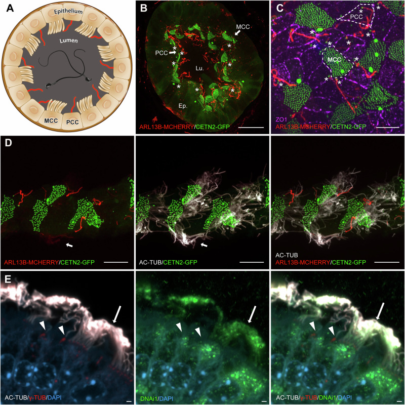
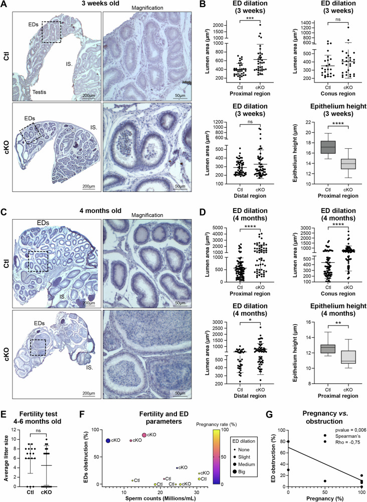
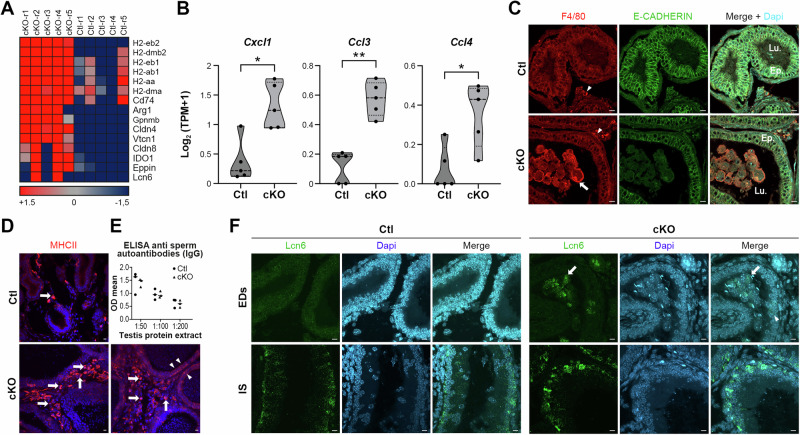
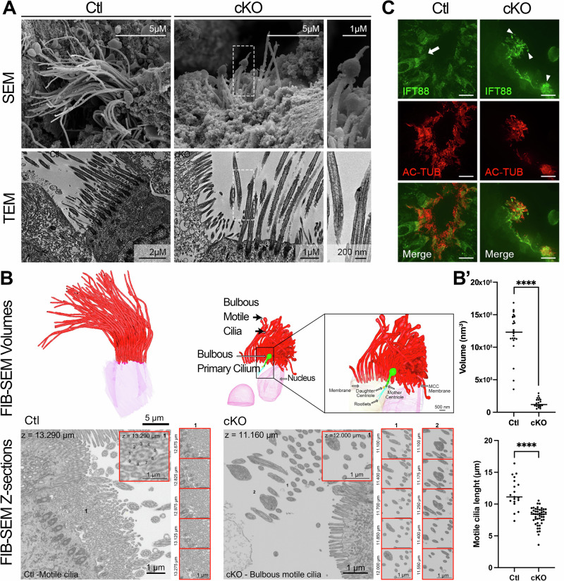
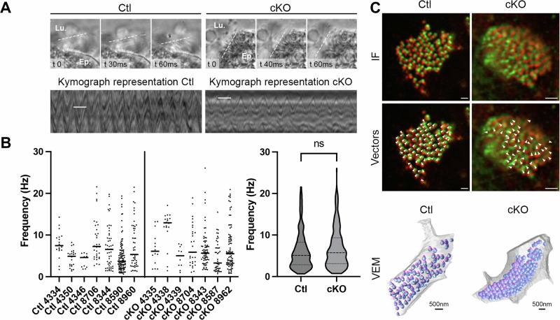
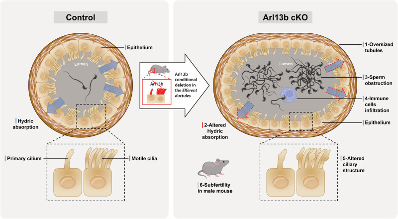
ARL13B is a small regulatory GTPase that controls ciliary membrane composition in both motile cilia and non-motile primary cilia. In this study, we investigated the role of ARL13B in the efferent ductules, tubules of the male reproductive tract essential to male fertility in which primary and motile cilia co-exist. We used a genetically engineered mouse model to delete Arl13b in efferent ductule epithelial cells, resulting in compromised primary and motile cilia architecture and functions. This deletion led to disturbances in reabsorptive/secretory processes and triggered an inflammatory response. The observed male reproductive phenotype showed significant variability linked to partial infertility, highlighting the importance of ARL13B in maintaining a proper physiological balance in these small ducts. These results emphasize the dual role of both motile and primary cilia functions in regulating efferent duct homeostasis, offering deeper insights into how cilia related diseases affect the male reproductive system.


Conflict of interest statement

The authors declare no competing interests.

Figures

Fig. 1
Fig. 1. Both multiple motile cilia and solitary primary cilia are present at the surface of the efferent ductules.
A Cross sectional representation of one mouse efferent ductule (ED) displaying the main cells populating the ED epithelium: the primary ciliated cells (PCC) and multiciliated cells (MCC). BD Confocal imaging microscopy of EDs from adult Arl13b-mCherry; Cetn2-GFP double transgenic mice. B Long Arl13b-positive cilia extend from PCC towards the lumen of the EDs (stars). Multiciliated cells are characterized by the apical alignment of numerous CETN2-positive basal bodies. Scale bar=15 µm. C Individual PCC and MCC delineation through ZO1 tight junction marker labeling (dotted lines) indicates that solitary ARL13B-positive cilia originate from CETN2-positive basal bodies of PCC (stars). Scale bar = 10 µm. D Exogenous labeling for alpha-acetylated tubulin cilia marker (AC-TUB). In situ discrimination of solitary primary cilia from multiple motile cilia is facilitated in Arl13b-mCherry; Cetn2-GFP double transgenic mice in which ARL13B-MCHERRY positive motile cilia are faintly detected (arrow). E Immunostaining for the alpha-acetylated tubulin cilia marker (AC-TUB, white), centrioles (γ-TUB, red), and DNAi1 (green) shows that DNAi1 labels the dynein intermediate chain in motile cilia (indicated by arrows) but is absent in primary cilia (indicated by arrowheads) in ED from wild-type mice. Scale bar 1 µm. Ep. Epithelium, Lu. Lumen, PCC Primary ciliated cells, MCC Multiciliated cells.
Fig. 2
Fig. 2. ARL13B is lost in the efferent ductules of Arl13b conditional KO mice.
A Schematic representation of the Arl13b conditional knock-out by the Cre-loxP system. Cell lineage specificity was provided by Villin-Cre mediated excision of the loxP-flanked Arl13b exon 2 to generate VillinCre; Arl13bFlox/Flox mice (cKO) and VillinCre; Arl13bFlox/+ control littermates (Ctl). B ARL13B Western blot detection in testis, EDs and epididymis protein extracts from Ctl and cKO mice. Box & whiskers plot showed protein quantification performed on ED extracts from n = 5 mice per genotype and normalized to GAPDH (right panel, center line: median, box limits: min to max); differences between Ctl and cKO were analyzed using unpaired t test. **p < 0.01. C Immunofluorescent detection of ARL13B (gray), alpha-acetylated tubulin (AC-TUB, red) and gamma tubulin (γ-TUB, green) in primary (arrowhead) and motile cilia (arrow) of EDs from Ctl and cKO adult mice. Note the absence of detection of ARL13B in both cilia in cKO mice. Scale bars = 10 µm. D Immunofluorescent detection of ARL13B (gray), alpha-acetylated tubulin (AC-TUB, red) and gamma tubulin (γ-TUB, green) in the EDs from 3, 8 weeks and 4 months old Ctl and cKO mice. While AC-TUB-positive cilia are observed in both Ctl and cKO mice (arrowheads), ARL13B-positive cilia are exclusively detected in EDs from Ctl mice (arrows). Scale bars = 10 µm; Ep. Epithelium; Lu. Lumen.
Fig. 3
Fig. 3. Loss of ARL13B results in efferent ductules dilation and sperm accumulation that correlates with reduced pregnancy rate.
A, C Hematoxylin staining of EDs from Ctl and cKO at 3 weeks (A) and 4 months old (C). 20× magnifications are shown on the right. EDs: Efferent ductules. IS: Initial segment. B, D Luminal area and epithelial cell thickness were measured in distinct regions of EDs from Ctl and cKO mice at 3 weeks (B) and 4 months old (D). Unpaired-t test was used to analyze luminal area (the data are presented as mean ± standard deviation (SD)) and Mann-Whitney test was used to analyze epithelial cell thickness (data presented in box plot, center line: median, box limits: min to max), *p < 0.05, **p < 0.01, ***p < 0.001, ****p < 0.0001. E Average litter size obtained after mating Ctl or cKO male mice with wild-type females is presented in mean with SD (ratio 2 females per 1 male). Difference between Ctl and cKO were analyzed with unpaired t-test. F Correlation matrix between the percentage of ED obstruction (in percent, 0% no obstruction observed; 100% all tubules obstructed with sperm clog), sperm counts (in millions/ml obtained from CASA analysis), tubule dilation (arbitrary units) and pregnancy rate (100%: 2 out of 2 females got pregnant, 50%: 1 out of 2 females got pregnant, 0%: none of the 2 females got pregnant) found in Ctl (n = 5) and cKO mice (n = 6). G Spearman’s Rho was employed to assess the strength and direction of the relationship between the levels of ED obstruction (number of tubules obstructed in percent, 0%: no tubules obstructed, 100%: all tubules obstructed) and the pregnancy success rate of wild type females mated with cKO males (100%: 2 out of 2 females got pregnant, 50%: 1 out of 2 females got pregnant, 0%: none of the 2 females got pregnant).
Fig. 4
Fig. 4. Loss of ARL13B is associated with unbalanced immunological environment in the efferent ductules.
A Heat map representation of immune and inflammatory related coding genes whose expression is altered in cKO mice. Color range indicates Log2FC ≥ 1.5 (upregulated genes); Log2FC ≤-1.5 (downregulated genes). B Cxcl1, Ccl3 and Ccl4 chemokines RNA expression level based on Log2(TPM + 1) showed in truncated violin plot (center line: median, small doted lines: first and third quartile) and analyzed with Mann-Whitney test. *p < 0.05, **p < 0.01. C Immunofluorescent detection of mononuclear phagocytes (F4/80, red) in EDs of Ctl and cKO mice. Mononuclear phagocytes are present around the tubules (arrowheads) and infiltrate the lumen of cKO mice (arrow). Scale bars = 10 µm. D Immunofluorescent detection of antigen presenting MHCII (red) in EDs of Ctl and cKO mice. While MHCII positive cells are present in the stroma of both Ctl and cKO (arrows), some epithelial cells also express MHCII marker in the EDs from cKO mice (arrowheads). Scale bars = 10 µm. E Detection of IgG in the serum of Ctl and cKO mice (serum dilution 1:50, 1:100 and 1:200) against testicular proteins extracts measured with optical density (OD) mean. F In situ fluorescent detection of Lcn6 transcripts in EDs and initial segment (IS) from Ctl and cKO mice using RNA scope technology. While Lcn6 is only expressed in IS from Ctl mice, Lcn6 is found expressed in cells populating the lumen (arrows) and in epithelial cells of the EDs in cKO mice (arrowhead). Scale bars = 10 µm.
Fig. 5
Fig. 5. Motile cilia are structurally affected by the loss of ARL13B.
A Scanning Electron Microscopy (SEM) and Transmission Electron Microscopy (TEM) acquisitions of motile cilia from the EDs of Ctl and cKO mice. Motile cilia appear shorter in cKO and some of them exhibit a bulging morphology (shown in magnified images on the right). B 3D segmentation of volume electron microscopy (VEM) data acquired by FIB-SEM of EDs in both Ctl and cKO mice. Motile cilia (red) in cKO mice exhibit bulbous extremities, similar to those observed in the primary cilium (green). In the motile cilia of Ctl mice (inset), note the presence of 9 peripheral microtubule doublets and a central pair which are also present in the normal motile cilia of cKO mice (inset 1), but absent in the bulbous extremities (inset 2). B’ Motile cilia in cKO mice are significantly shorter than those in Ctl mice, as quantified by volumetric measurements from FIB-SEM data (center line: median; top panel) and fluorescent image analyses (center line: median; bottom panel). C Immunofluorescent detection of IFT88 in EDs from Ctl and cKO mice. While IFT88 is present all along alpha-acetylated tubulin (AC-TUB) motile cilia in Ctl EDs (arrow), it is found accumulated in the bulge of some motile cilia from cKO models (arrowheads). Scale bars = 10 µm.
Fig. 6
Fig. 6. Cilia motility is not significantly impaired following the loss of ARL13B.
A Beating frequency of motile cilia was measured by high speed videomicroscopy (100 fps) on live efferent ductules (Supplementary videos 1 and 2). Beating frequency is illustrated by kymographs (bottom panel) obtained with Fiji software, with white lines showing a single wavelength. B The beating frequencies recorded within each group of mice (n = 7 per genotype) were heterogeneous and did not significantly differ between groups, as shown in the violin plot (center line: median; small dotted lines: first and third quartile) using the Mann-Whitney statistical test. C Airyscan images for immunofluorescent detection of basal feet (CENTRIOLIN, green) and basal bodies (POC1B, red) orientation in Ctl and cKO EDs. Vectors (Arrowheads) indicate beating directionality based on each cilium basal body/basal foot orientation (bottom panels). 3D reconstruction from VEM data of EDs of both Ctl and cKO showing basal bodies (blue) and basal feet (pink). Top panels scale bar=1 µm.
Fig. 7
Fig. 7. The loss of ARL13B disrupts the regulation of factors involved in solute transport and fluid reabsorption.
A Volcano plot representation of ED RNA seq data from Ctl (n = 5) and cKO (n = 5) mice. Although certain less-studied genes encoding water channels, such as AQP2 and AQP5, were downregulated, the primary characterized channels in EDs, such as AQP1 and NHE3, did not show significant alterations following the loss of Arl13b. B Heat map representation of solute carriers and aquaporins encoding genes whose expression are downregulated (Log2FC ≤ -1.5, blue) or upregulated (Log2FC > 1.5, red) in cKO compared to Ctl mice. C AQP5 (gray) staining in EDs from Arl13b-mcherry; Cetn2-GFP transgenic mice showing the water channel at the apical pole of primary ciliated cells. Scale bars = 10 µm. D Immunofluorescent detection of AQP5 (green) in EDs from Ctl and cKO mice. Scale bars=10 µm. D’ Plot depicting the average fluorescence intensity of AQP5 at the apical pole of ED epithelial cells (center line: median), normalized through background subtraction. Difference between Ctl and cKO were analyzed using Mann-Whitney statistical test. ****p < 0.0001. E Dot plot quantification of AQP5 in two-fold serial dilutions of ED protein extracts from Ctl and cKO mice. To control for specificity, the antibody was pre-incubated with a 10-fold excess of the AQP5 immune peptide. E’ AQP5 dot blot quantification was performed on samples from n = 5 mice per genotype. Data were analyzed using Two-way ANOVA with Tukey’s multiple comparisons test on serial dilutions (see Supplementary Fig. 5). Only quantification for the 3 µg dilution is shown (Center line: median). ***p < 0.001.
Fig. 8
Fig. 8. ARL13B controls efferent ductules physiology.
The absence of ARL13B led to disturbances in reabsorptive/secretory processes characterized by enlarged tubules and dysregulation of water channels and ionic exchangers and triggered an inflammatory response. In the absence of ARL13B, both motile and primary ciliogenesis are impaired, resulting in bulbous cilia and increased in cilia beating. Furthermore, subfertility in cKO male mice correlates with the severity of affection in the efferent ductules.

Similar articles

Cited by

References

    1. Hess, R. A. The efferent ductules:Stucture and functions. The Epidid (2002).
    1. Hess, R. A., Sharpe, R. M. & Hinton, B. T. Estrogens and development of the rete testis, efferent ductules, epididymis and vas deferens. Differentiation118, 41–71 (2021). - PMC - PubMed
    1. Newcombe, N., Clulow, J., Man, S. Y. & Jones, R. C. pH and bicarbonate in the ductuli efferentes testis of the rat. Int J. Androl.23, 46–50 (2000). - PubMed
    1. Sullivan, R., Légaré, C., Lamontagne-Proulx, J., Breton, S. & Soulet, D. Revisiting structure/functions of the human epididymis. Andrology7, 748–757 (2019). - PubMed
    1. Hess, R. A. & Cooke, P. S. Estrogen in the male: a historical perspective. Biol. Reprod. 99, 27–44 (2018). - PMC - PubMed

Substances

LinkOut - more resources