. 2024 Oct 13;14(1):23940.
doi: 10.1038/s41598-024-75275-7.

Importance of OCT-derived biomarkers for the recurrence of central serous chorioretinopathy using statistics and predictive modelling

Affiliations

Importance of OCT-derived biomarkers for the recurrence of central serous chorioretinopathy using statistics and predictive modelling

Emilien Seiler et al. Sci Rep. .

Abstract

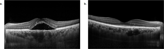
Central serous chorioretinopathy (CSCR) is a retinal disease characterised by the accumulation of subretinal fluid, which often resolves spontaneously in acute cases. However, approximately one-third of patients experience recurrences that may cause severe and irreversible vision. This study aimed to identify parameters derived from optical coherence tomography (OCT) that are associated with CSCR recurrence. Our dataset included 5211 OCT scans from 344 eyes of 255 patients diagnosed with CSCR. 178 eyes were identified as recurrent, 109 as non-recurrent, and 57 were excluded. We extracted parameters using artificial intelligence algorithms based on U-Nets, convolutional kernels, and morphological operators. We applied inferential statistics to evaluate differences between the recurrent and non-recurrent groups, and we used a logistic regression predictive model, reporting the coefficients as a measure of biomarker importance. We identified nine predictive biomarkers for CSCR recurrence: age, intraretinal fluid, subretinal fluid, pigment epithelial detachments, choroidal vascularity index, integrity of photoreceptors and retinal pigment epithelium layer, choriocapillaris and choroidal stroma thickness, and thinning of the outer nuclear layer, and of the inner nuclear layer combined with the outer plexiform layer. These results could enable future developments in the automatic detection of CSCR recurrence, paving the way for translational medical applications.

Keywords: Biomarker; Central serous chorioretinopathy; Choroid; Optical coherence tomography; Predictive modelling; Retina.


Conflict of interest statement

The authors declare no competing interests.

Figures

Fig. 1
Fig. 1
Manifestations of Central serous chorioretinopathy (CSCR) signs in OCT-Line scans with enhanced depth imaging (OCT-EDI). CSCR is a posterior segment disease characterised by subretinal fluid (SRF) accumulation and pachychoroid. SRF spontaneously resolves after the first episode. a SD-OCT B scan of a 45-year-old patient shows pathological accumulation of SRF in the foveal area and pachychoroid. b Follow-up SD-OCT B scan shows complete spontaneous resolution two months after the first manifestation.
Fig. 2
Fig. 2
Correlation between OCT-derived parameters. Based on the findings of the literature, an expert ophthalmologist suggested a list of parameters that might play a role in detecting recurrent CSCR events. We then extracted these parameters from the imaging dataset and correlated them, obtaining several clusters. A first cluster was composed of parameters related to the choroid: thickness of Choriocapillaris and Choroidal Stroma, Choroidal Vascularity Index and an estimation of the area of the largest Choroidal Pachyvessel. A second cluster comprised thicknesses of inner retina layers: the Retinal Nerve Fibre Layer, the Ganglion Cell Layer and Inner Plexiform Layer, and the Inner Nuclear Layer and Outer Plexiform Layer. The third cluster contained outer retinal layer thicknesses, namely the Outer Nuclear Layer and Photo-Receptors and Retinal Pigment Epithelium, as well as parameters related to their integrity (Pigment Epithelium Detachment and Disruption Score of the Photo-Receptors and Retinal Pigment Epithelium Layers) and measures of Subretinal Fluid and Intraretinal Fluid. SRF: Subretinal Fluid; IRF: Intraretinal Fluid; PED: Pigment Epithelium Detachment; RNFL: Retinal Nerve Fibre Layer; GCL + IPL: Ganglion Cell Layer and Inner Plexiform Layer; INL + OPL: Inner Nuclear Layer and Outer Plexiform Layer; ONL: Outer Nuclear Layer; PR + RPE: Photo-Receptors and Retinal Pigment Epithelium; CC + CS: Choriocapillaris and Choroidal Stroma; CVI: the Choroidal Vascularity Index; DSCORE: a score estimating disruption or the PR + RPE layer; PV_AREA: and an estimation of the area of the largest Choroidal Pachyvessel.
Fig. 3
Fig. 3
Importance of predictive biomarkers for CSCR, across analysis methods (ordered according to the Regression Analysis). The length of each bar in the graph represents the biomarker’s importance. Darker blue indicates biomarkers that were statistically significant (as reported in the column “P-value (Regression analysis)” of Table 1). a Cohen’s d effect sizes were obtained from the statistical analysis (T-test) of biomarkers extracted during the first visit. b Cohen’s d effect sizes were obtained from the statistical analysis (KS test) of longitudinal data (i.e., averaging all visits of the initial acute CSCR episode, disregarding any subsequent recurrent episodes). c Coefficients of the logistic regression model obtained by fitting one biomarker at a time. SRF: Subretinal Fluid; IRF: Intraretinal Fluid; PED: Pigment Epithelium Detachment; RNFL: Retinal Nerve Fibre Layer; GCL + IPL: Ganglion Cell Layer and Inner Plexiform Layer; INL + OPL: Inner Nuclear Layer and Outer Plexiform Layer; ONL: Outer Nuclear Layer; PR + RPE: Photo-Receptors and Retinal Pigment Epithelium; CC + CS: Choriocapillaris and Choroidal Stroma; CVI: the Choroidal Vascularity Index; DSCORE: a score estimating disruption or the PR + RPE layer; PV_AREA: and an estimation of the area of the largest Choroidal Pachyvessel.
Fig. 4
Fig. 4
Longitudinal dataset comprising OCT-derived parameters in CSCR patients (N = 337). X-Axis = time. Y Axis = SRF accumulation. Blue Line: a non-recurrent case characterised by a single acute episode (i.e., accumulation of subretinal fluid). Orange Line: a recurrent case characterised by a second CSCR episode following a first acute episode.
Fig. 5
Fig. 5
Data extraction and processing pipeline. We extracted the list of patients who visited the medical retina unit at Jules-Gonin Eye Hospital between March 2017 and September 2022. We crossed this list with information extracted from the electronic medical records (EMR) indicating a diagnosis of Central Serous Chorioretinopathy (CSCR). We extracted the corresponding optical coherence tomography (OCT) scans for the totality of their visits, generating a longitudinal data set. We only considered those patients who had signed the General Consent to reuse their data. We extracted parameters from the OCT scans using artificial intelligence (AI)-based methods. An expert ophthalmologist labelled patients as recurrent or non-recurrent. Finally, we performed statistical tests and evaluated the efficacy of a logistic regression model to study each parameter and its link to recurrence.

References

    1. Liew, G., Quin, G., Gillies, M. & Fraser-Bell, S. Central serous chorioretinopathy: a review of epidemiology and pathophysiology. Clin. Exp. Ophthalmol. 41, 201–214 (2013). - PubMed
    1. Wang, M., Munch, I. C., Hasler, P. W., Prünte, C. & Larsen, M. Central serous chorioretinopathy. Acta Ophthalmol. 86, 126–145 (2008). - PubMed
    1. Kitzmann, A. S., Pulido, J. S., Diehl, N. N., Hodge, D. O. & Burke, J. P. The incidence of central serous chorioretinopathy in Olmsted County, Minnesota, 1980–2002. Ophthalmology. 115, 169–173 (2008). - PubMed
    1. Sartini, F., Figus, M., Nardi, M., Casini, G. & Posarelli, C. Non-resolving, recurrent and chronic central serous chorioretinopathy: available treatment options. Eye. 33, 1035–1043 (2019). - PMC - PubMed
    1. Piccolino, F. C., Borgia, L., Zinicola, E. & Zingirian, M. Indocyanine green angiographic findings in central serous chorioretinopathy. Eye. 9 (Pt 3), 324–332 (1995). - PubMed

MeSH terms

LinkOut - more resources