. 2025 Feb 4;24(2):200-213.
doi: 10.1158/1535-7163.MCT-24-0225.

Targeting NAD+ Metabolism Vulnerability in FH-Deficient Hereditary Leiomyomatosis and Renal Cell Carcinoma with the Novel NAMPT Inhibitor OT-82

Affiliations

Targeting NAD+ Metabolism Vulnerability in FH-Deficient Hereditary Leiomyomatosis and Renal Cell Carcinoma with the Novel NAMPT Inhibitor OT-82

Susana S Najera et al. Mol Cancer Ther. .

Abstract

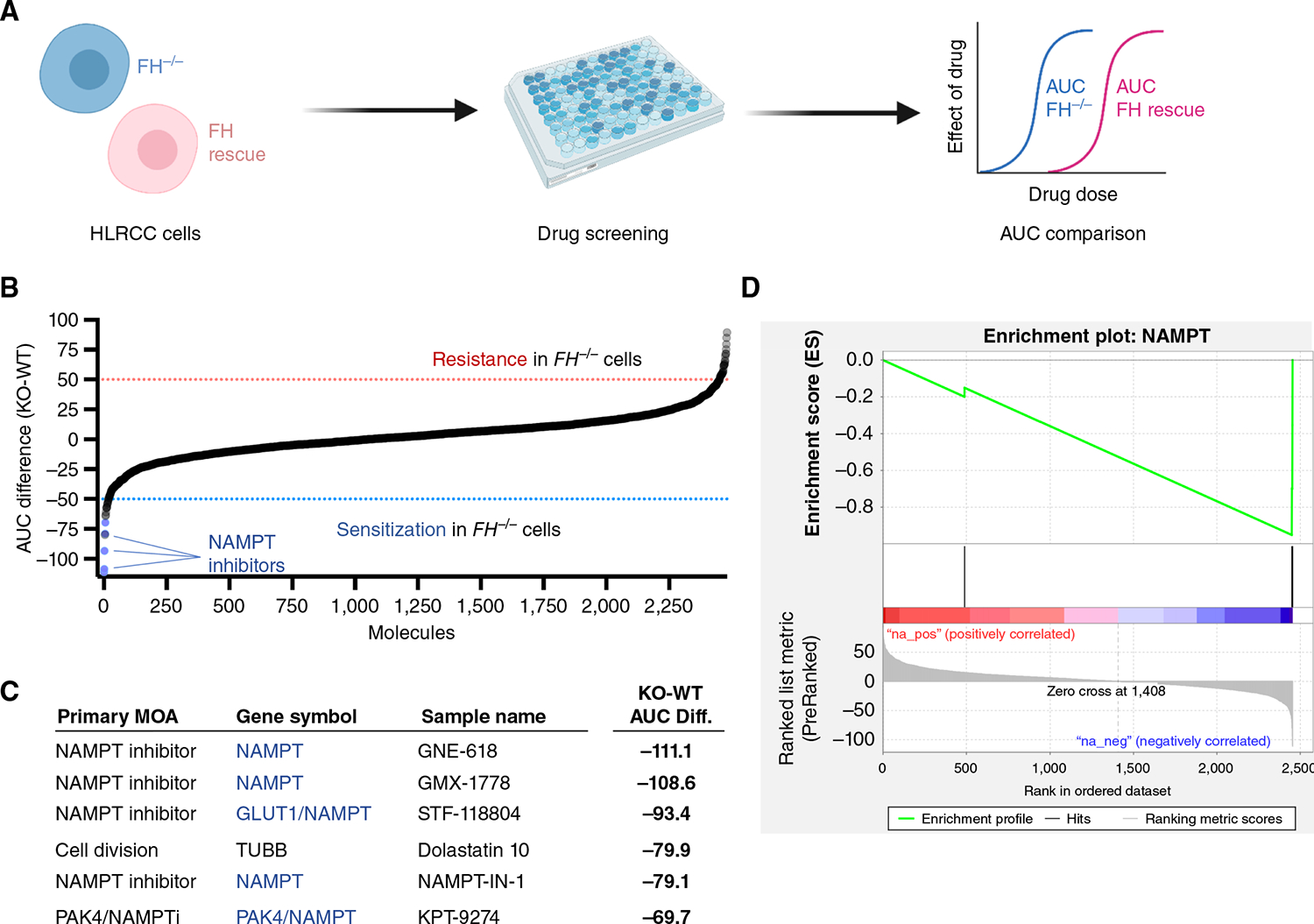
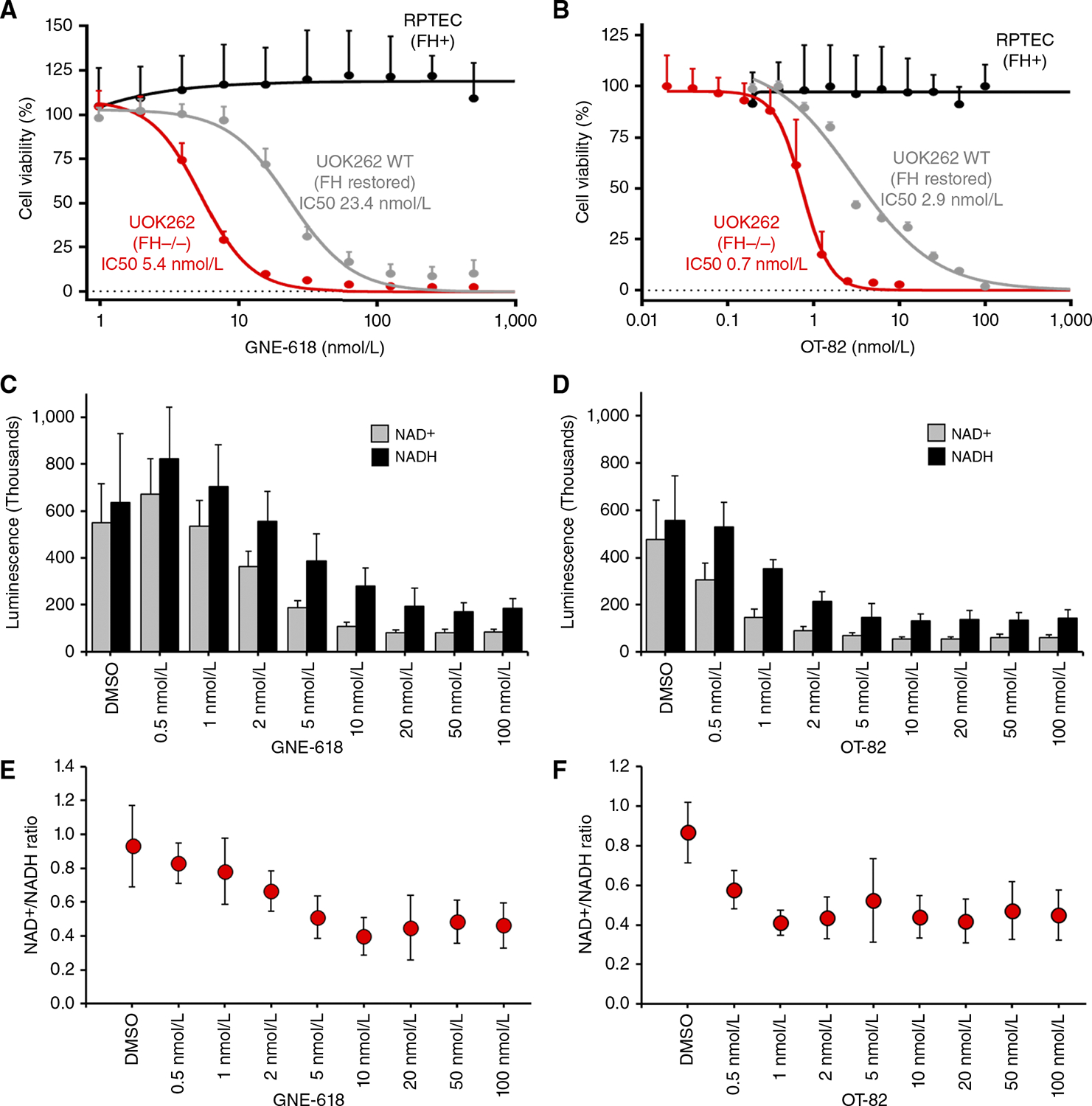
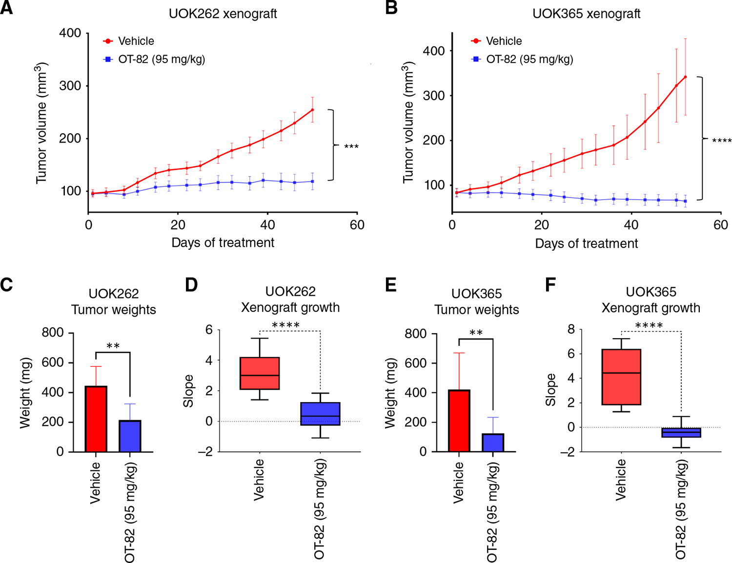
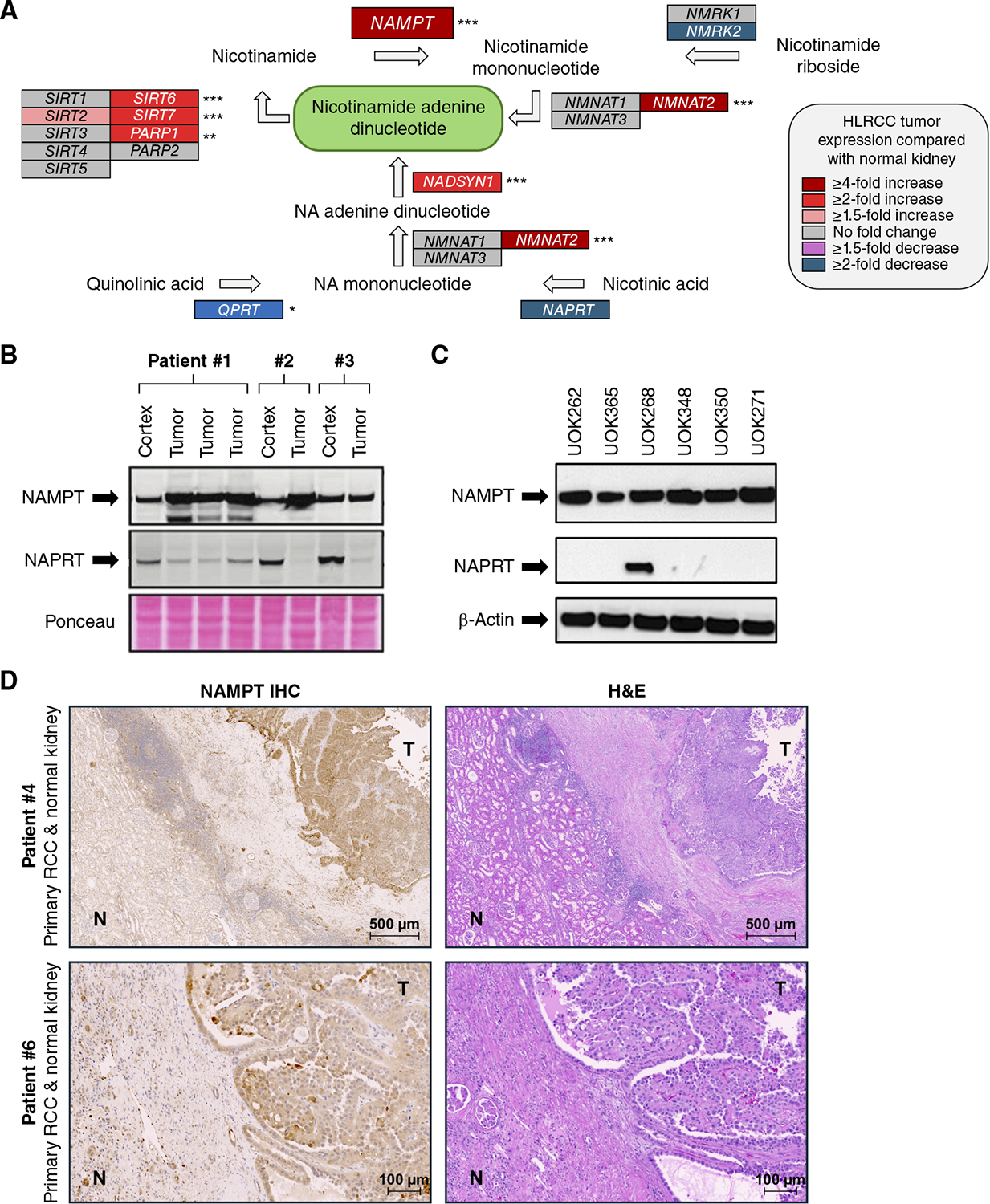
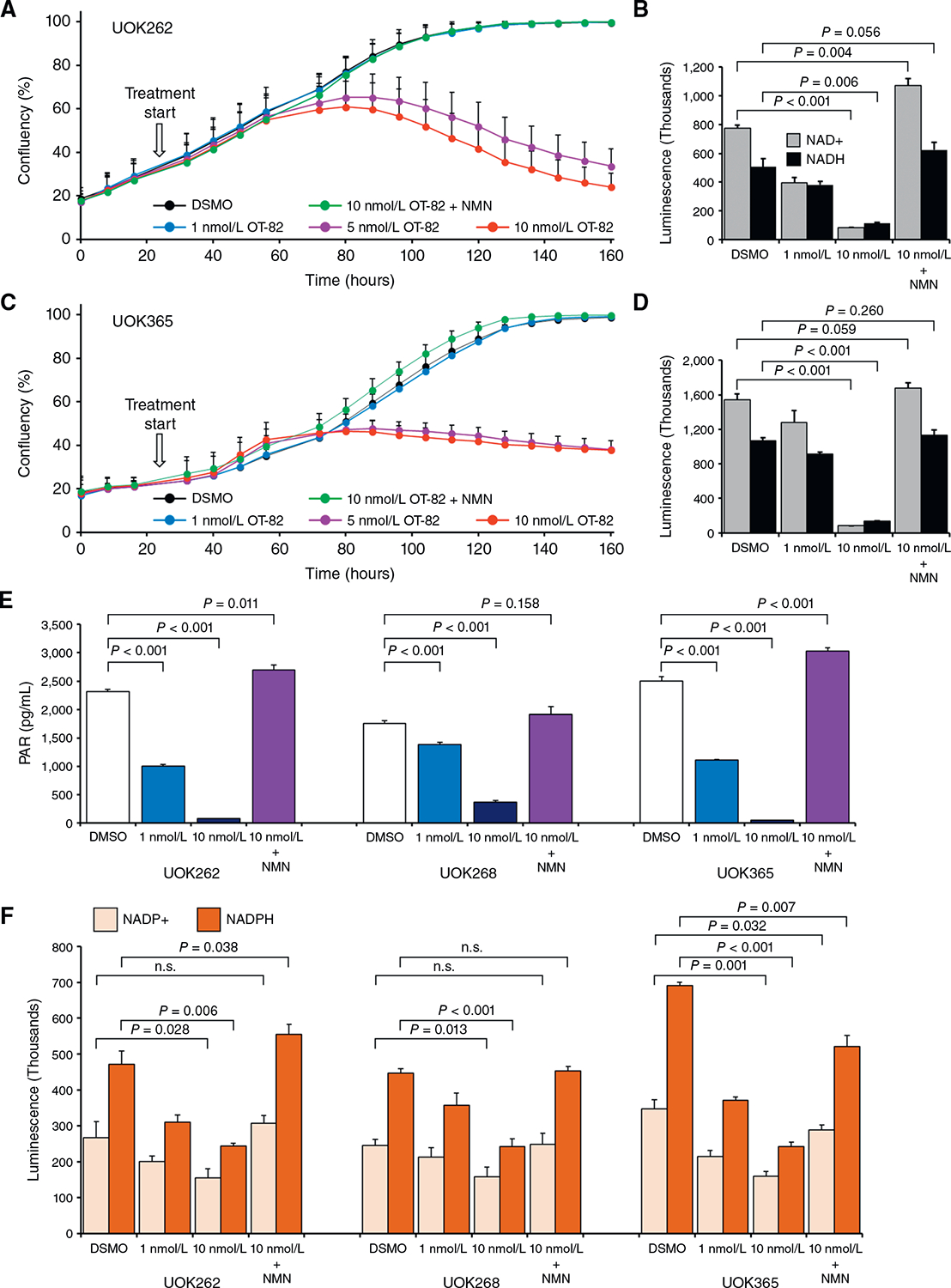
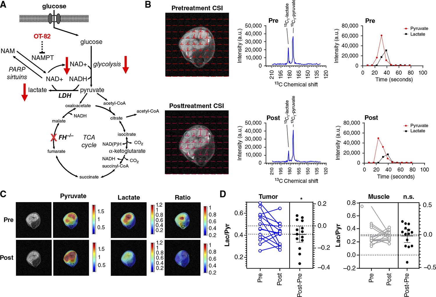
Hereditary leiomyomatosis and renal cell cancer (HLRCC) is an inherited cancer syndrome caused by germline pathogenic variants in the fumarate hydratase (FH) gene. Affected individuals are at risk for developing cutaneous and uterine leiomyomas and aggressive FH-deficient renal cell carcinoma (RCC) with a papillary histology. Due to a disrupted tricarboxylic acid cycle, FH-deficient kidney cancers rely on aerobic glycolysis for energy production, potentially creating compensatory metabolic vulnerabilities. This study conducted a high-throughput drug screen in HLRCC cell lines, which identified a critical dependency on nicotinamide adenine dinucleotide (NAD), a redox cofactor produced by the biosynthetic enzyme nicotinamide phosphoribosyltransferase (NAMPT). Human HLRCC tumors and HLRCC-derived cell lines exhibited elevated NAMPT expression compared with controls. FH-deficient HLRCC cells, but not FH-restored HLRCC or normal kidney cells, were sensitive to NAMPT inhibition. HLRCC cell line viability was significantly decreased in both 2D and 3D in vitro cultures in response to the clinically relevant NAMPT inhibitor OT-82. NAMPT inhibition in vitro significantly decreased the total amount of NAD+, NADH, NADP, NADPH, and poly-ADP-ribose levels, and the effects of NAMPT inhibition could be rescued by the downstream NAD precursor nicotinamide mononucleotide (NMN), confirming the on-target activity of OT-82. Moreover, NAMPT inhibition by OT-82 in two HLRCC xenograft models resulted in severely reduced tumor growth. OT-82 treatment of HLRCC xenograft tumors in vivo inhibited glycolytic flux as demonstrated by reduced lactate/pyruvate ratio in hyperpolarized 13C-pyruvate magnetic resonance spectroscopic imaging experiments. Overall, our data define NAMPT inhibition as a potential therapeutic approach for FH-deficient HLRCC-associated RCC.


Figures

Figure 1.
Figure 1.
A mechanism-of-action-focused drug screen highlights the susceptibility of a UOK262 HLRCC model to NAMPT inhibitors. A, Schematic of overall screening strategy. UOK262 cells (FH/) and a rescue cell line in which WT FH has been reintroduced (FH+, also referred to as UOK262WT) were screened in quantitative dose response against a manually curated panel of >2,000 mechanistically annotated drugs and small molecules (MIPE 5.0 library). AUC measurements of cell viability were used to distinguish responses that were specific to one of the two cell lines. B, Plot of differential AUC for each library member as a function of cellular FH status. Small molecules targeting NAMPT constituted five of the six most potent hits. C, AUC values for NAMPT inhibitors showing selective activity in the FH/ UOK262 cell line. D, Drug set enrichment analysis indicating enrichment of NAMPT inhibitors in the set of molecules showing greater activity in UOK262 FH/ cell line.
Figure 2.
Figure 2.
Validation studies reveal potent inhibition of UOK262 HLRCC cells by NAMPT inhibitors. A, Effects of the NAMPT inhibitor GNE-618 on cell viability in FH/ UOK262 (red), FH-restored UOK262 WT (gray), and nontransformed kidney epithelial cells RPTEC (black). Cell viability was assessed by CellTiter-Glo assay at 96 hours. B, Effects of the NAMPT inhibitor OT-82 on cell viability in FH/ UOK262 (red), FH-restored UOK262 WT (gray), and nontransformed kidney epithelial cells RPTEC (black). C–F, Analysis of the effects in UOK262 cells on the relative levels of NAD+ and NADH and their ratio after 24-hour treatment with a range of concentrations (0.5–100 nmol/L) of either GNE-618 or OT-82. All levels were compared with cells treated with DMSO alone.
Figure 3.
Figure 3.
In vivo effects of NAMPT inhibitor OT-82 in HLRCC xenograft models. Tumor growth curves of xenografts in which UOK262 (A) and UOK365 (B) cells were injected s.q. in the flanks of female NSG mice and treated with OT-82 (95 mg/kg) or vehicle (p.o.; n = 10 mice per arm, 8-week duration; two repeats per cell line). Tumors were measured weekly using calipers, and tumor volume was calculated as V (mm3) = (D × d2)/6 × 3.14. C and D, Tumor weights at the study endpoint and tumor growth rate for UOK262. E and F, Tumor weights at the study endpoint and tumor growth rate for UOK365. **, P < 0.005; ***, P < 0.001; ****, P < 0.0001.
Figure 4.
Figure 4.
Altered expression of NAD biosynthetic pathway genes in patient-derived HLRCC tumors and cell lines. A, RNA-seq analysis summary of NAD+ biosynthetic and utilization pathways in HLRCC tumors compared with expression in renal cortex. *, P < 0.05; **, P < 0.01; ***, P < 0.001. B, Assessment of NAMPT and NAPRT protein levels in HLRCC tumor samples and adjacent normal kidney parenchyma. Ponceau-S staining of total protein is used as a loading control. C, Assessment of NAMPT and NAPRT protein levels in HLRCC kidney tumor cell lines by Western blot. Β-actin and total protein staining were used as a loading control. D, Representative images of NAMPT immunohistochemical staining at the boundary between tumor and normal kidney tissue in tumors excised from two patients with HLRCC. The accompanying H&E images for the section are shown. N = normal kidney tissue, T = HLRCC tumor.
Figure 5.
Figure 5.
Assessing the effects of NAMPT inhibitors in HLRCC cells and rescue with NMN. Measurement of effects of the NAMPT inhibitor OT-82 on UOK262 (A) and UOK365 (C) cell proliferation treated for 5 days at 1, 5, and 10 nmol/L. Measurement of levels of NAD+ and NADH in UOK262 (B) and UOK365 (D) cells treated for 24 hours with 1 nmol/L OT-82, 10 nmol/L OT-82, and 10 nmol/L OT-82 plus NMN (1 mmol/L). E, Measurement of PAR levels in UOK262, UOK268, and UOK365 cells treated with 1 nmol/L OT-82, 10 nmol/L OT-82, and 10 nmol/L OT-82 plus NMN (1 mmol/L). F, Measurement of NADP+ and NADPH levels in UOK262, UOK268, and UOK365 cells treated with 1 nmol/L OT-82, 10 nmol/L OT-82, and 10 nmol/L OT-82 plus NMN (1 mmol/L). n.s., not significant.
Figure 6.
Figure 6.
Impact of NAMPT inhibition on glycolysis in FH-deficient tumor xenografts. A, Summary of the potential effects of NAMPT inhibitor OT-82 on glycolysis in preclinical HLRCC tumor models. B, Representative chemical shift imaging of pyruvate and lactate in a UOK262 xenograft pre- and posttreatment with OT-82 overlaid on T2W MRI images at the same time point after injection of hyperpolarized [1-13C]-pyruvate. The chemical shift spectra obtained from the 2 mm × 2 mm × 8 mm representative voxel circled in blue is shown. Kinetic intensity curves of the total tumor for 13C-pyruvate (13C1-pyruvate) and 13C-lactate (13C1-lactate) detected in a UOK262 xenograft pre and post OT-82 treatment after hyperpolarized [1-13C]-pyruvate injection. C, Parametric (false-color) images of the total 13C-pyruvate signal (left), total 13C-lactate signal (middle), and the 13C-Lac/Pyr ratio (right), before (top) and after (bottom) OT-82 treatment for a representative UOK262 xenograft. The parametric images were generated from the chemical shift data in B. D, Summary of lactate/pyruvate ratios in UOK262 tumors vs. leg muscle pre- and posttreatment with OT-82. n.s., not significant.

Similar articles

Cited by

References

    1. Toro JR, Nickerson ML, Wei M-H, Warren MB, Glenn GM, Turner ML, et al. Mutations in the fumarate hydratase gene cause hereditary leiomyomatosis and renal cell cancer in families in North America. Am J Hum Genet 2003;73:95–106. - PMC - PubMed
    1. Tomlinson IP, Alam NA, Rowan AJ, Barclay E, Jaeger EE, Kelsell D, et al. Germline mutations in FH predispose to dominantly inherited uterine fibroids, skin leiomyomata and papillary renal cell cancer. Nat Genet 2002;30:406–10. - PubMed
    1. Linehan WM, Schmidt LS, Crooks DR, Wei D, Srinivasan R, Lang M, et al. The metabolic basis of kidney cancer. Cancer Discov 2019;9:1006–21. - PubMed
    1. Sourbier C, Ricketts CJ, Matsumoto S, Crooks DR, Liao P-J, Mannes PZ, et al. Targeting ABL1-mediated oxidative stress adaptation in fumarate hydratase-deficient cancer. Cancer Cell 2014;26:840–50. - PMC - PubMed
    1. Yang Y, Valera VA, Padilla-Nash HM, Sourbier C, Vocke CD, Vira MA, et al. UOK 262 cell line, fumarate hydratase deficient (FH−/FH−) hereditary leiomyomatosis renal cell carcinoma: in vitro and in vivo model of an aberrant energy metabolic pathway in human cancer. Cancer Genet Cytogenet 2010;196:45–55. - PMC - PubMed

MeSH terms

Supplementary concepts