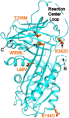
Improving the diffraction quality of heat-shock protein 47 crystals
- PMID: 39397789
- PMCID: PMC11533366
- DOI: 10.1107/S2053230X24009233
Improving the diffraction quality of heat-shock protein 47 crystals
Abstract
Heat-shock protein 47 (HSP47) is a potential target for inhibitors that ameliorate fibrosis by reducing collagen assembly. In an effort to develop a structure-based drug-design system, it was not possible to replicate a previous literature result (PDB entry 4au4) for apo dog HSP47; instead, crystal forms were obtained in which pairs of dog HSP47 molecules interacted through a noncleavable C-terminal His-tag to build up tetramers, all of which had multiple molecules of HSP47 in the asymmetric unit and none of which diffracted as well as the literature precedent. To overcome these difficulties, a two-pronged approach was followed: (i) the His-tag was moved from the C-terminus to the N-terminus and was made cleavable, and (ii) Adnectin (derived from the tenth domain of human fibronectin type III) crystallization chaperones were developed. Both approaches provided well diffracting crystals, but the latter approach yielded crystal forms with only one or two HSP47 complexes per asymmetric unit, which made model building less onerous.
Keywords: C- and N-terminal His-tags; HSP47; crystallization chaperones.
Figures


References
-
- Agirre, J., Atanasova, M., Bagdonas, H., Ballard, C. B., Baslé, A., Beilsten-Edmands, J., Borges, R. J., Brown, D. G., Burgos-Mármol, J. J., Berrisford, J. M., Bond, P. S., Caballero, I., Catapano, L., Chojnowski, G., Cook, A. G., Cowtan, K. D., Croll, T. I., Debreczeni, J. É., Devenish, N. E., Dodson, E. J., Drevon, T. R., Emsley, P., Evans, G., Evans, P. R., Fando, M., Foadi, J., Fuentes-Montero, L., Garman, E. F., Gerstel, M., Gildea, R. J., Hatti, K., Hekkelman, M. L., Heuser, P., Hoh, S. W., Hough, M. A., Jenkins, H. T., Jiménez, E., Joosten, R. P., Keegan, R. M., Keep, N., Krissinel, E. B., Kolenko, P., Kovalevskiy, O., Lamzin, V. S., Lawson, D. M., Lebedev, A. A., Leslie, A. G. W., Lohkamp, B., Long, F., Malý, M., McCoy, A. J., McNicholas, S. J., Medina, A., Millán, C., Murray, J. W., Murshudov, G. N., Nicholls, R. A., Noble, M. E. M., Oeffner, R., Pannu, N. S., Parkhurst, J. M., Pearce, N., Pereira, J., Perrakis, A., Powell, H. R., Read, R. J., Rigden, D. J., Rochira, W., Sammito, M., Sánchez Rodríguez, F., Sheldrick, G. M., Shelley, K. L., Simkovic, F., Simpkin, A. J., Skubak, P., Sobolev, E., Steiner, R. A., Stevenson, K., Tews, I., Thomas, J. M. H., Thorn, A., Valls, J. T., Uski, V., Usón, I., Vagin, A., Velankar, S., Vollmar, M., Walden, H., Waterman, D., Wilson, K. S., Winn, M. D., Winter, G., Wojdyr, M. & Yamashita, K. (2023). Acta Cryst. D79, 449–461.
-
- Berman, H. M., Henrick, K. & Nakamura, H. (2003). Nat. Struct. Mol. Biol.10, 980. - PubMed
-
- Bricogne, G., Blanc, E., Brandl, M., Flensburg, C., Keller, P., Paciorek, W., Roversi, P., Smart, O., Vonrhein, C. & Womack, T. (2019). BUSTER–TNT 2.11.7. Global Phasing Ltd, Cambridge, United Kingdom.
MeSH terms
Substances
LinkOut - more resources
Full Text Sources
Miscellaneous

