. 2024 Sep 26;10(19):e38539.
doi: 10.1016/j.heliyon.2024.e38539. eCollection 2024 Oct 15.

Assessing the efficacy of Stemona collinsiae roots extract against third-stage larvae of Gnathostoma spinigerum and its safety profiles

Affiliations

Assessing the efficacy of Stemona collinsiae roots extract against third-stage larvae of Gnathostoma spinigerum and its safety profiles

Norinee Arlee et al. Heliyon. .

Abstract

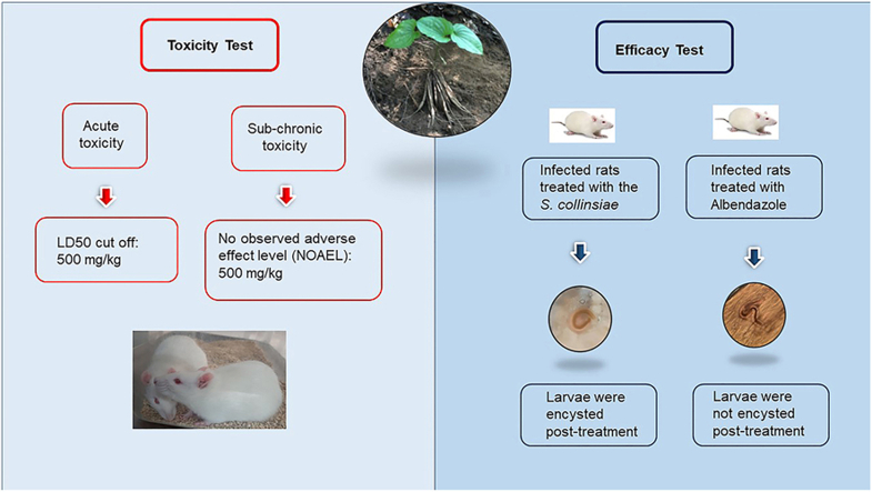
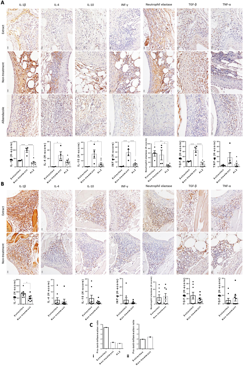
Gnathostomiasis, caused by the advanced third-stage larvae of Gnathostoma spinigerum, demands novel treatment avenues. The ethanolic root extract of Stemona collinsiae has been postulated to have anthelminthic properties, suggesting its potential as an alternative remedy. In this study, S. collinsiae roots were collected, identified, and extracted with 95 % ethanol. The crude extracts were standardized using didehydrostemofoline as chemical marker. The efficacy of the S. collinsiae root extract against third-stage larvae of G. spinigerum and its toxicity to Wistar rats were evaluated. Both in vitro and in vivo tests were performed, where the in vitro tests assessed the anthelminthic potential of S. collinsiae extract against G. spinigerum larvae, while in vivo tests examined the extract's efficacy against G. spinigerum larvae in infected Wistar rats and the efficacy was compared with albendazole. Parallelly, Wistar rats underwent acute and sub-chronic toxicity tests to establish the safe dosage of the extract. The in vitro tests showcased significant anthelminthic activity, marked by discernible morphological alterations in the exposed larvae. Acute toxicity proved fatal at 2000 mg/kg body weight, while a dose of 300 mg/kg proved non-toxic. Using the Globally Harmonized Classification System, an LD50 of 500 mg/kg was determined. In vivo trials revealed a pronounced decline in G. spinigerum larvae among rats treated with the S. collinsiae extract. The larvae were also observed to be encysted post-treatment, while those treated with albendazole were not encysted. The S. collinsiae extract, with its noteworthy in vitro efficacy and favorable safety metrics in rodents, can be a potential anthelminthic agent. The diminished inflammatory response compared to albendazole hints at S. collinsiae being a safer gnathostomiasis treatment alternative. The promising results in these preliminary trials warrant a deeper investigation to determine the root extract's optimal dosing, suitable delivery methods, and its broader clinical implications.

Keywords: Acute toxicity; Alkaloids; Anthelminthic drug; Didehydrostemofoline; Gnathostomiasis; Stemona collinsiae; Stemonaceae; Sub-chronic toxicity; Wistar rats.


Conflict of interest statement

The authors declare that they have no known competing financial interests or personal relationships that could have appeared to influence the work reported in this paper.

Figures

Image 1
Graphical abstract
Fig. 1
Fig. 1
LC-DAD-MS chromatograms of S. collinsiae extract detected at UV 290 nm (A) full scan in positive mode (B) and full scan in negative mode (C). Peak identification: (1) didehydrostemofoline. Peak indicated with + showing the presence of stilbenoids.
Fig. 2
Fig. 2
Morphological changes observed in L3 G. spinigerum. Light microscopy photograph of the third stage larvae of G. spinigerum: (A) worm treated with S. collinsiae crude extract; (B) worm treated with albendazole; (C) Untreated worm. Scanning electron micrograph of the third stage larvae of G. spinigerum: (E) cephalic bulb of the third stage larvae of G. spinigerum treated with S. collinsiae crude extract; (F) cephalic bulb of the third stage larvae of G. spinigerum treated with albendazole; (G) cephalic bulb of untreated third stage larvae of G. spinigerum; (H) spines in the middle region of the body of the third stage larvae of G. spinigerum treated with S. collinsiae crude extract; (I) spines in the middle region of the body of the third stage larvae of G. spinigerum treated with albendazole; (J) spines in the middle region of the body of untreated worm; (K) posterior part of the third stage larvae of G. spinigerum treated with S. collinsiae crude extract; (L) posterior part of the third stage larvae of G. spinigerum treated with albendazole; (M) posterior part of untreated worm.
Fig. 3
Fig. 3
Histopathological appearances and scores in L3 gnathostomiasis rats with any treatment: (A) 3 days post treatment; fibrotic granuloma in rats-treated with extraction (i) and non-treated rats (ii) exhibited the tissue interfacing area between host and parasite with thickening fibrosis formation (v) and the accumulation of several types of white blood cells majority with macrophage, neutrophil, and lymphocyte (vi-vii) and minority with eosinophil (vi) and giant cell (vii). Non-fibrotic granuloma in rats-treated with albendazole (iii) demonstrated only white cells infiltration on tissue interfacing area (ix). Bar graph of pathological score at day 3 post treatment (iv). (B) 10 days post treatment; fibrotic granuloma in rats-treated with extraction (i) and non-treated rats (ii) characterized the tissue interfacing area deposition with white cells majority with eosinophil, macrophage and giant cell. Bar graph of pathological score at day 10 post treatment (iii). ∗ p-value <0.05.
Fig. 4
Fig. 4
Pro- and anti-inflammatory cytokine levels in L3 gnathostomiasis rats with any treatment: Immunological expression of IL-1β, IL-4, IL-10, INF-γ, neutrophil elastase, TGF-β, and TNF-α in L3 migratory tissue on 3 (A) and 10 (B) days post treatment with or without extraction and albendazole. Bar graph represented the cytokine expression score with ∗ p-value <0.05, ∗∗ p-value <0.01, and ∗∗∗ p-value <0.001. (C) Pro- and anti-inflammatory cytokine ratio in any treatment on both 3- and 10-days post treatment.

Similar articles

References

    1. Liu G.H., Sun M.M., Elsheikha H.M., Fu Y.T., Sugiyama H., Ando K., et al. Human gnathostomiasis: a neglected food-borne zoonosis. Parasit Vectors. 2020;13:616. doi: 10.1186/s13071-020-04494-4. - DOI - PMC - PubMed
    1. Diaz J. Gnathostomiasis: an emerging infection of raw fish consumers in Gnathostoma-endemic and non-endemic countries. J Travel Med. 2015;16:352–364. doi: 10.1111/jtm.12212. - DOI - PubMed
    1. Herman J.S., Chiodini P.L. Gnathostomiasis, another emerging imported disease. Clin. Microbiol. Rev. 2009;22:484–492. doi: 10.1128/CMR.00003-09. - DOI - PMC - PubMed
    1. Chai J., Jung B., Lee S. Gnathostomiasis: an emerging infectious disease relevant to all countries. Acta Trop. 2013;126:79–88. doi: 10.1590/abd1806-4841.20187498. - DOI
    1. Ramirez-Avila L., Slome S., Schuste F.L., Gavali S., Schantz P.M., Sejvar J., et al. Eosinophilic meningitis due to Angiostrongylus and Gnathostoma species. Clin. Infect. Dis. 2009;48:322–327. doi: 10.1086/595852. - DOI - PubMed

LinkOut - more resources