. 2024 Sep 27;10(19):e38647.
doi: 10.1016/j.heliyon.2024.e38647. eCollection 2024 Oct 15.

Cabozantinib prevents the progression of metabolic dysfunction-associated steatohepatitis by inhibiting the activation of hepatic stellate cell and macrophage and attenuating angiogenic activity

Affiliations

Cabozantinib prevents the progression of metabolic dysfunction-associated steatohepatitis by inhibiting the activation of hepatic stellate cell and macrophage and attenuating angiogenic activity

Takuya Matsuda et al. Heliyon. .

Abstract

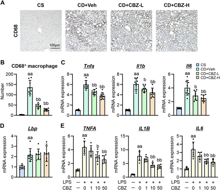
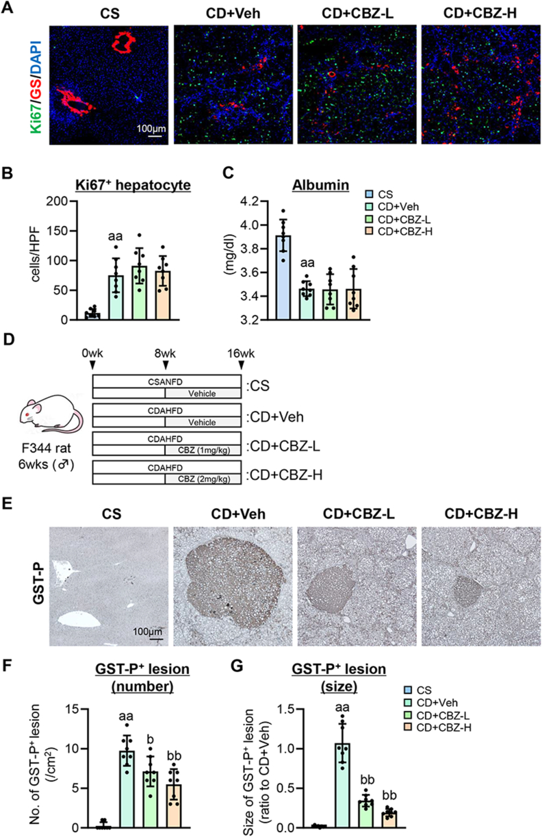
Cabozantinib, a multiple tyrosine kinase inhibitor targeting AXL, vascular endothelial growth factor receptor (VEGFR), and MET, is used clinically to treat certain cancers, including hepatocellular carcinoma. This study aimed to assess the impact of cabozantinib on liver fibrosis and hepatocarcinogenesis in a rat model of metabolic dysfunction-associated steatohepatitis (MASH). MASH-based liver fibrosis and hepatocarcinogenesis were induced in rats by feeding them a choline-deficient, L-amino acid-defined, high-fat diet (CDAHFD) for eight and 16 weeks, respectively. Cabozantinib (1 or 2 mg/kg, daily) was administered concurrently with the diet in the fibrosis model and after eight weeks in the carcinogenesis model. Treatment with cabozantinib significantly attenuated hepatic inflammation and fibrosis without affecting hepatocyte steatosis and ballooning in CDAHFD-fed rats. Cabozantinib-treated rats exhibited a marked reduction in α-smooth muscle actin+ activated hepatic stellate cell (HSC) expansion, CD68+ macrophage infiltration, and CD34+ pathological angiogenesis, along with reduced hepatic AXL, VEGF, and VEGFR2 expression. Consistently, cabozantinib downregulated the hepatic expression of profibrogenic markers (Acta2, Col1a1, Tgfb1), inflammatory cytokines (Tnfa, Il1b, Il6), and proangiogenic markers (Vegfa, Vwf, Ang2). In a cell-based assay of human activated HSCs, cabozantinib inhibited Akt activation induced by GAS6, a ligand of AXL, leading to reduced cell proliferation and profibrogenic activity. Cabozantinib also suppressed lipopolysaccharide-induced proinflammatory responses in human macrophages, VEGFA-induced collagen expression and proliferation in activated HSCs, and VEGFA-stimulated proliferation in vascular endothelial cells. Meanwhile, administration of cabozantinib did not affect Ki67+ hepatocyte proliferation or serum albumin levels, indicating no negative impact on regenerative capacity. Treatment with cabozantinib also reduced the placental glutathione transferase+ preneoplastic lesions in CDAHFD-fed rats. In conclusion, cabozantinib shows promise as a novel option for preventing MASH progression.

Keywords: Angiogenesis; Hepatocarcinogenesis; Inflammation; Liver fibrosis; MASH.


Conflict of interest statement

The authors declare that they have no known competing financial interests or personal relationships that could have appeared to influence the work reported in this paper.

Figures

Fig. 1
Fig. 1
Effect of cabozantinib on hepatic inflammation in CDAHFD-fed rats. (A) Experimental design of CDAHFD-induced liver fibrosis. (B) Changes in the body weights during the experimental period. (C) Serum levels of aspartate aminotransferase (AST) and alanine aminotransferase (ALT). (D) Liver/body weight at the end of experiment. (E) Representative microphotographs of hematoxylin and eosin (H&E) of the livers in the experimental rats. (F–H) Pathological scores for (F) steatosis, (G) ballooning and (H) inflammation at a 400-fold magnification. Data are the mean ± SD (n = 8; B−D, F−H). a, aa: P < 0.05, 0.01 vs CS group, b, bb: P < 0.05, 0.01 vs CD + Veh group, significant difference between groups by Student's t-test. CS, CSANFD-fed and vehicle-treated group; CD + Veh, CDAHFD-fed and vehicle-treated group; CD + CBZ-L, CDAHFD-fed and cabozantinib (1 mg/kg)-treated group; CD + CBZ-H, CDAHFD-fed and cabozantinib (2 mg/kg)-treated group.
Fig. 2
Fig. 2
Effect of cabozantinib on hepatic fibrosis in CDAHFD-fed rats. (A) Representative microphotographs of Sirius-Red and α-smooth muscle actin (SMA) staining of the livers in the experimental rats. (B and C) Quantification of (B) Sirius-Red stained fibrotic area and (C) α-SMA-positive area in high-power field. (D) Hepatic concentration of hydroxyproline. (E) Hepatic mRNA level of profibrotic markers (Acta2, Col1a1 and Tgfb1). Gapdh was used as an internal control for qRT-PCR. Quantitative values are indicated as fold changes to the values of CD + Veh group (B and C) or CS group (E). Data are the mean ± SD (n = 8; B−E). a, aa: P < 0.05, 0.01 vs CS group, b, bb: P < 0.05, 0.01 vs CD + Veh group, significant difference between groups by Student's t-test. CS, CSANFD-fed and vehicle-treated group; CD + Veh, CDAHFD-fed and vehicle-treated group; CD + CBZ-L, CDAHFD-fed and cabozantinib (1 mg/kg)-treated group; CD + CBZ-H, CDAHFD-fed and cabozantinib (2 mg/kg)-treated group. (For interpretation of the references to colour in this figure legend, the reader is referred to the Web version of this article.)
Fig. 3
Fig. 3
Effect of cabozantinib on GAS6/AXL signaling in LX-2 cells. (A) Serum Gas6 level in the experimental rats. (B) Hepatic mRNA expression of Axl in the experimental rats. (C) Western blotting for the effect of cabozantinib (CBZ) (0−50 nM) on the phosphorylation of AXL and AKT in rGAS6 (500 ng/mL)-stimulated LX-2 cells. Whole images of blotting were shown in Supplementary Fig. 2 in Supplementary Material. (D) Effect of CBZ (0−50 nM) on rGAS6 (500 ng/mL)-induced LX-2 proliferation. (E) Inducible effect of rGAS6 (500 ng/mL) on the mRNA expression of profibrogenic markers (ACTA2, COL1A1, TIMP1 and CTGF) in LX-2 cells. (F) Effect of CBZ (0−50 nM) on the mRNA expression of profibrogenic markers in rGAS6 (500 ng/mL)-stimulated LX-2 cells. Gapdh/GAPDH was used as an internal control for qRT-PCR (B, E and F). Actin was used as an internal control for western blotting (C). Quantitative values are indicated as fold changes to the values of CS group (B), the value of each group at 0 h (D), the value at 0 h (E) and the value of rGAS6 (−)/CBZ (−) group (F). Data are the mean ± SD (n = 8; A and B, n = 6; D−F). a, aa: P < 0.05, 0.01 vs CS group (A and B), rGAS6 (−)/CBZ (−) group at 72 h (D), 0 h group (E), rGAS6 (−)/CBZ (−) group (F), b, bb: P < 0.05, 0.01 vs CD + Veh group (A and B), rGAS6 (+)/CBZ (−) group at 72 h (D), rGAS6 (+)/CBZ (−) group (F) significant difference between groups by Student's t-test. CS, CSANFD-fed and vehicle-treated group; CD + Veh, CDAHFD-fed and vehicle-treated group; CD + CBZ-L, CDAHFD-fed and cabozantinib (1 mg/kg)-treated group; CD + CBZ-H, CDAHFD-fed and cabozantinib (2 mg/kg)-treated group.
Fig. 4
Fig. 4
Effect of cabozantinib on macrophage activation in CDAHFD-fed rats and THP cells. (A) Representative microphotographs of CD68 staining of the livers in the experimental rats. (B) Quantification of CD68-positive macrophage in high-power field. (C) Hepatic mRNA expression of proinflammatory cytokines (Tnfa, Il1b and Il6) in the experimental rats. (D) Hepatic mRNA expression of Lbp in the experimental rats. (E) Effect of cabozantinib (CBZ) (0−50 nM) on the mRNA expression of proinflammatory cytokines in LPS (50 ng/mL)-stimulated THP-1 cells. Gapdh/GAPDH was used as an internal control for qRT-PCR (C–E). Quantitative values are indicated as fold changes to the values of CS group (C and D), and the value of LPS (−)/CBZ (−) group (E). Data are the mean ± SD (n = 8; B−D, n = 6; E). a, aa: P < 0.05, 0.01 vs CS group (B–D), LPS (−)/CBZ (−) group (E), b, bb: P < 0.05, 0.01 vs CD + Veh group (B–D), and LPS (+)/CBZ (−) group (E) significant difference between groups by Student's t-test. CS, CSANFD-fed and vehicle-treated group; CD + Veh, CDAHFD-fed and vehicle-treated group; CD + CBZ-L, CDAHFD-fed and cabozantinib (1 mg/kg)-treated group; CD + CBZ-H, CDAHFD-fed and cabozantinib (2 mg/kg)-treated group.
Fig. 5
Fig. 5
Effect of cabozantinib on pathological angiogenesis and VEGF signaling in LX-2 and HUEhT-1 cells. (A) Hepatic VEGF-A content in the experimental rats. (B and C) Hepatic mRNA expression of (B) Vegfa and (C) Vegfr2 in the experimental rats. (D and E) Effect of cabozantinib (CBZ) (0−50 nM) on (D) COL1A1 expression and (E) proliferation in VEGF-A (10 ng/mL)-stimulated LX-2 cells. (F) Representative microphotographs of CD34 staining of the livers in the experimental rats. (G) Quantification of CD34-positive neovascularization in high-power field. (H) Hepatic mRNA expression of Vwf and Ang2 in the experimental rats. (I) Effect of CBZ (0−50 nM) on cell proliferation in VEGF-A (10 ng/mL)-stimulated HUEhT-1 cells. Gapdh/GAPDH was used as an internal control for qRT-PCR (B−D and H). Quantitative values are indicated as fold changes to the values of CS group (B, C, G and H), and the value of VEGF-A (−)/CBZ (−) group (D), the value of each group at 0 h (E and I). Data are the mean ± SD (n = 8; A−C, G and H, n = 6; D, E and I). a, aa: P < 0.05, 0.01 vs CS group (A−C, G and H), VEGF-A (−)/CBZ (−) group (D), VEGF-A (−)/CBZ (−) group at 72 h (E and I), b, bb: P < 0.05, 0.01 vs CD + Veh group (A−C, G and H), and VEGF-A (+)/CBZ (−) group (D), VEGF-A (+)/CBZ (−) group at 72 h (E and I), significant difference between groups by Student's t-test. CS, CSANFD-fed and vehicle-treated group; CD + Veh, CDAHFD-fed and vehicle-treated group; CD + CBZ-L, CDAHFD-fed and cabozantinib (1 mg/kg)-treated group; CD + CBZ-H, CDAHFD-fed and cabozantinib (2 mg/kg)-treated group.
Fig. 6
Fig. 6
Effect of cabozantinib on hepatic regeneration and hepatocarcinogenesis in CDAHFD-fed rats. (A) Representative microphotographs of double-immunofluorescence with GS/Ki67/DAPI staining in the CDAHFD-fed fibrosis model. (B) Quantitation of Ki67-positive hepatocytes in high-power field (HPF). (C) Serum albumin level in the CDAHFD-fed fibrosis model. (D) Experimental design of CDAHFD-induced hepatocarcinogenesis. (E) Representative microphotographs of placental glutathione transferase (GST-P)-positive preneoplastic foci in the experimental rats. (F) Number of GST-P-positive neoplastic lesions per square centimeter. (G) Relative size of GST-P-positive neoplastic lesions. Data are the mean ± SD (n = 8; B, C, F and G). Quantitative values are indicated as fold changes to the values of CD + Veh group (G). a, aa: P < 0.05, 0.01 vs CS group, b, bb: P < 0.05, 0.01 vs CD + Veh group (B, C, F and G), significant difference between groups by Student's t-test. CS, CSANFD-fed and vehicle-treated group; CD + Veh, CDAHFD-fed and vehicle-treated group; CD + CBZ-L, CDAHFD-fed and cabozantinib (1 mg/kg)-treated group; CD + CBZ-H, CDAHFD-fed and cabozantinib (2 mg/kg)-treated group.
Fig. 7
Fig. 7
Graphic representation of the effect of cabozantinib on MASH-related liver fibrosis and hepatocarcinogenesis.

References

    1. Angulo P. Nonalcoholic fatty liver disease. N. Engl. J. Med. 2002;346:1221–1231. doi: 10.1056/NEJMra011775. - DOI - PubMed
    1. Rinella M.E., Lazarus J.V., Ratziu V., et al. A multisociety Delphi consensus statement on new fatty liver disease nomenclature. Hepatology. 2023;78:1966–1986. doi: 10.1097/HEP.0000000000000520. - DOI - PMC - PubMed
    1. Rinella M.E., Lazarus J.V., Ratziu V., et al. A multisociety Delphi consensus statement on new fatty liver disease nomenclature. J. Hepatol. 2023;79:1542–1556. doi: 10.1016/j.jhep.2023.06.003. - DOI - PubMed
    1. Friedman S.L., Neuschwander-Tetri B.A., Rinella M., et al. Mechanisms of NAFLD development and therapeutic strategies. Nat Med. 2018;24:908–922. doi: 10.1038/s41591-018-0104-9. - DOI - PMC - PubMed
    1. Simon T.E., Roelstraete B., Khalili H., et al. Mortality in biopsy-confirmed nonalcoholic fatty liver disease. Gut. 2021;70:1375–1382. doi: 10.1136/gutjnl-2020-322786. - DOI - PMC - PubMed

LinkOut - more resources