

A 6-Month Follow-Up Comparative Study of Single-Step Transepithelial Photorefractive Keratectomy (Trans-PRK) Using the StreamLight Software with and without Epithelial Thickness Customization
- PMID: 39398468
- PMCID: PMC11471074
- DOI: 10.2147/OPTH.S487627
A 6-Month Follow-Up Comparative Study of Single-Step Transepithelial Photorefractive Keratectomy (Trans-PRK) Using the StreamLight Software with and without Epithelial Thickness Customization
Abstract
Purpose: To compare corneal aberrometry, densitometry, and refractive outcomes of single-step Transepithelial Photorefractive Keratectomy (Trans-PRK) with and without epithelial thickness customization.
Patients and methods: This was a prospective, interventional, randomized controlled study. Patients undergoing Trans-PRK using the WaveLight EX500 laser with StreamLight software (Alcon Laboratories, Forth Worth, TX, USA) were randomly assigned to control (55 µm standard epithelial thickness) or customized (thinnest point of epithelial thickness for each patient) groups. MS-39 (CSO, Italy) anterior segment optical coherence tomography was used to measure the epithelial thickness. Inclusion criteria were spherical equivalent <6 diopters (D), astigmatism <4D, and CDVA 20/25 or better. The assessments were at baseline and 6 months post-op: visual acuity, refraction, aberrometry, and corneal densitometry.
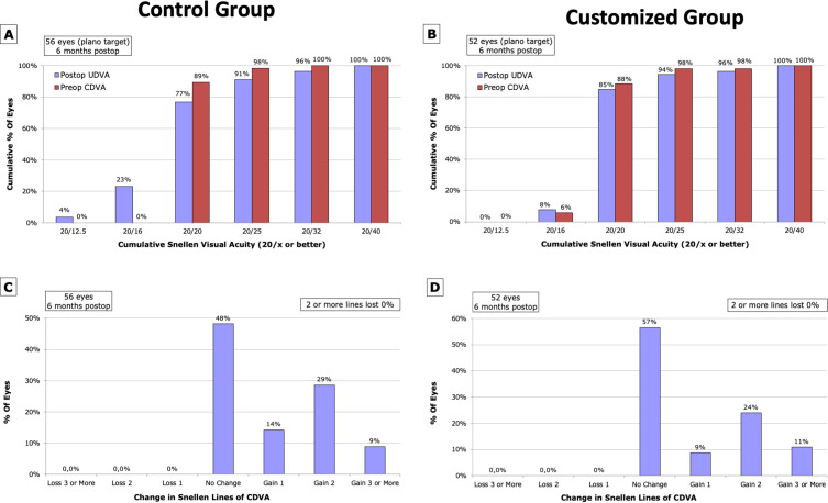
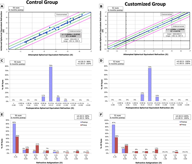
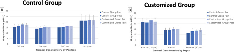
Results: 108 eyes were enrolled, [control group (n=56) and customized group (n=52)]. Mean epithelial ablation thickness in the customized group was 54.81±3.56µm (p=0.470 vs control group). Both groups experienced significant postoperative increases in higher-order aberrations (HOA) and spherical aberrations, with no significant intergroup differences. Mean HOA RMS (µm) of the frontal cornea and total cornea increased by 0.27, and 0.29, respectively, in the control group, and 0.26 and 0.28, respectively, in the customized group (p<0.001 for all). Mean change in spherical aberrations in the frontal cornea and total cornea was 0.23µm (p<0.001) and 0.25µm (p<0.001), in the control group, and 0.19µm (p<0.001) and 0.20µm (p<0.001), in the customized group. Mean corneal densitometry in anterior cornea decreased by 0.63GSU (p=0.021) and 1.18GSU (p<0.001) in the control and customized groups. In the posterior cornea, it increased by 1.67GSU (p=0.004) and 0.38GSU (p=0.006).
Conclusion: No significant differences in refractive and aberrometry outcomes between control and customized Trans-PRK groups, with corneal densitometry changes not affecting visual acuity.
Keywords: Trans-PRK; corneal aberrometry; corneal densitometry; corneal epithelial thickness; corneal refractive surgery.
© 2024 Aramberri et al.
Conflict of interest statement
Jaime Aramberri and Javier Mendicute are consultants to Alcon. Jaime Aramberri also reports personal fees from Alcon, Staar Surgical, Johnson and Johnson, Heidelberg Eng., and Schwind, outside the submitted work. The remaining authors have nothing to declare for this work. In addition, the authors have no financial or proprietary interests in the medical field or products involved in this manuscript.
Figures
References
LinkOut - more resources
Full Text Sources
Miscellaneous
