. 2024 Oct;18(5):617-632.
doi: 10.4162/nrp.2024.18.5.617. Epub 2024 Jul 10.

Dietary ellagic acid blocks inflammation-associated atherosclerotic plaque formation in cholesterol-fed apoE-deficient mice

Affiliations

Dietary ellagic acid blocks inflammation-associated atherosclerotic plaque formation in cholesterol-fed apoE-deficient mice

Sin-Hye Park et al. Nutr Res Pract. 2024 Oct.

Abstract

Background/objectives: Atherosclerosis particularly due to high circulating level of low-density lipoprotein is a major cause of cardiovascular diseases. Ellagic acid is a natural polyphenolic compound rich in pomegranates and berries. Our previous study showed that ellagic acid improved functionality of reverse cholesterol transport in murine model of atherosclerosis. The aim of this study is to investigate whether ellagic acid inhibited inflammation-associated atherosclerotic plaque formation in cholesterol-fed apolipoprotein E (apoE)-knockout (KO) mice.

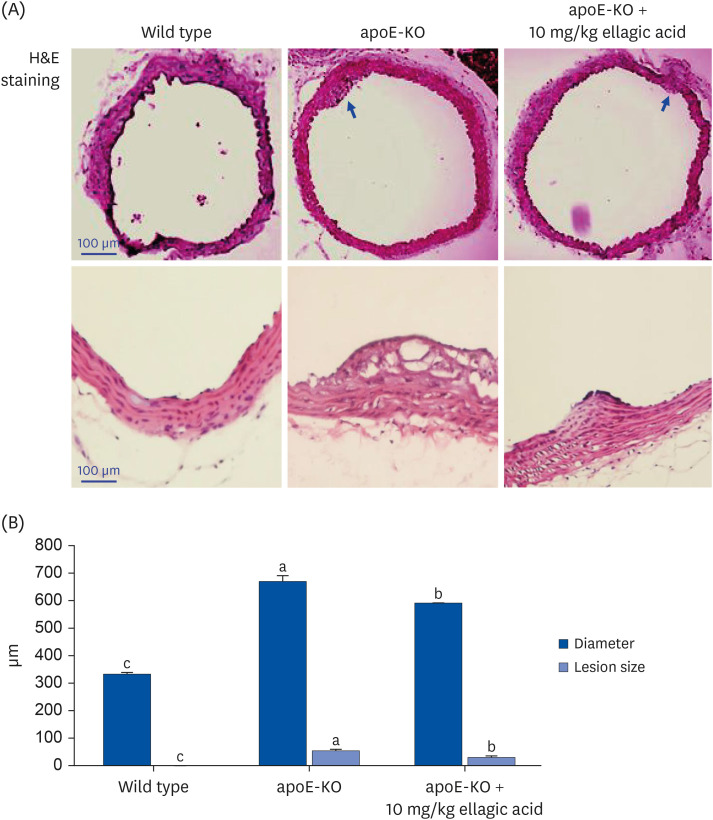
Materials/methods: Wild type mice and apoE-KO mice were fed a cholesterol-rich Paigen diet for 10 weeks to induce severe atherosclerosis. Concurrently, 10 mg/kg ellagic acid was orally administered to the apoE-KO mice. Plaque lesion formation and lipid deposition were examined by staining with hematoxylin and eosin, Sudan IV and oil red O.

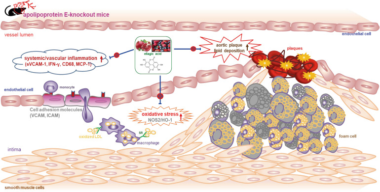
Results: The plasma leukocyte profile of cholesterol-fed mice was not altered by apoE deficiency. Oral administration of ellagic acid attenuated plaque lesion formation and lipid deposition in the aorta tree of apoE-KO mice. Ellagic acid substantially reduced plasma levels of soluble vascular cell adhesion molecule and interferon-γ in Paigen diet-fed apoE-KO mice. When 10 mg/kg ellagic acid was administered to cholesterol-fed apoE-KO mice, the levels of CD68 and MCP-1 were strongly reduced in aorta vessels. The protein expression level of nitric oxide synthase-2 (NOS2) in the aorta was highly enhanced by supplementation of ellagic acid to apoE-KO mice, but the expression level of heme oxygenase-1 (HO-1) in the aorta was reduced. Furthermore, ellagic acid diminished the increased aorta expression of the inflammatory adhesion molecules in cholesterol-fed apoE-KO mice. The treatment of ellagic acid inhibited the scavenger receptor-B1 expression in the aorta of apoE-KO mice, while the cholesterol efflux-related transporters were not significantly changed.

Conclusion: These results suggest that ellagic acid may be an atheroprotective compound by attenuating apoE deficiency-induced vascular inflammation and reducing atherosclerotic plaque lesion formation.

Keywords: Apolipoproteins E; atherosclerosis; atherosclerotic plaque; ellagic acid; inflammation.


Conflict of interest statement

Conflict of Interest: The authors declare that they have no potential conicts of interests.

Figures

Fig. 1
Fig. 1. Wild-type mice and apoE-deficient (apoE-KO) mice were fed an atherogenic Paigen diet for 10 weeks daily with and without oral administration of 10 mg/kg ellagic acid. Cells in blood were counted using a Hemavet HV950 Multispecies Hematologic Analyzer (Drew Scientific). (A) Chemical structure of ellagic acid, (B) schematic illustration of animal experimental design/timeline, and (C) leukocyte profile in blood.
apoE, apolipoprotein E; KO, knockout.
Fig. 2
Fig. 2. Inhibition of lipid accumulation in aorta by ellagic acid. After apoE-deficient (apoE-KO) mice were orally treated with 10 mg/kg ellagic acid for 10 weeks, aortas were extracted. Aortas of wild type C57BL/6 mice and untreated apoE-KO mice were also obtained. Aorta was longitudinally opened from the aorta arch to the iliac bifurcation. Aorta was fixed with 4% buffered formalin overnight and dehydrated with 30% sucrose. (A) To determine whether lipid-enriched plaques were formed, Sudan IV staining was performed within whole aortas. For lipid accumulation in aorta, oil red O staining was done with aorta tissues cut in 5 μm thickness. (B) Counter-staining was conducted with hematoxylin. Magnification: ×200 (n = 6, each of groups). Scale bar = 100 μm.
apoE, apolipoprotein E; KO, knockout.
Fig. 3
Fig. 3. Effects of ellagic acid on atherosclerotic lesion formation within aortas. After apoE-deficient (apoE-KO) mice were orally treated with 10 mg/kg ellagic acid for 10 weeks, aortas were extracted. Aortas of wild type C57BL/6 mice and untreated apoE-KO mice were also obtained. Aorta was longitudinally opened from the aorta arch to the iliac bifurcation and was fixed with 4% buffered formalin overnight and dehydrated with 30% sucrose. (A) To examine atherosclerotic lesion formation on aorta wall, H&E staining was performed with aorta tissues cut in 5–6 μm thickness. Stained tissues were observed by microscopy with 200× magnification (n = 6, each of groups). (B) The extent of atherosclerotic areas was expressed as lesion diameter and size in the entire aortic surface area. Scale bar = 100 μm.
apoE, apolipoprotein E; KO, knockout; H&E, hematoxylin and eosin. a-cValues in bar graphs not sharing a same lower case indicate significant different at P < 0.05.
Fig. 4
Fig. 4. Effects of ellagic acid on systemic and vascular inflammation. After apoE-deficient (apoE-KO) mice were orally treated with 10 mg/kg ellagic acid for 10 weeks, blood was collected and centrifugated at 3,000 rpm for 10 min to obtain plasma. Blood samples of wild type C57BL/6 mice and untreated apoE-KO mice were also collected. Plasma levels of CRP, sVCAM-1, TNF-α, IFN-γ, and MCP-1 were measured by using ELISA kits. All the ELISA procedures were followed according to the manufacturer’s instructions.
apoE, apolipoprotein E; KO, knockout; CRP, C-reactive protein; sVCAM-1, soluble vascular cell adhesion molecule-1; TNF-α, tumor necrosis factor-α; IFN-γ, interferon-γ; MCP-1, monocyte chemoattractant protein-1; ELISA, enzyme-linked immunosorbent assay; SEM, standard error of the mean; WT, wild type. a,bData (mean ± SEM, n = 3–6) in respective bar graphs without a common letter differ at P < 0.05.
Fig. 5
Fig. 5. Effects of ellagic acid on induction of induction of inflammatory biomarkers in aortas. After apoE-deficient (apoE-KO) mice were orally treated with 10 mg/kg ellagic acid for 10 weeks, aortas were extracted (n = 6, each of groups). Aortas of wild type C57BL/6 mice and untreated apoE-deficient mice were also obtained. (A) For the measurement of protein induction of CD68 and MCP-1 in aorta vessels, the CD68 localization was identified as FITC-green staining and the MCP-1 was visualized with Cy3-red staining. Nuclear staining was done with DAPI (blue). Each photograph is representative of 3–6 mice. Magnification: 200-fold. Scale bar = 100 μm. Total tissue extracts were prepared for Western blot analysis with a primary antibody against (B) NOS2, (C) HO-1, and (D) TF. The bar graphs (mean ± SEM, n = 3) represent quantitative results of the upper bands obtained from a densitometer. β-Actin protein was used as an internal control.
apoE, apolipoprotein E; KO, knockout; MCP-1, monocyte chemoattractant protein-1; FITC, fluorescein isothiocyanate; DAPI, 4′,6-diamidino-2-phenylindole; NOS2, nitric oxide synthase-2; HO-1, heme oxygenase-1; TF, tissue factor; SEM, standard error of the mean; WT, wild type. a-cValues in bar graphs not sharing a same lower case indicate significant different at P < 0.05.
Fig. 6
Fig. 6. Blockade of induction of cell adhesion molecules by oral treatment of ellagic acid. After apoE-deficient (apoE-KO) mice were orally treated with 10 mg/kg ellagic acid for 10 weeks, aortas were extracted (n = 6, each of groups). Aortas of wild type C57BL/6 mice and untreated apoE-deficient mice were also obtained. Tissue extracts were prepared for Western blot analysis with a primary antibody against (A) VCAM-1, (B) ICAM-1, and (C) P-selectin. The bar graphs (mean ± SEM, n = 3) represent quantitative results of the upper bands obtained from a densitometer. β-Actin protein was used as an internal control. (D) Aorta tissue level of VCAM-1 was determined by visualizing with brown DAB and being counterstained with hematoxylin. In addition, PECAM-1 was immunohistologically stained with red Cy3-conjugated second antibody. Counterstaining was performed with blue DAPI. Each photograph is representative of 3–6 mice. Magnification: 200-fold. Scale bar = 100 μm.
apoE, apolipoprotein E; KO, knockout; VCAM-1, vascular cell adhesion molecule-1; ICAM-1, intracellular adhesion molecule-1; SEM, standard error of the mean; DAB, 3,3′-diaminobenzidine; PECAM-1, platelet endothelial cell adhesion molecule-1; DAPI, 4′,6-diamidino-2-phenylindole; WT, wild type. a,bValues in bar graphs not sharing a same lower case indicate significant different at P < 0.05.
Fig. 7
Fig. 7. Effects of ellagic acid on cholesterol trafficking in aortas. After apoE-deficient (apoE-KO) mice were orally treated with 10 mg/kg ellagic acid for 10 weeks, aortas were extracted (n = 6, each of groups). Aortas of wild type C57BL/6 mice and untreated apoE-deficient mice were also obtained. Tissue extracts were prepared for Western blot analysis with a primary antibody against (A) ABCA1, (B) ABCG1, and (C) SR-B1. The bar graphs (mean ± SEM, n = 3) represent quantitative results of the upper bands obtained from a densitometer. β-Actin protein was used as an internal control.
apoE, apolipoprotein E; KO, knockout; ABC, ATP-binding cassette transporter; SR-B1, scavenger receptor class type B1; SEM, standard error of the mean; WT, wild type. a,bValues in bar graphs not sharing a same lower case indicate significant different at P < 0.05.
Fig. 8
Fig. 8. Diagram illustrating effects of ellagic acid on atherosclerosis in apoE-deficient (apoE-KO) mice. Ellagic acid reduced atherosclerosis by alleviating aortic plaque formation and lipid deposition. Furthermore, ellagic acid diminished vascular inflammation by down-regulating expression of sVCAM-1, IFN-γ, CD68, and MCP-1.
apoE, apolipoprotein E; KO, knockout; sVCAM-1, soluble vascular cell adhesion molecule-1; IFN-γ, interferon-γ; MCP-1, monocyte chemoattractant protein-1; VCAM, vascular cell adhesion molecule; ICAM, intracellular adhesion molecule; NOS2, nitric oxide synthase 2; HO-1, heme oxygenase-1; LDL, low-density lipoprotein; SR, scavenger receptor.

References

    1. Fazio S, Linton MF. Mouse models of hyperlipidemia and atherosclerosis. Front Biosci. 2001;6:D515–D525. - PubMed
    1. Meir KS, Leitersdorf E. Atherosclerosis in the apolipoprotein-E-deficient mouse: a decade of progress. Arterioscler Thromb Vasc Biol. 2004;24:1006–1014. - PubMed
    1. Nakashima Y, Plump AS, Raines EW, Breslow JL, Ross R. ApoE-deficient mice develop lesions of all phases of atherosclerosis throughout the arterial tree. Arterioscler Thromb. 1994;14:133–140. - PubMed
    1. Lee YT, Lin HY, Chan YW, Li KH, To OT, Yan BP, Liu T, Li G, Wong WT, Keung W, et al. Mouse models of atherosclerosis: a historical perspective and recent advances. Lipids Health Dis. 2017;16:12. - PMC - PubMed
    1. Mahdinia E, Shokri N, Taheri AT, Asgharzadeh S, Elahimanesh M, Najafi M. Cellular crosstalk in atherosclerotic plaque microenvironment. Cell Commun Signal. 2023;21:125. - PMC - PubMed