

Isolated spermatic cord metastasis of prostate cancer after radiotherapy detected with 18F-PSMA PET/CT: a case report and literature review
- PMID: 39398910
- PMCID: PMC11464869
- DOI: 10.1007/s13691-024-00692-4
Isolated spermatic cord metastasis of prostate cancer after radiotherapy detected with 18F-PSMA PET/CT: a case report and literature review
Abstract
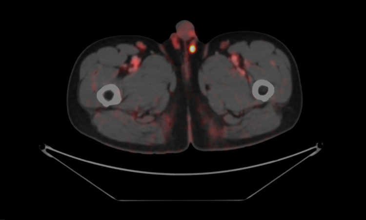
We report the case of a 65 year-old male with prostate cancer previously treated with external beam radiotherapy and 2 years of androgen deprivation therapy. His nadir PSA reached undetectable level but gradually increased to 0.89 ng/dL. 18F-PSMA PET/CT demonstrated a PSMA-avid lesion at left spermatic cord. Left groin exploration revealed an 8 mm left vas deferens mass. Mass excision was performed and pathology result showed prostatic adenocarcinoma. The metastatic route is unknown but the possible routes are intraluminal route via ejaculatory duct, hematogenous route and lymphatic route. This case also highlights the role of 18F-PSMA PET/CT to detect a recurrent lesion at an atypical site in biochemical failure patients even at the low PSA level.
Keywords: 18F-PSMA PET/CT; Oligometastasis; Prostate cancer; Radiotherapy; Spermatic cord metastasis.
© The Author(s) under exclusive licence to The Japan Society of Clinical Oncology 2024. Springer Nature or its licensor (e.g. a society or other partner) holds exclusive rights to this article under a publishing agreement with the author(s) or other rightsholder(s); author self-archiving of the accepted manuscript version of this article is solely governed by the terms of such publishing agreement and applicable law.
Conflict of interest statement
Conflict of interestPD has received a speaker honorarium from Elekta in FARO-KOSRO Meeting 2023. Other authors declare that they have no conflict of interest.
Figures

References
LinkOut - more resources
Full Text Sources
Research Materials
Miscellaneous