[Preprint]. 2024 Sep 27:2024.09.26.24313895.
doi: 10.1101/2024.09.26.24313895.

Loss-of-function of RNA-binding protein PRRC2B causes translational defects and congenital cardiovascular malformation

Affiliations

Loss-of-function of RNA-binding protein PRRC2B causes translational defects and congenital cardiovascular malformation

Debojyoti Das et al. medRxiv. .

Abstract

Alternative splicing generates variant forms of proteins for a given gene and accounts for functional redundancy or diversification. A novel RNA-binding protein, Pro-rich Coiled-coil Containing Protein 2B (PRRC2B), has been reported by multiple laboratories to mediate uORF-dependent and independent regulation of translation initiation required for cell cycle progression and proliferation. We identified two alternative spliced isoforms in human and mouse hearts and HEK293T cells, full-length (FL) and exon 16-excluded isoform ΔE16. A congenital heart disease-associated human mutation-mimicry knock-in of the equivalent variant in the mouse genome leads to the depletion of the full-length Prrc2b mRNA but not the alternative spliced truncated form ΔE16, does not cause any apparent structural or functional disorders. In contrast, global genetic inactivation of the PRRC2B gene in the mouse genome, nullifying both mRNA isoforms, caused patent ductus arteriosus (PDA) and neonatal lethality in mice. Bulk and single nucleus transcriptome profiling analyses of embryonic mouse hearts demonstrated a significant overall downregulation of multiple smooth muscle-specific genes in Prrc2b mutant mice resulting from reduced smooth muscle cell number. Integrated analysis of proteomic changes in Prrc2b null mouse embryonic hearts and polysome-seq and RNA-seq multi-omics analysis in human HEK293T cells uncover conserved PRRC2B-regulated target mRNAs that encode essential factors required for cardiac and vascular development. Our findings reveal the connection between alternative splicing regulation of PRRC2B, PRRC2B-mediated translational control, and congenital cardiovascular development and disorder. This study may shed light on the significance of PRRC2B in human cardiovascular disease diagnosis and treatment.

Keywords: PRRC2B; RNA-binding protein; congenital heart disease; human mutation; translation.


Conflict of interest statement

Competing interests None of the authors declares any competing interests.

Figures
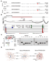
Figure 1.
Figure 1.. Alternative splicing isoforms of PRRC2B mRNA in humans and mice.
A. Schematic of two alternative splicing isoforms of PRRC2B mRNA in humans and mice. B. RNA-seq reads mapped to exon-exon junction regions at the exons 15-17. Adult hearts from male C57BL/6J WT mice were used for RNA-seq. C. RT-PCR validation of alternative splicing isoforms of PRRC2B mRNA in HEK293T and AC16 cells. P1-6, primers 1-6. D. Predicted protein structure of full-length and ΔE16 PRRC2B in humans and mice by Alpha-Fold.
Figure 2.
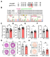
Figure 2.. prrc2bR1128X/R1128X knock-in (FL-Prrc2bgKO) mice with human genetic mutation of a premature termination codon show inactivation of full-length Prrc2b mRNA without causing apparent cardiac phenotypes.
A. Schematic of Prrc2bR1128X/R1128X (FL-Prrc2bgKO) mouse model. B. Sanger sequencing confirms genotyping of the Prrc2bR1128X/R1128X (FL-Prrc2bgKO) mouse model. C-F. LV mass, ejection fraction (EF), fractional shortening (FS), and cardiac output showed no significant difference between WT control and homozygous FL-Prrc2bgKO 3 months after birth. WT: N=5M+8F; KO: N=7M+7F. G. H&E images of WT and KI mice hearts at 5 months old. Heart size was quantified using Image J. WT: N=3M+2F; KO: N=3M+3F. H. Picrosirius red staining of WT and KI mice hearts at 5 months old. The quantification of the fibrotic area for comparison was performed using Image J. WT: N=3M+2F; KO: N=3M+3F. Data are represented as mean ± SD. An unpaired two-tailed Student t-test was performed to compare two groups for C-H. ns: not significant; * P < 0.05.
Figure 3.
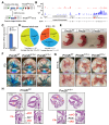
Figure 3.. Mice lacking two Prrc2b alternative splicing isoforms manifest patent ductus arteriosus (PDA) and die perinatally.
A. Schematic representation of the generation of Prrc2b global KO mouse model (Prrc2btm1b−/−) by crossing Prrc2btm1a with CMV-Cre mice. Both full-length and ΔE16 Prrc2b isoforms are knocked out. B. Integrative Genomics Viewer (IGV) plot of RNA-seq measurement of Prrc2b mRNA expression across all the exons. C. RT-qPCR analysis of Prrc2b mRNA expression in whole hearts of WT and Prrc2btm1b−/− mice. D. Number of weaned adult Prrc2btm1b mice and prenatal Prrc2btm1b mice (E12.5 – E18.5) during heterozygous breeding indicated by their respective pie charts. E. Images of mice at P0. Prrc2b−/− pups were found dead as early as 7 hours postnatally, while Prrc2b+/− remained alive. Scale bar: 1 cm. F. Images of hearts with Coomassie blue dye injection from WT, Prrc2b+/− and Prrc2b−/− at P0. Scale bar: 1 mm. PA, pulmonary artery; DA, ductus arteriosus; LA, ligamentum arteriosum; PDA, patent ductus arteriosus. G. Images of hearts in the bright field from WT, Prrc2b+/− and Prrc2b−/− at E17.5. Scale bar: 1 mm. H. H&E images of WT and homozygous tm1b KO hearts at P0. LV: left ventricle, RV: Right ventricle, LA: left atrium, RA: right atrium, aAO: ascending aorta, dAO: descending aorta, DA: ductus arteriosus, PA: pulmonary artery, PDA: patent ductus arteriosus.
Figure 4.
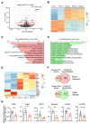
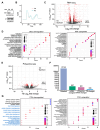
Figure 4.. Bulk RNA-seq analysis of Prrc2b global knockout hearts shows reduced smooth muscle cell contraction and mitochondrial respiration-related gene expression.
A. Volcano plot of RNA-seq of WT and Prrc2btm1b−/− hearts at E18.5. Log2 Fold Change (Prrc2btm1b−/− / WT) is plotted as the X-axis, while −Log10 P-values are plotted as the Y-axis. Genes with |Log2 Fold Change| > 1 and P-value < 0.05 are colored red. WT: N=2; Prrc2btm1b−/−: N=3. B. Heatmap of RNA-seq of WT and Prrc2btm1b−/− hearts. TPM (transcript per million) is plotted. Values are scaled for each row. C and D. Gene ontology analysis of upregulated and downregulated genes in RNA-seq of WT and Prrc2btm1b−/− hearts. E. A zoomed heatmap showing the top 20 dysregulated genes in (B). TPM (transcript per million) is plotted. Values are scaled for each row. F. Venn diagrams showing the overlaps between PDA genes and dysregulated genes in Prrc2btm1b−/− hearts. Genes with Log2 FC > 1 and P < 0.05 are considered dysregulated. G. RT-qPCR validation of dysregulated genes related to smooth muscle cell contraction and mitochondrial respiratory chain complex in Prrc2btm1b−/− hearts. 18S rRNA is used as the normalizer. Values are plotted as relative values to WT hearts. Data is shown as mean ± SD. Technical replicates for each biological sample are plotted (WT: N=2; Prrc2btm1b−/−: N=3). An unpaired two-tailed Student t-test was performed to compare two groups for G. * P < 0.05; ** P < 0.01.
Figure 5.
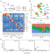
Figure 5.. snRNA-seq analysis of Prrc2b global knockout hearts.
A. A sketch showing the workflow of snRNA-seq for homozygous Prrc2btm1b−/− gKO, heterozygous Prrc2btm1b+/−, and WT control hearts at E18.5. B. A UMAP presentation of clustering results based on the top 3000 variant features. 19 clusters were identified and labeled with cell type and top marker genes. C. Heatmap showing the expression of well-established marker genes in each cluster. Scaled and normalized expressions were plotted. D. A grouped bar plot showing the relative abundance of WT, Het, and Homo cells in each cell type. The Y-axis represents the fraction of total cells in each cell type. E. Volcano plots showing the DEGs in smooth muscle cells. Genes with significantly differential expression (Log2 FC > 0.2 and Bonferroni correction adjusted P < 0.05) were red-colored. Ven.CM, ventricular cardiomyocytes; Atri.CM, atrial cardiomyocytes; FB, fibroblasts; EC, endothelial cells; SMC, smooth muscle cells; BC, blood cells; VEC, vascular EC; Epi, epicardial cells; PC, pericytes; ProCM, proliferating CM; Endo, endocardial; Lym, lymphatic.
Figure 6.
Figure 6.. Mass spectrometry analysis of Prrc2b global knockout hearts.
A. Volcano plot of differentially expressed proteins identified by mass spectrometry in Prrc2btm1b−/− gKO compared with WT control hearts. 33 downregulated and 18 upregulated proteins with statistical significance (P < 0.05) were listed. B. Gene ontology analysis of significantly downregulated proteins (P < 0.05). C. Gene ontology analysis of drastically downregulated proteins (Log2FC < −0.75).
Figure 7.
Figure 7.. Transcriptomic and translatomic profiling of PRRC2B knockout human cells.
A. Immunoblot validation of PRRC2B gene knockout in HEK293T cells. B. Polysome profiling of control and KO HEK293T cells. C. Volcano plot of differentially expressed genes identified by RNA-seq. D. GO analysis of significantly downregulated (left) and upregulated (right) genes. E. Volcano plot of differentially translated mRNAs (DTM) identified by polysome-seq. F. Number of genes significantly dysregulated at the RNA and translation efficiency (TE) levels in KO cells. G. Gene ontology analysis of significantly TE-downregulated (left) and -upregulated (right) mRNAs. DTM-downregulated pathways are highlighted in blue.
Figure 8.
Figure 8.. Conserved PRRC2B-regulated target mRNAs across cell types and species.
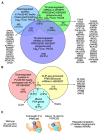
A. Venn diagram of shared PRRC2B-regulated genes from Prrc2btm1b−/− gKO mouse hearts and PRRC2B KO and KD human HEK293T cells. Conserved downregulated genes at the protein translation level are listed. B. Venn diagram of overlapping reduced proteins in Prrc2btm1b−/− KO mouse hearts with PRRC2B-bound mRNAs from human cells and well-known mouse PDA genes. Conserved downregulated genes at the protein translation level are listed. C. Schematic model showing the two alternative spliced PRRC2B isoforms (FL and ΔE16) and phenotypes of the Prrc2b genetic mouse models.

References

    1. Gao C. and Wang Y. (2020) mRNA Metabolism in Cardiac Development and Disease: Life After Transcription. Physiol Rev, 100, 673–694. - PMC - PubMed
    1. Kalsotra A. and Cooper T.A. (2011) Functional consequences of developmentally regulated alternative splicing. Nat Rev Genet, 12, 715–729. - PMC - PubMed
    1. Tahmasebi S., Khoutorsky A., Mathews M.B. and Sonenberg N. (2018) Translation deregulation in human disease. Nat Rev Mol Cell Biol, 19, 791–807. - PubMed
    1. Castello A., Fischer B., Eichelbaum K., Horos R., Beckmann B.M., Strein C., Davey N.E., Humphreys D.T., Preiss T., Steinmetz L.M. et al. (2012) Insights into RNA biology from an atlas of mammalian mRNA-binding proteins. Cell, 149, 1393–1406. - PubMed
    1. Baltz A.G., Munschauer M., Schwanhausser B., Vasile A., Murakawa Y., Schueler M., Youngs N., Penfold-Brown D., Drew K., Milek M. et al. (2012) The mRNA-bound proteome and its global occupancy profile on protein-coding transcripts. Mol Cell, 46, 674–690. - PubMed

Publication types

LinkOut - more resources