. 2024 Sep 27:15:1478917.
doi: 10.3389/fphar.2024.1478917. eCollection 2024.

Notoginsenoside R1, a metabolite from Panax notoginseng (Burkill) F.H.Chen, stimulates insulin secretion through activation of phosphatidylinositol 3-kinase (PI3K)/Akt pathway

Affiliations

Notoginsenoside R1, a metabolite from Panax notoginseng (Burkill) F.H.Chen, stimulates insulin secretion through activation of phosphatidylinositol 3-kinase (PI3K)/Akt pathway

Altaf Al-Romaiyan et al. Front Pharmacol. .

Abstract

Background: For ages, botanical medicine has been used in the treatment of diabetes mellitus (DM). Notoginsenoside R1 (NGR1), a Panax notoginseng (Burkill) F.H.Chen metabolite, has been documented to possess antidiabetic action in vivo. However, its precise molecular mechanism of action is not clear.

Objectives: We evaluated NGR1's effects on blood glucose in vivo and then evaluated in vitro whether NGR1 has effects on insulin secretion and the probable molecular pathways involved in NGR1-induced insulin secretion.

Methods: Diabetes was induced in mice by streptozotocin. Glucose tolerance test was performed before and after NGR1 was administered intraperitoneally to diabetic animals for 4 weeks. Static and perifusion experiments were performed using isolated female BALB/c mouse islets. Preproinsulin (Ins) mRNA expression was measured using q-PCR. Protein expression of PI3K/Akt pathway was assessed using the fully automated Wes™ capillary-based protein electrophoresis.

Results: Treatment of diabetic mice with NGR1 improved their glucose intolerance. In vitro, NGR1 increased insulin secretion in a concentration-dependent manner. NGR1 initiated the secretion of insulin at 2 mM glucose and augmented glucose-stimulated insulin secretion which was sustained throughout NGR1 perifusion. NGR1-induced insulin secretion was not altered by a voltage gated calcium channel blocker or protein kinase A inhibitor. NGR1 did not significantly modulate Ins mRNA expression. However, NGR1 significantly increased the levels of phospho-Akt and phopho-p-85.

Conclusion: In conclusion, this study has shown that NGR1 ameliorates hyperglycemia in diabetic mice. NGR1 has a direct insulin secretagogue activity on mouse islets, stimulates insulin secretion at both basal and postprandial glucose concentrations, and activates PI3K/Akt pathway to induce insulin secretion. These results suggest that NGR1 may provide an alternative therapy to manage DM.

Keywords: Panax notoginseng; diabetes; insulin secretion; mouse islets; plant extract.


Conflict of interest statement

The authors declare that the research was conducted in the absence of any commercial or financial relationships that could be construed as a potential conflict of interest. The author(s) declared that they were an editorial board member of Frontiers, at the time of submission. This had no impact on the peer review process and the final decision.

Figures
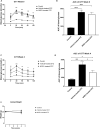
FIGURE 1
FIGURE 1
The effect of NGR1 on glucose tolerance test (GTT) from diabetic mice in vivo. NGR1 or its vehicle was administered daily by intraperitoneal (IP) injection to diabetic mice for 4 weeks. (A,C) Time course of GTT and (B,D) area under the curve of the GTT were measured before and after 4 weeks of NGR1 administration. (E) Weight of animals. The bars and points represent mean ± SEM. Each experimental group had 5-8 animals per group (B,D): *p < 0.05, ***p < 0.001, ****p < 0.0001 (One-way ANOVA followed by Tukey’s multiple comparisons test and (A,C,E): *p < 0.05, ***p < 0.001, ****p < 0.0001 vehicle-treated STZ vs. control; ##p < 0.01, ####p < 0.0001 NGR1-treated STZ vs. control; §p < 0.05 vehicle-treated STZ vs. NGR1-treated STZ (Two-way ANOVA followed by Tuckey multiple comparisons).
FIGURE 2
FIGURE 2
Effect of acute NGR1 incubation on mouse islets insulin secretion. (A) Mouse islets were incubated with NGR1 (1–100 µM) at 2 mM glucose for 1 h in a static insulin secretion setting. Insulin concentration was determined from the supernatant. NGR1 caused a concentration-dependent increase in insulin section from mouse islets at 2 mM glucose. The bars represent mean ± SEM for at least three separate experiments. Each experiment had 5–8 replicates per treatment group. *p < 0.05, ***p < 0.001, ****p < 0.0001 vs. 0 µM NGR1 (One way ANOVA followed by Bonferroni’s multiple comparisons test). (B) The EC50 of NGR1-induced insulin secretion at 2 mM glucose concentrations. Each point represents mean ± SEM for at least three separate experiments. Each experiment had 5-8 replicates per treatment group.
FIGURE 3
FIGURE 3
Effect of NGR1 on the rate and pattern of mouse islets insulin secretion. Fifty islets were loaded into perifusion chambers and pre-perifused with 2 mM glucose physiological buffer for 64 min and the perfusate was discarded. (A, B) The islets were perifused with 2 mM glucose or 2 mM glucose + 100 µM NGR1 or 20 mM glucose in a timely fashion (0–62 min) and perfusate was collected every 2 min at a flow rate of 0.1 mL/min. The insulin content was determined from the perfusate. The data was plotted as a treatment-time graph (A) or as area under the curve of each treatment slot graph (B). NGR1 caused a rapid and sustained increase in insulin secretion. The points and bars represent mean ± SEM, n = 3. Each replicate represents 50 islets from a pool of 3 mice. Data are representative of two separate experiments. ****p < 0.0001 vs. 2 mM glucose (One-way ANOVA followed by Dunnett’s multiple comparisons test). (C, D) The islets were perifused with 2 mM glucose (0–10 min) before being challenged with 11.1 mM glucose (black circle) or 11.1 mM glucose + 100 µM NGR (white circle) (10–30 min) and perfusate was collected every 2 min at a flow rate of 0.1 mL/min. The insulin content was determined from the perfusate. The data was plotted as a treatment-time graph (C) or as area under the curve of each treatment slot graph (D). The points and bars represent mean ± SEM, n = 3. Each replicate represents 50 islets from a pool of 6 mice. **p < 0.01 vs. 11.1 mM glucose (unpaired Student’s t-test or Two-way ANOVA followed by Bonferroni’s multiple comparisons test).
FIGURE 4
FIGURE 4
Effect of chronic NGR1 treatment on insulin secretion, preproinsulin mRNA expression, total intra-islet insulin content and mouse islets cell viability. Mouse islets were incubated with NGR1 (100 µM) for 24 h, (A) Insulin concentration was determined from the supernatant and (B) Ins mRNA expression was determined. (C) Mouse islets were sonicated in acidified alcohol and total intra-islet insulin content was determined. The bars represent mean ± SEM for at least three separate experiments. Each experiment had 3 replicates per treatment group for mRNA expression experiments and 6-8 replicates per treatment group for insulin secretion and intra-islet insulin content experiments. **p < 0.01, ****p < 0.0001 vs. 2 mM glucose (One-way ANOVA followed by Dunnett’s multiple comparisons test). (D) Mouse islets (3 islets/well) were seeded in a white 96-well plate and treated with NGR1 (1–100 µM) for 24 h. ATP content as an indicator of cell viability was determined by CellTiter-Glo® Luminescent Cell Viability Assay (Promega, United States). The bars represent mean ± SEM of three separate experiments. Each experiment had 6-8 replicates per treatment group. p > 0.05 (One-way ANOVA followed by Dunnett’s multiple comparisons test).
FIGURE 5
FIGURE 5
The effect of NGR1 on voltage gated calcium channels (VGCC) and protein kinase A (PKA) pathway. Fifty islets were loaded into perifusion chambers and pre-perifused with 2 mM glucose physiological buffer for 64 min and the perfusate was discarded. The islets were then perifused with 100 µM NGR1 in the presence or absence of nifedipine (A, B) and H-89 (C, D) and perfusate was collected every 2 min at a flow rate of 0.1 mL/min. The insulin content was determined from the perfusate. The data was plotted as a treatment-time graph (A, C) or as area under the curve of each treatment slot graph (B, D). NGR1 caused a rapid and sustained increase in insulin secretion that is not affected by blocking Ca2+ influx through VGCC or inhibiting PKA activation. The points and bars represent mean ± SEM, n = 6. Each replicate represents 50 islets from a pool of 6 mice. p > 0.05 (unpaired Student’s t-test or Two-way ANOVA followed by Bonferroni’s multiple comparisons test).
FIGURE 6
FIGURE 6
The effect of NGR1 on PI3K/Akt pathway. (A) Mouse islets (3 islets/tube) were pre-incubated with 2 mM glucose for 2 h before being incubated with 100 µM NGR1 in the presence or absence of 50 µM LY29004 (a PI3K inhibitor) for 1 h at 37°C. Insulin content was determined from the supernatant. Data are mean ± SEM of at least three separate experiments. Each experiment had 5–12 replicates per treatment group. ****p < 0.0001 (One-way ANOVA followed by Bonferroni’s multiple comparisons test). (B–D) INS-1 832/13 cells were incubated for 5 min with NGR1 (100 µM) for the measurement of Akt and p85 phosphorylation. Cells were lysed and 3 µg of protein was used. Expression of phosphorylated and total protein of interest was detected by Wes™ capillary-based protein electrophoresis and normalized to β-actin (and total protein; inset) and expressed relative to control. The bars represent mean ± SEM of 3–4 separate experiments. Each experiment had 3 replicates per treatment group. *p ˂ 0.05, **p < 0.01, ***p < 0.001 (Unpaired t-test).

Similar articles

References

    1. Al-Romaiyan A., Jayasri M. A., Mathew T. L., Huang G. C., Amiel S., Jones P. M., et al. (2010). Costus pictus extracts stimulate insulin secretion from mouse and human islets of Langerhans in vitro . Cell. Physiology Biochem. 26, 1051–1058. 10.1159/000324007 - DOI - PubMed
    1. Al-Romaiyan A., King A. J., Persaud S. J., Jones P. M. (2013). A novel extract of Gymnema sylvestre improves glucose tolerance in vivo and stimulates insulin secretion and synthesis in vitro . Phytotherapy Res. 27, 1006–1011. 10.1002/ptr.4815 - DOI - PubMed
    1. Al-Romaiyan A., Huang G.-C., Jones P., Persaud S. (2020a). Commiphora myrrha stimulates insulin secretion from mouse and human islets of Langerhans. J. Ethnopharmacol. 264, 113075. 10.1016/j.jep.2020.113075 - DOI - PubMed
    1. Al-Romaiyan A., Liu B., Persaud S., Jones P. (2020b). A novel Gymnema sylvestre extract protects pancreatic beta-cells from cytokine-induced apoptosis. Phytotherapy Res. 34, 161–172. 10.1002/ptr.6512 - DOI - PubMed
    1. Al-Romaiyan A., Masocha W., Oyedemi S., Marafie S. K., Huang G. C., Jones P. M., et al. (2023a). Commiphora myrrha stimulates insulin secretion from β-cells through activation of atypical protein kinase C and mitogen-activated protein kinase. J. Ethnopharmacol. 302, 115937. 10.1016/j.jep.2022.115937 - DOI - PubMed

LinkOut - more resources