. 2024 Sep 27:15:1426975.
doi: 10.3389/fimmu.2024.1426975. eCollection 2024.

Dysregulated C1q and CD47 in the aging monkey brain: association with myelin damage, microglia reactivity, and cognitive decline

Affiliations

Dysregulated C1q and CD47 in the aging monkey brain: association with myelin damage, microglia reactivity, and cognitive decline

Sarah A DeVries et al. Front Immunol. .

Abstract

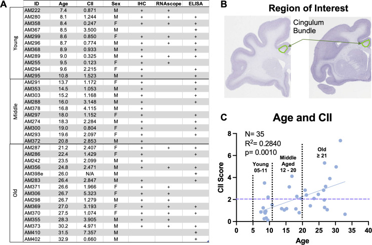
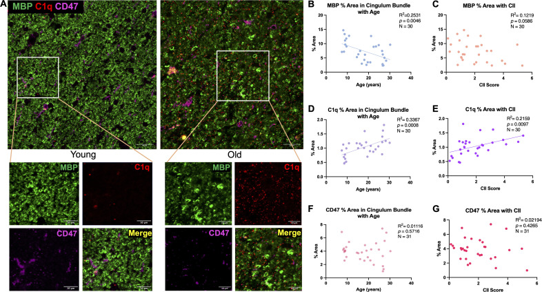
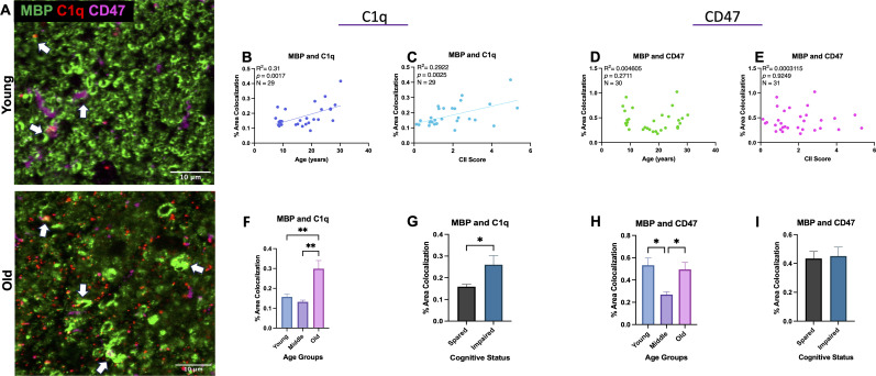
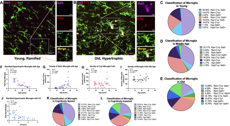
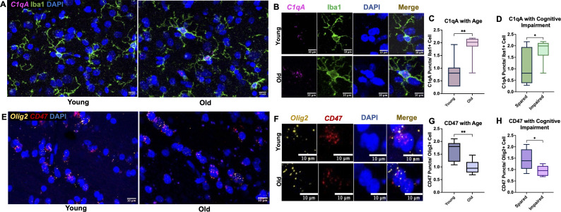
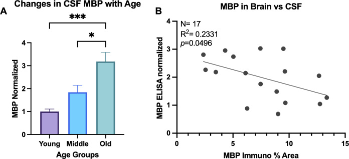
Normal aging, though lacking widespread neurodegeneration, is nevertheless characterized by cognitive impairment in learning, memory, and executive function. The aged brain is spared from neuron loss, but white matter is lost and damage to myelin sheaths accumulates. This myelin damage is strongly associated with cognitive impairment. Although the cause of the myelin damage is not known, microglia dysregulation is a likely contributor. Immunologic proteins interact with microglial receptors to modulate microglia-mediated phagocytosis, which mediates myelin damage clearance and turn-over. Two such proteins, "eat me" signal C1q and "don't eat me" signal CD47, act in opposition with microglia. Both C1q and CD47 have been implicated in Multiple Sclerosis, a demyelinating disease, but whether they play a role in age-related myelin pathology is currently unknown. The present study investigates C1q and CD47 in relation to age-related myelin degeneration using multilabel immunofluorescence, RNAscope, and confocal microscopy in the cingulum bundle of male and female rhesus monkeys across the lifespan. Our findings showed significant age-related elevation in C1q localized to myelin basic protein, and this increase is associated with more severe cognitive impairment. In contrast, CD47 localization to myelin decreased in middle age and oligodendrocyte expression of CD47 RNA decreased with age. Lastly, microglia reactivity increased with age in association with the changes in C1q and CD47. Together, these results suggest disruption in the balance of "eat me" and "don't eat me" signals during normal aging, biasing microglia toward increased reactivity and phagocytosis of myelin, resulting in cognitive deficits.

Keywords: C1q; CD47; complement system; innate immune system; microglia; neurodegeneration; white matter.


Conflict of interest statement

The authors declare that the research was conducted in the absence of any commercial or financial relationships that could be construed as a potential conflict of interest.

Figures

Figure 1
Figure 1
Summary of subject information and experimental parameters. (A) Table detailing subject ID, age, sex, and CII score for this study’s cohort of monkeys and defining which samples from each monkey were used for experiments. (B) Histological sections showing the cingulum bundle region of interest. (C) Graph showing the distribution of cognitive impairment index (CII) score for all 36 animals. Linear regression analysis revealed significant cognitive impairment with age. Stratification of age groups is shown on the graph, where young are aged 5-11, middle age 12-20, and old are >21 years old. Purple dashed line indicates the cut off for cognitive impairment, with scores above 2.0 considered severely impaired and scores below considered cognitively spared.
Figure 2
Figure 2
Changes in MBP, C1q, and CD47 in the cingulum bundle with age and CII score. (A) Representative immunofluorescent images for MBP, C1q, and CD47 in a young and old animal. (B) Significant decrease in % area of MBP was found with age that also correlated with (C) cognitive impairment. C1q was significantly increased with (D) age and (E) CII score but % area of CD47 remained stable with both (F) age and (G) higher CII score. Scale bars represent 20µm in zoomed out images and 10µm for close-up images.
Figure 3
Figure 3
Colocalization of C1q and CD47 with MBP during aging and related cognitive impairment. (A) Representative immunofluorescent images of C1q, CD47, and MBP. Colocalization is marked with white arrows. (B) C1q and MBP colocalization increased with age and (C) CII score but (D, E) CD47 colocalized with MBP did not change linearly with age or CII score. (F, G) Analysis into age and cognition group differences revealed increased C1q-MBP colocalization in old age compared to young and middle age as well as in cognitively impaired compared to spared. (H) Analysis into age groups revealed significant decrease in CD47 localized with MBP in middle age compared to young and old age. (I) CD47-MBP colocalization did not correlate with cognitive impairment. Young <10, Old >21; cognitively spared <2.0, cognitively impaired >2.0.
Figure 4
Figure 4
Increased proportion and density of phagocytic and inflammatory microglia with age and cognitive impairment. (A) Representative image of Iba1+ microglia in a young animal largely expressing ramified morphology without C1q or Gal-3, as shown on zoomed in image, in contrast to (B) old animals with more Iba1+ cells exhibiting hypertrophic morphology with C1q and Gal-3 expression. The proportion of microglia cells classified according to ramified or hypertrophic morphology and expression of C1q and Gal-3 are displayed in (C) young (D) middle aged (E) and old animals. The proportion of ramified: hypertrophic microglia significantly decreased with (F) age (J) and CII score, indicating an increase in hypertrophic microglia. (G) Density of Gal3+ cells increased with age, (H) while there were no significant age-related changes in the density of C1q+ cells. (I) There was no change in density of total microglia. Proportion of microglia morphology and C1q/Gal3 expression in (K) cognitively spared animals and in (L) cognitively impaired are also shown. Scale bars represent 20µm in zoomed out images and 10µm for close-up images.
Figure 5
Figure 5
C1qA RNA expression increases in microglia cells while CD47 RNA expression decreases in oligodendrocytes. (A) Representative RNAscope images of C1qA and DAPI with IF for Iba1 captured in young vs old animals with (B) zoomed examples show C1qA RNA puncta within single microglia cells. Results show significant increase in C1qA RNA expression with (C) age and (D) with cognitive impairment. (E) Example RNAscope images of Olig2 and CD47 probes with DAPI in the cingulum bundle of young and old animals. (F) Zoomed in images of Olig2+ oligodendrocytes expressing CD47 puncta within DAPI labeled nuclei. (G) Decreased CD47 RNA expression in oligodendrocytes was found in old animals and (H) in cognitively impaired animals. *p<0.05; **p<0.01; N=16.
Figure 6
Figure 6
MBP in CSF increases with age. (A) ELISA was run for MBP in CSF samples and concentrations normalized to the young age group. One-way ANOVA analysis revealed MBP increases in old animals compared to both young and middle-aged animals. (B) Linear regression comparing the % area of MBP in the brain compared to the normalized concentration of MBP in the CSF of the same animals. *p<0.05; *** p<0.001.

Similar articles

References

    1. Hung CW, Chen YC, Hsieh WL, Chiou SH, Kao CL. Ageing and neurodegenerative diseases. Ageing Res Rev. (2010) 9:S36–46. doi: 10.1016/j.arr.2010.08.006 - DOI - PubMed
    1. Park DC, Reuter-Lorenz P. The adaptive brain: aging and neurocognitive scaffolding. Annu Rev Psychol Annu Rev Psychol 60. Palo Alto: Annu Reviews. (2009) 60:173–96. doi: 10.1146/annurev.psych.59.103006.093656 - DOI - PMC - PubMed
    1. Harada CN, Love MCN, Triebel KL. Normal cognitive aging. Clinics Geriatr Med. (2013) 29:737–+. doi: 10.1016/j.cger.2013.07.002 - DOI - PMC - PubMed
    1. Salthouse TA. When does age-related cognitive decline begin? Neurobiol Aging. (2009) 30:507–14. doi: 10.1016/j.neurobiolaging.2008.09.023 - DOI - PMC - PubMed
    1. Salthouse TA. Selective review of cognitive aging. J Int Neuropsychol Society. (2010) 16:754–60. doi: 10.1017/S1355617710000706 - DOI - PMC - PubMed