. 2024 Sep 27:15:1451005.
doi: 10.3389/fpls.2024.1451005. eCollection 2024.

Unveiling the diversification and dispersal of the Lewinskya firma complex (Orthotrichaceae, Bryophyta) across Africa and India

Affiliations

Unveiling the diversification and dispersal of the Lewinskya firma complex (Orthotrichaceae, Bryophyta) across Africa and India

Francisco Lara et al. Front Plant Sci. .

Abstract

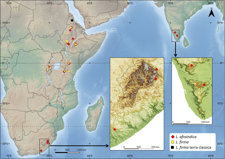
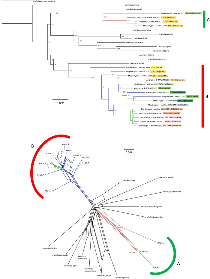
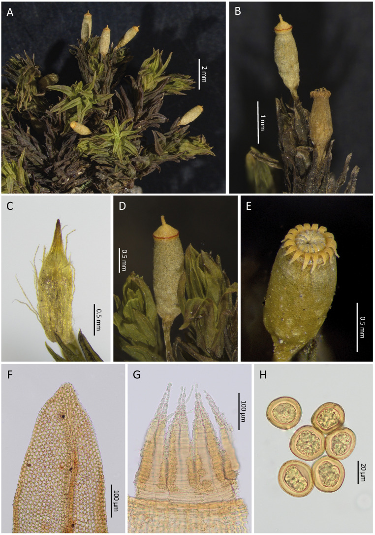
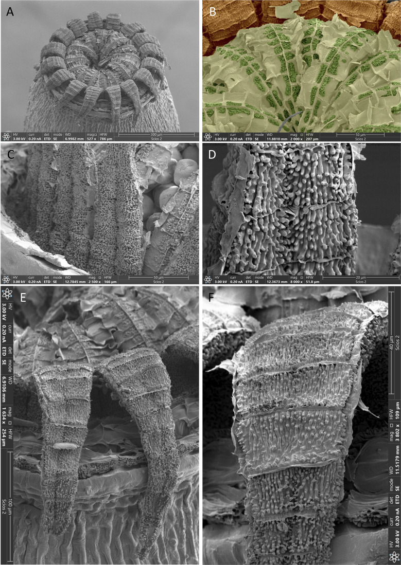
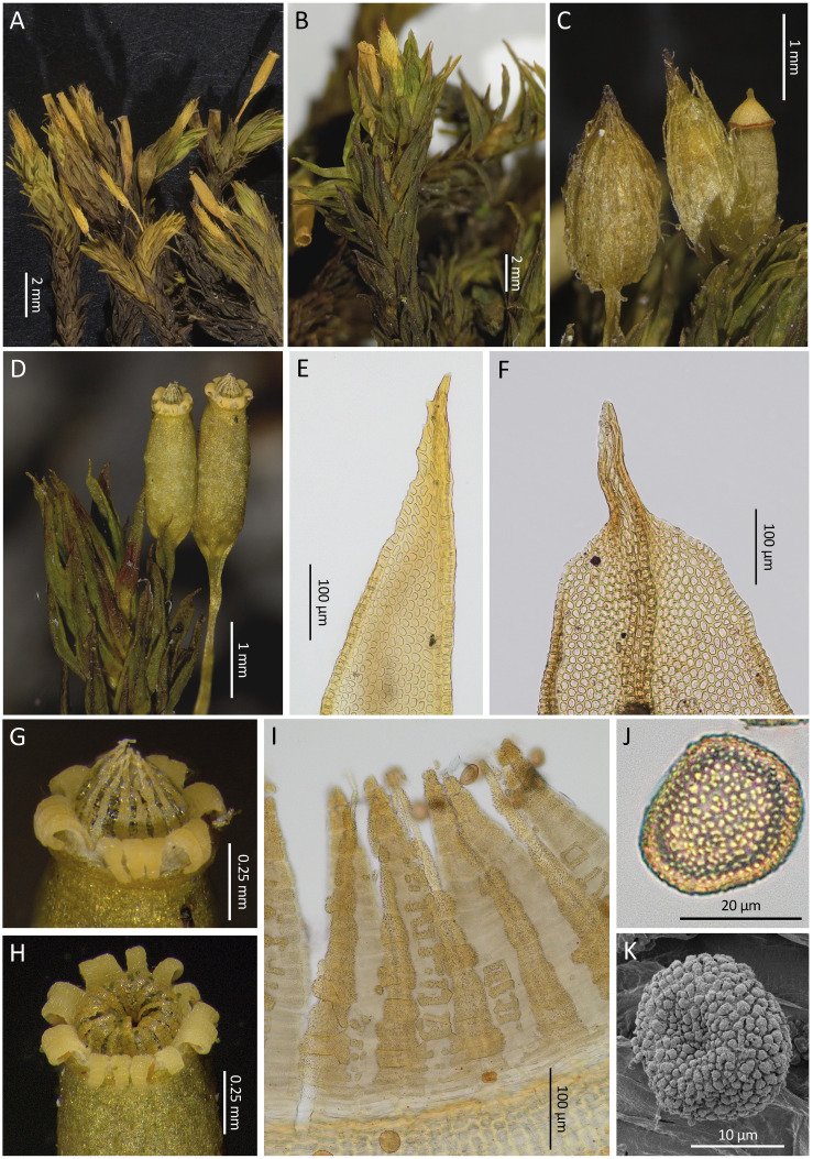
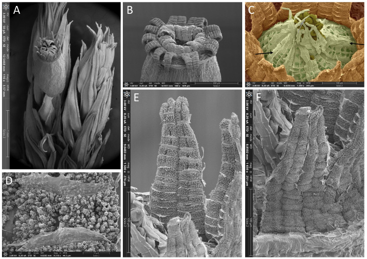
Intercontinental disjunctions are one of the most attractive and interesting biogeographical patterns. Bryophytes often exhibit such distributions, which is partly explained by their great ability to disperse over long distances. However, such intercontinental ranges are sometimes a distorted reality caused by the existence of unnoticed species. This study investigates whether the disjunction between East Africa and southern India of the moss Lewinskya firma reflects the genuine distribution of a single species or implies pseudo-cryptic species (whose morphological differentiation is subtle and have therefore been masked). An integrative taxonomic approach combining morphological and molecular methods (genotyping by sequencing, GBS) was used, based on a representation of samples specifically collected from all the major mountainous regions where this moss occurs. Two species, L. firma s. str. and L. afroindica sp. nov. are involved, whose ranges completely overlap in East Africa, although genetic distance and morphological differences in leaf apex shape, vaginula hairs shape and papillosity, spore ornamentation and peristome constitution and ornamentation allow distinguishing both. In addition, the range of L. afroindica extends into both southern Africa and southern India. The phylogenetic reconstruction obtained shows a certain degree of differentiation of the Indian populations, although they are yet morphologically indistinguishable from African populations. The results thus highlight both the existence of overlooked species and the complexity of bryophyte biogeography.

Keywords: Orthotrichum firmum; biogeography; intercontinental disjunction; mosses; speciation; taxonomy.


Conflict of interest statement

The authors declare that the research was conducted in the absence of any commercial or financial relationships that could be construed as a potential conflict of interest. The handling editor MTB declared a past co-authorship with the author JACA.

Figures

Figure 1
Figure 1
Lewinskya firma complex distribution range. The distribution of Lewinskya firma and Lewinskya afroindica is based on the studied specimens cited in the text. (Map created from base map downloaded from https://www.naturalearthdata.com, and regional DEM downloaded from https://gdemdl.aster.jspacesystems.or.jp).
Figure 2
Figure 2
Lewinskya firma complex phylogenetic reconstruction, TOP: phylogenetic tree inferred by Maximum Likelihood, based on concatenated DNA sequences of 7,086 loci obtained by genotyping by sequencing (c0.95m4 dataset), with clade bootstrap support indicated beside the nodes. Branch colors indicate the morphotypes (1, red; 2 and 4, blue; and 3, green). ETH, Ethiopia; IND, India; RWA, Rwanda; TZA, Tanzania; SAF, South Africa. BOTTOM: Splits network showing potential phylogenetic conflicts among the samples, with the same branch colors to indicate the morphotypes.
Figure 3
Figure 3
Lewinskya firma. (A) Habit. (B), Detail of two capsules in different stages of development. (C), Calyptra. (D), Operculate capsule with a conspicuous thin red basal ring in the lid. (E), A typical peristome when dry, with 16 recurved exostome teeth and 16 incurved endostome segments; note the hyaline and shine layer connecting the endostome segments. (F), vegetative leaf apex. (G), Detail of the peristome; the endostome segments seem not to be connected to the capsule mouth since their basal parts are hyaline and lack any ornamentation. (H), Spores. Source of images: (A, C, D), MAUAM 3632; (B, E, G, H), MAUAM 3670; (F), MAUAM 3623. Photographs by R. Garilleti & R. D. San Román.
Figure 4
Figure 4
Lewinskya firma, SEM micrographs. (A) General view of peristome. (B) Endostome external side (endostome Primary Peristomial Layer, PPL) in false color, where it can be seen that it is a continuous layer (light green) extending both on the segments and over the space between them; each segments can be identified by a median vertical line separating two rows of cell areas, most of which are partially ornamented with papillae (highlighted in dark green); the layer (PPL) between the segments have no papillae although its cell areas are clearly delimited by horizontal lamellae; in the upper part of the picture, the inner side (exostome Primary Peristome Layer) of the basal portion of some exostome teeth (in brown) can be seen. (C) External side of exostome (OPL, Outer Peristomial Layer); note the almost smooth spores in the background. (D) Detail of the OPL ornamentation. (E) General view of two recurved exostome teeth; the endostome can be seen in the upper part of the image. (F) Detail of the ornamentation of exostome internal side (exostome Primary Peristome Layer). Source of images: (A–C, F), MAUAM 3670; (D, E), MAUAM 3650. Photographs by R. Garilleti.
Figure 5
Figure 5
Lewinskya afroindica. (A) Habit of a specimen with exserted capsules. (B) Detail of the habit of a specimen with immersed to hemiemergent capsules. (C) Calyptrae and an operculate capsule. (D) Two mature capsules. (E, F) Variability in the apices of vegetative leaves. (G, H) Aspect of peristomes when dry, with the revolute exostome teeth in 8 pairs, some of them split, and the endostome segments erect or incurved; the hyaline layer connecting the segments can be seen in both cases thanks to its brightness. (I) Detail of the peristome from the inside showing in the foreground the Inner Peristome Layer (IPL) which is densely papillose; the IPL develops along the segments and in a fragmentary manner, irregularly pectinate, in the spaces between segments; the endostome Primary Peristome Layer (PPL) is not visible, because although continuous it is a thin and transparent layer that is only partially ornamented in some central areas of the segments, being hidden by the papillosity of the IPL. (J) LM image of a spore. (K) SEM image of a spore. Source of images: (A), MAUAM 3628; (B), MAUAM 3627; (C), MAUAM 5590; (D, G), MAUAM 5603; (E), MAUAM 3643; (F), MAUAM 5584; (H, J), MAUAM 3647; (I), MAUAM 3638; (K), MAUAM 5600. Photographs by R. Garilleti & R. D. San Román.
Figure 6
Figure 6
Lewinskya afroindica, SEM micrographs. (A) Upper part of a gametophore with one capsule; note the long cuspidate apices of the upper leaves and the faintly ribbed upper part of the capsule. (B) General view of the peristome. (C) Detail of a peristome in false color, centered in the endostome (light green); only the external side (Primary Peristomial Layer, PPL) of the endostome can be seen, with the segments traversed by a median vertical line from which transversal lines delimit two rows of cell areas, some of which are partially ornamented with papillae (highlighted in dark green); this layer (PPL) also extends between the segments although it is only visible in some spaces where it is not folded (arrows), being the cellular areas barely marked and devoid of ornamentation by papillae; the exostome teeth (in brown) have become semi-erect after removal of the operculum; the spores (dark olive green) remain retained by the tips of the segments in this initial phase of the spore release. (D) Detail of the ornamentation of the exostome internal side (exostome Primary Peristome Layer). (E) General view of the internal side (exostome PPL) of two paired teeth. (F) External side (OPL, Outer Peristome Layer) of two paired teeth. Source of images: (A, B), MAUAM 3647; (C–F), MAUAM 5585. Photographs by R. Garilleti.

Similar articles

References

    1. Alonso A., Gallego-Narbón A., Coca-de-la-Iglesia M., Monjas D., Medina N. G., Fernández-Mazuecos M., et al. . (2022). Climatic niche pre-adaptation facilitated island colonization followed by budding speciation in the Madeiran ivy (Hedera maderensis, Araliaceae). Front. Plant Sci. 13. doi: 10.3389/fpls.2022.935975 - DOI - PMC - PubMed
    1. Bickford D., Lohman D. J., Sodhi N. S., Ng P. K. L., Meier R., Winker K., et al. . (2007). Cryptic species as a window on diversity and conservation. Trends Ecol. Evol. 22, 148–155. doi: 10.1016/j.tree.2006.11.004 - DOI - PubMed
    1. Breinholt J. W., Carey S. B., Tiley G. P., Davis E. C., Endara L., McDaniel S. F., et al. . (2021). A target enrichment probe set for resolving the flagellate land plant tree of life. Appl. Pl. Sci. 9, e11406. doi: 10.1002/aps3.11406 - DOI - PMC - PubMed
    1. Brinda J. C., Atwood J. J. (2023). “A synopsis of Orthotrichaceae,” in The bryophyte nomenclator. Available at: https://www.bryonames.org/nomenclator?group=orthotrichaceae.
    1. Bryant D., Moulton V. (2004). Neighbor-Net: an agglomerative method for the construction of phylogenetic networks. Mol. Biol. Evol. 21, 255–265. doi: 10.1093/molbev/msh018 - DOI - PubMed

LinkOut - more resources