[Preprint]. 2024 Sep 23:rs.3.rs-5002222.
doi: 10.21203/rs.3.rs-5002222/v1.

MECHANISMS OF TECOVIRIMAT ANTIVIRAL ACTIVITY AND POXVIRUS RESISTANCE

Affiliations

MECHANISMS OF TECOVIRIMAT ANTIVIRAL ACTIVITY AND POXVIRUS RESISTANCE

Riccardo Vernuccio et al. Res Sq. .

Update in

  • Structural insights into tecovirimat antiviral activity and poxvirus resistance.
    Vernuccio R, Martínez León A, Poojari CS, Buchrieser J, Selverian CN, Jaleta Y, Meola A, Guivel-Benhassine F, Porrot F, Haouz A, Chevreuil M, Raynal B, Mercer J, Simon-Loriere E, Chandran K, Schwartz O, Hub JS, Guardado-Calvo P. Vernuccio R, et al. Nat Microbiol. 2025 Mar;10(3):734-748. doi: 10.1038/s41564-025-01936-6. Epub 2025 Feb 12. Nat Microbiol. 2025. PMID: 39939832 Free PMC article.

Abstract

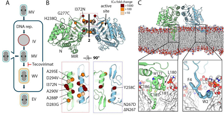
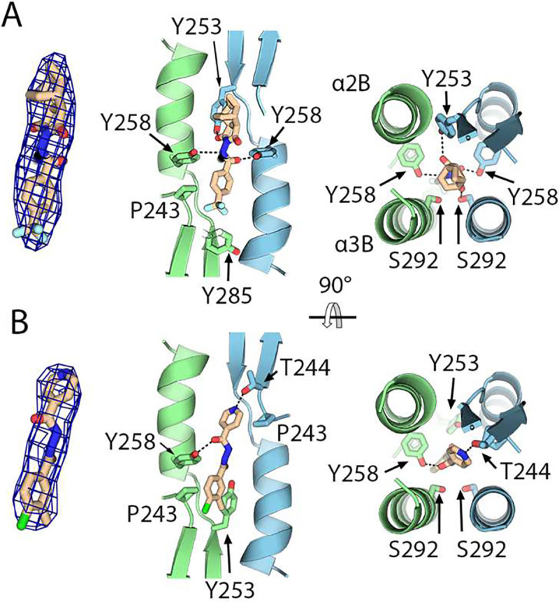
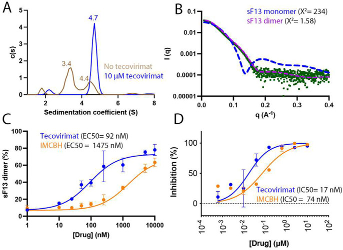
Mpox is a zoonotic disease endemic in central and west Africa. However, since 2022, human-adapted mpox virus (MPXV) strains are causing large outbreaks spreading outside these regions, leading the World Health Organization to declare public health emergency twice. Tecovirimat, the most widely used drug to treat these infections, blocks viral egress through a poorly understood mechanism. Tecovirimat-resistant strains, all with mutations in the viral phospholipase F13, pose public health concerns. Herein, we report the structure of an F13 homodimer, both alone and in complex with tecovirimat. We demonstrate that tecovirimat acts as a molecular glue, inducing the dimerization of the phospholipase. F13 escape mutations in MPXV clinical isolates are at the dimer interface and prevent drug-induced dimerization in solution and cells. These findings, which decipher tecovirimat's mode of action, will allow better monitoring of poxvirus outbreaks and pave the way for developing more potent and resilient therapeutics.


Conflict of interest statement

Competing interests: Authors declare that they have no competing interests.

Figures

Fig. 1.
Fig. 1.. F13 forms a homodimer that can be inserted into a membranes surface.
(A) Schematic representation of the replication cycle of OPXVs. Mature viruses (MV) enter cell fusing their membrane (in blue) with the cellular one. After DNA replication, immature particles are formed (IV, membrane in red), which give rise to intracellular MV particles in the cytoplasm of the infected cell. MVs can either be released by lysis or wrapping. In the latter, MVs acquire two additional membranes (in yellow) from the Golgi apparatus or endosomal vesicles to form wrapped virions (WVs), fuse the outermost with the plasma membrane and release enveloped viruses (EVs). Tecovirimat blocks wrapping, as indicated. (B) Crystal structure of the sF13 homodimer represented in cartoon. One protomer is colored blue and the other green. The N-termini and the membrane-interacting region (MIR) are indicated on one protomer, and the phospholipase active site is indicated on the other. Bottom panels provide close-up views of the two regions forming the dimer interface, indicated by colored rectangles in the upper panel. All single escape mutants identified to date are shown as spheres, colored according to their potency, reported as IC50 fold change. (C) Side view of the F13 homodimer interacting with a lipid membrane that mimics Golgi membrane composition, as observed from MD simulations. For clarity, water molecules and lipids in the foreground of the membrane are not shown. sF13 chains are colored as in (B), with palmitoylated cysteines and hydrophobic residues in the MIR and N-termini depicted as sticks. The bottom panel provides close-up views to show lipid-protein interactions, with the protein residues involved in the interaction depicted as sticks and labeled. Protein carbons are colored according to the chain, membrane carbons in white. Nitrogen, oxygen, sulphur, and phosphate atoms are colored blue, red, yellow, and orange, respectively.
Fig. 2.
Fig. 2.. Tecovirimat binding site.
(A) Crystal structure of the sF13/tecovirimat complex. The left panel shows a Fo-Fc omit map contoured at 3σ showing the density found at the dimer interface in the soaked crystal with the tecovirimat molecule modeled. The central and right panels provide orthogonal views of the dimerization interface with the tecovirimat molecule modeled and the residues contacting the drug represented as sticks and labeled. sF13 chains are colored as in Fig 1. (B) Crystal structure of the sF13/IMCBH complex. As in panel B, the left panel shows an omit map showing the electron density at the dimer interface in the soaked crystal, the central and right panels provide orthogonal views showing the sF13/IMCBH contacts.
Figure 3.
Figure 3.. Tecovirimat induces sF13 dimerization in solution.
(A) Analytical ultracentrifugation (AUC) analysis of sF13 without tecovirimat (brown line) and with 10 μM tecovirimat (black line). Experimentally derived sedimentation coefficient values (Svedberg units [S]) are shown above each peak. (B) Experimental SAXS profile (green dots) and theoretical profiles (dashed) calculated using CRYSOL for one monomer of sF13 (blue line) and the dimer shown in Fig. 1 (pink line). (C) Dose-response curve used to estimate tecovirimat effect in solution. The Y-axis represents the proportion of dimers in a dilute solution of F13 measured by mass photometry (MP). The X-axis represents the concentration of drug (tecovirimat or IMCBH) present in the solution. The EC50 values were determined from a dose-response curve fitted using GraphPad Prism. (D) Tecovirimat (blue line) and IMCBH (orange) inhibits plaque formation of MPXV. Vero cells were infected with MPXV clade IIb and treated with the indicated concentrations of tecovirimat or IMCBH. Plaque inhibition is expressed as a percentage, normalized to control conditions. Data are presented as mean and standard deviation.
Figure 4.
Figure 4.. Escape mutants identified in mpox patients prevent tecovirimat-induced dimerization.
(A) Mass-photometry-based dose-response curve showing tecovirimat activity against different escape mutants, as indicated. (B) Analytical ultracentrifugation (AUC) analysis of sF13A295E (left panel) and sF134MUT (right panel) without tecovirimat (brown line) and with tecovirimat (black line). Experimentally derived sedimentation coefficient (S) values are shown above each peak. (C) and (D) are two orthogonal views showing the dimer interface of sF13A295E (cyan, panel C) and sF13A295E/tecovirimat (green, panel D) superimposed on sF13WT (orange). Residues E295, R291, Y285, and N300 are represented as sticks and labeled.
Figure 5.
Figure 5.. Tecovirimat induces F13 dimerization in cells.
(A) Schematic model representing the PLA experiment. F13 protomers are colored green and cyan with the approximate location of the flag tag indicated with a blue sphere. The three steps of the assay: dimerization, ligation and amplification, are indicated. (B) The left panel are representative fluorescence microscopy images with the nuclei colored in blue and the PLA signal in red. Scale bars: 100 μm. The right panel is the quantification of the PLA signal as the average area of PLA fluorescence per cell. 7000 to 12000 cells were analyzed per data point. Data are mean±sd of two independent experiments performed in triplicat (n=6). Statistical analysis: Two-Way ANOVA. ns: non-significant, ****p < 0.0001.
Figure 6.
Figure 6.. Structure-based escape mutations do not generate viable viruses.
(A) Close view of the dimerization interface across the two-fold axis showing the designed mutations S292F, S292K and L296Y and the mutation identified in VARV, R291E. The circle indicates the localization of the tecovirimat-binding site. (B) MP-based dose-response curve showing tecovirimat activity against different mutants, as indicated. (C) Viral titers in PFU/mL (left panel) and plaque size (right panel) in the presence (+) and absence (−) of 10 μM tecovirimat calculated from plaque assays. Each bar represents the means ± standard deviation (SD) from replicate experiments. The limit of detection (LOD) is marked by the dashed horizontal line.

References

    1. Yang Z., Gray M. & Winter L. Why do poxviruses still matter? Cell Biosci 11, 96, doi:10.1186/s13578-021-00610-8 (2021). - DOI - PMC - PubMed
    1. Thornhill J. P. et al. Monkeypox Virus Infection in Humans across 16 Countries – April-June 2022. N Engl J Med 387, 679–691, doi:10.1056/NEJMoa2207323 (2022). - DOI - PubMed
    1. Cohen J. Africa steps up battle against mpox outbreaks. Science 384, 373–374, doi:10.1126/science.adq0315 (2024). - DOI - PubMed
    1. Moschetta N. et al. Mpox neutralising antibodies at 6 months from mpox infection or MVA-BN vaccination: a comparative analysis. Lancet Infect Dis 23, e455–e456, doi:10.1016/S1473-3099(23)00571-6 (2023). - DOI - PubMed
    1. Freyn A. W. et al. An mpox virus mRNA-lipid nanoparticle vaccine confers protection against lethal orthopoxviral challenge. Sci Transl Med 15, eadg3540, doi:10.1126/scitranslmed.adg3540 (2023). - DOI - PubMed

Publication types

LinkOut - more resources