Review
. 2024 Nov 4;223(11):e202403051.
doi: 10.1083/jcb.202403051. Epub 2024 Oct 14.

Migrasomes: Biogenesis, physiological roles, and therapeutic potentials

Affiliations
Review

Migrasomes: Biogenesis, physiological roles, and therapeutic potentials

Haifeng Jiao et al. J Cell Biol. .

Abstract

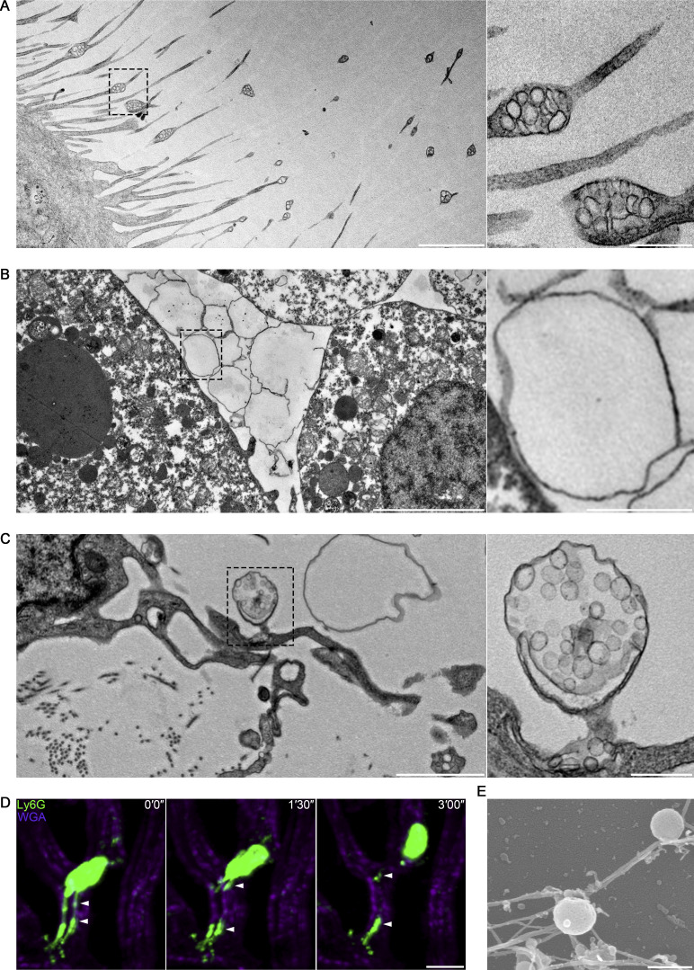
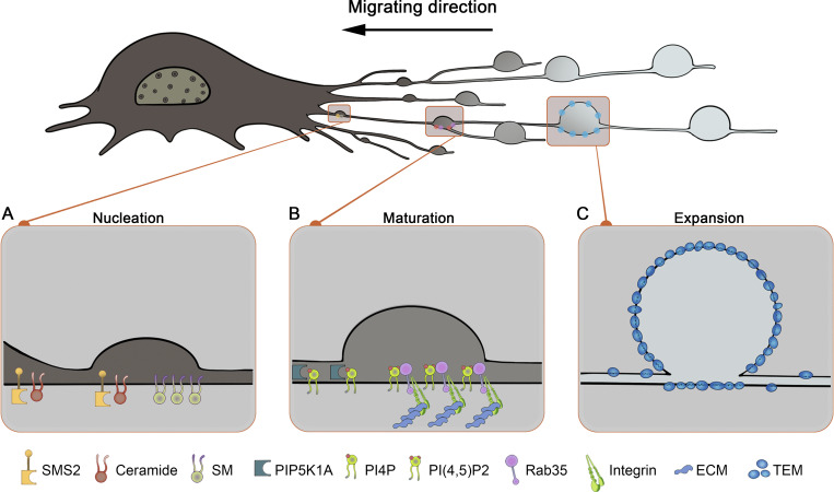
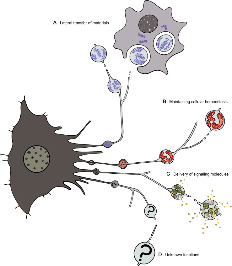
Migrasomes, vesicular structures discovered in migrating cells, arise from the junctions or tips of retraction fibers, and gradually grow to microscale vesicles. Migrasomes have garnered attention for their role in intercellular communication and potential therapeutic implications. This review presents an overview of recent advances in migrasome biology, covering the mechanisms of migrasome biogenesis, essential physiological roles, and their association with various diseases, alongside potential therapeutic applications. Furthermore, we share our perspectives on potential future directions in the study of migrasomes and highlight the challenges that remain in this developing area of research.


Conflict of interest statement

Disclosures: The authors declare no competing interests exist.

Figures

Figure 1.
Figure 1.
Migrasomes are widely distributed in many migrating cells in vitro and in vivo. (A) Representative transmission electron microscopy image of an L929 cell (mouse fibroblast cell line). The right panel shows enlarged pomegranate-like structures, which we named migrasomes. (B) Transmission electron microscopy image of a migrasome-enriched pocket between the yolk syncytial layer and mesendodermal cells in a zebrafish gastrula. The right panel shows an enlarged migrasome. (C) Transmission electron microscopy image of chicken embryo from day 9 in embryo CAM. The right panel shows an enlarged migrasome. (D) Intravital imaging of circulating neutrophil and neutrophil-derived migrasomes in mouse liver. Neutrophil and neutrophil-derived migrasomes are detected with Ly6G-PE antibody. WGA-F647 labels blood vessels and white arrowheads indicate Ly6G-positive migrasomes. (E) Representative scanning electron microscopy image of migrasomes isolated from blood neutrophils. Scale bars: 2 μm (left), 500 nm (right) (A); 5 μm (left), 1 μm (right) (B); 2 μm (left), 500 nm (right) (C); 10 μm (D); 500 nm (E).
Figure 2.
Figure 2.
Categorization of migrasome biogenesis into three phases. (A) Nucleation; the initial assembly of SMS2 foci marks the first step in migrasome biogenesis. SMS2 will assemble into the SMS2 foci on the basal membrane at the leading edges of migrating cells. These SMS2 foci become more concentrated and remain anchored as the cell migrates, eventually transitioning into RFs and forming MFSs, which determine the migrasome formation sites. (B) Maturation; the initial synthesis of PI(4,5)P2 and subsequent recruitment of Rab35 and integrin α5 indicate the maturation phase in migrasome development. PI(4,5)P2, synthesized de novo at MFSs by PIP5K1A, can recruit Rab35 to MFSs. Through the Rab35–integrin α5 interaction, Rab35 specifically targets and localizes integrin α5 to the bottom of MFSs. This recruitment initiates a tetraspanin-mediated expansion phase. (C) Expansion; the recruitment of tetraspanins to MFSs marks the beginning of the migrasome expansion phase. TSPAN4 primarily forms TEMs with cholesterol in the constricted regions on the RF tube. These TEMs are subsequently recruited to swelling regions of RFs, which can be further assembled into larger macrodomains, termed TEMAs. These TEMAs then transition into the vesicle-like structure, typical of migrasomes, to facilitate the final expansion of migrasomes.
Figure 3.
Figure 3.
Modes of migrasome functions. (A) Migrasomes act as vehicles for material and information exchange between adjacent cells. When engulfed by surrounding cells, the contents of the migrasome such as mRNA are transferred to the recipient cells, affecting neighboring cells. (B) Migrasomes mediate the maintenance of cellular homeostasis. Damaged mitochondria can be selectively transported into migrasomes and expelled from cells, in a process termed mitocytosis. Mitocytosis is an important mitochondrial quality-control process in migrating cells, which couples mitochondrial homeostasis with cell migration. (C) Migrasomes serve as efficient vehicles for targeted delivery of signaling molecules at specific sites. Signaling molecules such as cytokines, chemokines, morphogens, and growth factors are packaged into migrasomes from various routes. The release of signaling molecules from migrasomes can create migrasome-centered local gradients of these molecules, providing a mechanism to generate localized microgradients. The controlled release of these signaling molecules and spatially defined locations of migrasomes indicate that migrasomes can integrate spatial and biological information. (D) Unknown functions of migrasome still await discovery.

Similar articles

Cited by

References

    1. Ardalan, M., Hosseiniyan Khatibi S.M., Rahbar Saadat Y., Bastami M., Nariman-Saleh-Fam Z., Abediazar S., Khalilov R., and Zununi Vahed S.. 2022. Migrasomes and exosomes; different types of messaging vesicles in podocytes. Cell Biol. Int. 46:52–62. 10.1002/cbin.11711 - DOI - PubMed
    1. Arya, S.B., Collie S.P., and Parent C.A.. 2024. The ins-and-outs of exosome biogenesis, secretion, and internalization. Trends Cell Biol. 34:90–108. 10.1016/j.tcb.2023.06.006 - DOI - PMC - PubMed
    1. Charrin, S., le Naour F., Silvie O., Milhiet P.E., Boucheix C., and Rubinstein E.. 2009. Lateral organization of membrane proteins: Tetraspanins spin their web. Biochem. J. 420:133–154. 10.1042/BJ20082422 - DOI - PubMed
    1. Chen, L., Ma L., and Yu L.. 2019. WGA is a probe for migrasomes. Cell Discov. 5:13. 10.1038/s41421-018-0078-2 - DOI - PMC - PubMed
    1. Deniz, I.A., Karbanová J., Wobus M., Bornhäuser M., Wimberger P., Kuhlmann J.D., and Corbeil D.. 2023. Mesenchymal stromal cell-associated migrasomes: A new source of chemoattractant for cells of hematopoietic origin. Cell Commun. Signal. 21:36. 10.1186/s12964-022-01028-6 - DOI - PMC - PubMed

LinkOut - more resources