. 2024 Oct 14;148(1):53.
doi: 10.1007/s00401-024-02805-y.

Neuropathological spectrum of anti-IgLON5 disease and stages of brainstem tau pathology: updated neuropathological research criteria of the disease-related tauopathy

Affiliations

Neuropathological spectrum of anti-IgLON5 disease and stages of brainstem tau pathology: updated neuropathological research criteria of the disease-related tauopathy

Ellen Gelpi et al. Acta Neuropathol. .

Abstract

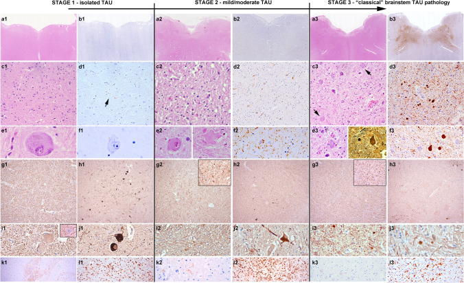
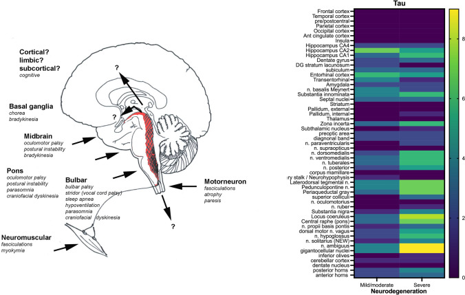
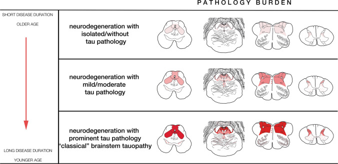
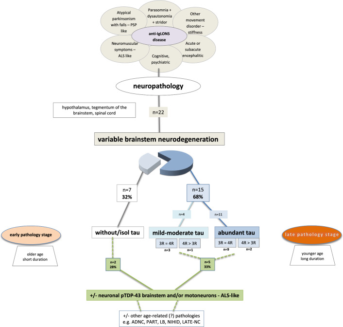
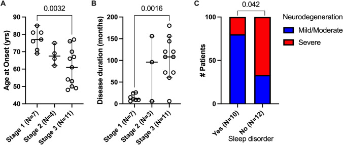
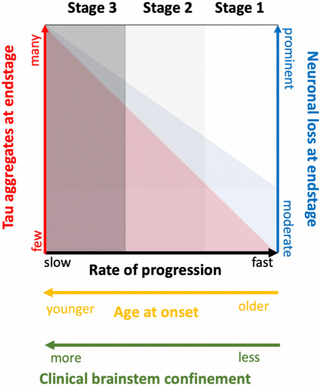
Anti-IgLON5 disease is a unique condition that bridges autoimmunity and neurodegeneration. Since its initial description 10 years ago, an increasing number of autopsies has led to the observation of a broader spectrum of neuropathologies underlying a particular constellation of clinical symptoms. In this study, we describe the neuropathological findings in 22 patients with anti-IgLON5 disease from 9 different European centers. In 15 patients (68%), we observed a hypothalamic and brainstem-predominant tauopathy of varying severity in which the original research neuropathological criteria were readily applicable. This pathology was observed in younger patients (median age at onset 61 years) with a long disease duration (median 9 years). In contrast, in 7 (32%) patients, the originally described brainstem tauopathy was nearly absent or only minimal in the form of delicate threads, despite mild-to-moderate neurodegenerative features, consistent clinical symptoms and the presence of anti-IgLON5 antibodies in CSF and serum. These patients were older at onset (median 79 years) and had shorter disease duration (median < 1 year). Overall, about one-third of the patients showed concomitant TDP-43 pathology within the regions affected by tau pathology and/or neurodegeneration. Based on these observations and in view of the spectrum of the tau burden in the core regions involved in the disease, we propose a simple staging system: stage 1 mild neurodegeneration without overt or only minimal tau pathology, stage 2 moderate neurodegeneration and mild/ moderate tauopathy and stage 3 prominent neurodegeneration and tau pathology. This staging intends to reflect a potential (age- and time-dependent) progression of tau pathology, supporting the current notion that tau accumulation is a secondary phenomenon related to the presence of anti-IgLON5 antibodies in the CNS. Finally, we adapt the original research criteria of the anti-IgLON5 disease-related tauopathy to include the spectrum of pathologies observed in this larger postmortem series.

Keywords: 3R; 4R tau; ALS; Anti-IgLON5 disease; Anti-IgLON5 tauopathy; Atypical; Brainstem tauopathy; Dementia; IgLON5; Motor neuron disease; Neuropathology; PSP; Stages; TDP-43.


Figures

Fig. 1
Fig. 1
Histological findings at the different stages of pathology. Representative histological images of the neuropathological features of anti-IgLON5 disease at different grades of severity. a1f1, a2f2 and a3f3: First row: HE and Tau immunohistochemistry at the level of the medulla oblongata. At low magnification (a1b3), extensive tau pathology can be clearly observed in the tegmentum at stage 3 (b3), but not at stage 1 (b1) and barely at stage 2 (b2). Second row: at higher magnification (c1d3), Tau pathology is already visible at stage 2 (d2) in form of neuropil threads and pretangles, while at stage 1 isolated delicate threads are visible (d1, arrow). In contrast, HE-stained sections in stage 1 (c1) and stage 2 (c2) already show reactive changes in the reticular formation. In addition, some enlarged neurons are observed at stage 1 and stage 2 (e1, e2), that are tau negative, while at stage 3, they are already containing tau-positive neurofibrillary tangles, but are overall reduced in numbers. These enlarged neurons show a slightly increased immunoreactivity for phosphorylated (SMI31; g1, i1) and particularly of non-phosphorylated (SMI32; h1, j1) neurofilaments, and have a regular axonal density and morphology. In stage 2 and stage 3, the number of enlarged neurons decreases (h2, j2 and h3, j3) but an increase in axonal spheroids is detected (i2, i3). k1, k2, k3: Immmunohistochemistry for IgG4 in the brainstem shows focally marked deposits at stage 1 (k1), mild deposits in single cases at stage 2 (k2) and no deposits at stage 3 (k3). i1, i2, i3: different grades and states of microglial activation at different disease stages (HLA-DR immunohistochemistry)
Fig. 2
Fig. 2
Left: graphical neuroanatomical representation of the main focus of anti-IgLON5 disease neuropathology and possible related clinical symptoms. Right: grouped analysis and heat map; source of variations: tau burden (0 = absent or mild; 1 = moderate or prominent) and neurodegeneration (mild/moderate vs severe) (two-way ANOVA, source of variation: anatomical region: p = 0.0004; degree of neurodegeneration: p < 0.0001)
Fig. 3
Fig. 3
Pathology burden. Graphical representation of the severity of neurodegeneration and tau burden in the brainstem regions observed at the different stages of pathology: midbrain (leftmost), pons (left middle), medulla oblongata (right middle), and upper cervical cord (rightmost), and their correlation with age at onset and duration of disease on the left. Color codes: upper line: the focus of neurodegeneration in the brainstem is represented as a pale-red area with minimal or even absent tau pathology; middle line: the mild-to-moderate deposition of tau is represented in softer red; lower line: prominent tau pathology is represented in intense red and areas with milder or occasional pathology in form of pale-red dots (s. nigra midbrain, pontine base, inferior olives). Figure adapted from the original criteria [23]. Stage and pathology burden. Stage 1 (upper row): mild/moderate neuronal loss/gliosis and isolated tau-positive neuropil threads in the lateral segments of the midbrain and pontine tegmentum and/or reticular formation/respiratory nuclei of the medulla oblongata and/or hypothalamic nuclei/preoptic area/pituitary stalk. Stage 2 (middle row): moderate neuronal loss/gliosis and moderate tau-positive neuropil threads, pretangles and NFT in the previous regions extending to adjacent tegmental nuclei and hypothalamic /prehypothalamic nuclei. Stage 3 (lower row): Prominent neuronal loss/gliosis and extensive tau pathology in the aforementioned regions + mild involvement of s. nigra, inferior olives, dentate nucleus of the cerebellum and glomerula of the granule cell layer of the cerebellar cortex
Fig. 4
Fig. 4
Summary of the neuropathological findings in 22 patients with anti-IgLON5 disease. Of these, 68% showed a brainstem tauopathy, while 32% did not. The latter group was represented by older patients with shorter disease duration, compared to the patients with brainstem tauopathy, who were younger at disease onset and had longer disease duration
Fig. 5
Fig. 5
Correlation analysis between the different pathology stages and A age of onset and B disease duration; and C between the presence of sleep disorder and degree of neurodegeneration (A, B one-way ANOVA with Tukey’s multiple comparisons test, C Fisher’s exact test)
Fig. 6
Fig. 6
Potential relationship between grades of severity of neurodegeneration of the brainstem (mild—severe), tau accumulation (few—prominent), age at onset (older—younger) and rate of disease progression (fast – slow)

References

    1. Acewicz A, Stępień T, Grzegorczyk M, Ostrowski RP, Tarka S, Felczak P et al (2024) TDP-43 pathology in subacute sclerosing panencephalitis. J Neuropathol Exp Neurol 83:251–257. 10.1093/jnen/nlae017 - PubMed
    1. Alkabie S, Murray BJ, Budhram A (2023) Incidental detection of anti-IgLON5: a diagnostic and therapeutic dilemma. Can J Neurol Sci 26:1–7. 10.1017/cjn.2023.239 - PubMed
    1. Al-Sarraj S, King A, Troakes C, Smith B, Maekawa S, Bodi I et al (2011) p62 positive, TDP-43 negative, neuronal cytoplasmic and intranuclear inclusions in the cerebellum and hippocampus define the pathology of C9orf72-linked FTLD and MND/ALS. Acta Neuropathol 122:691–702. 10.1007/s00401-011-0911-2 - PubMed
    1. Ahmed Z, Josephs KA, Gonzalez J, DelleDonne A, Dickson DW (2008) Clinical and neuropathologic features of progressive supranuclear palsy with severe pallido-nigro-luysial degeneration and axonal dystrophy. Brain 131:460–472. 10.1093/brain/awm301 - PubMed
    1. Askin B, Cordero Gómez C, Lan-Linh Duong S, Kilic C, Goihl A, Newman A et al (2024) Autoimmune antibody-induced neuronal hyperactivity triggers pathological Tau in IgLON5 disease. bioRxiv. 10.1101/2024.03.10.584272

Publication types

LinkOut - more resources