. 2024 Oct 14;81(1):434.
doi: 10.1007/s00018-024-05463-1.

Pharmacologically increasing cGMP improves proteostasis and reduces neuropathy in mouse models of CMT1

Affiliations

Pharmacologically increasing cGMP improves proteostasis and reduces neuropathy in mouse models of CMT1

Seth M Moore et al. Cell Mol Life Sci. .

Abstract

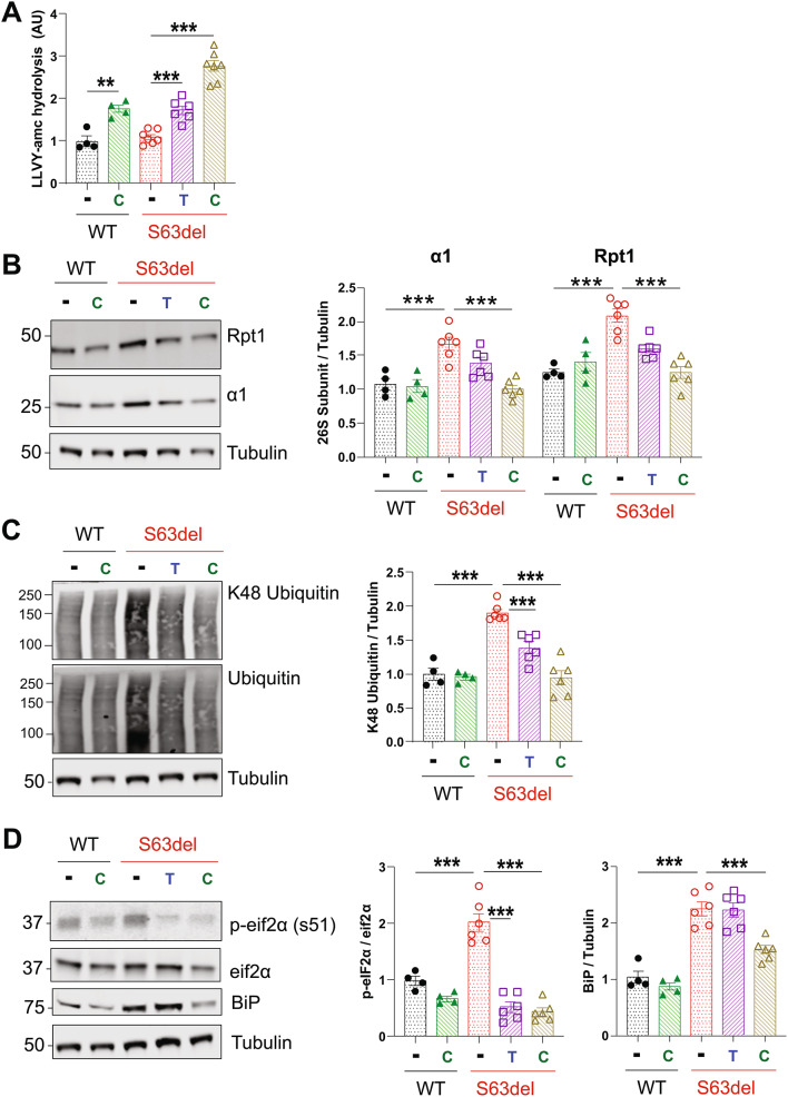
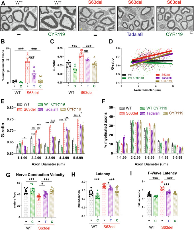
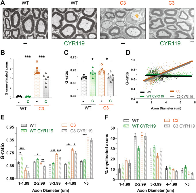
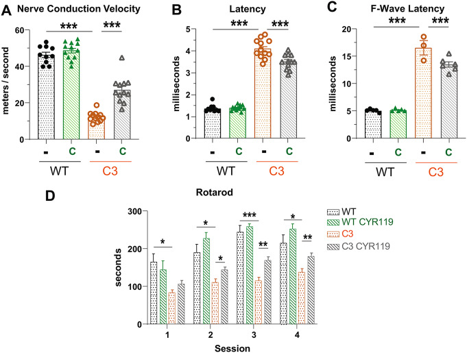
Increasing cyclic GMP activates 26S proteasomes via phosphorylation by Protein Kinase G and stimulates the intracellular degradation of misfolded proteins. Therefore, agents that raise cGMP may be useful therapeutics against neurodegenerative diseases and other diseases in which protein degradation is reduced and misfolded proteins accumulate, including Charcot Marie Tooth 1A and 1B peripheral neuropathies, for which there are no treatments. Here we increased cGMP in the S63del mouse model of CMT1B by treating for three weeks with either the phosphodiesterase 5 inhibitor tadalafil, or the brain-penetrant soluble guanylyl cyclase stimulator CYR119. Both molecules activated proteasomes in the affected peripheral nerves, reduced polyubiquitinated proteins, and improved myelin thickness and nerve conduction. CYR119 increased cGMP more than tadalafil in the peripheral nerves of S63del mice and elicited greater biochemical and functional improvements. To determine whether raising cGMP could be beneficial in other neuropathies, we first showed that polyubiquitinated proteins and the disease-causing protein accumulate in the sciatic nerves of the C3 mouse model of CMT1A. Treatment of these mice with CYR119 reduced the levels of polyubiquitinated proteins and the disease-causing protein, presumably by increasing their degradation, and improved myelination, nerve conduction, and motor coordination. Thus, pharmacological agents that increase cGMP are promising treatments for CMT1 neuropathies and may be useful against other proteotoxic and neurodegenerative diseases.

Keywords: Charcot Marie Tooth; Peripheral neuropathy; Phosphorylation; Proteasome; Protein degradation; cGMP.


Conflict of interest statement

GTM and ESB were employees of Cyclerion Therapeutics and may own stock. All other authors report no conflicts of interest.

Figures

Fig. 1
Fig. 1
Tadalafil and CYR119 increase proteasome peptidase activities and cGMP in sciatic nerves of WT and S63del mice. A.) Tadalafil per os for 7 days increased the chymotrypsin-like activity of proteasomes in sciatic nerve lysates from WT and S63del mice. Here and below, n = 3–5 mice per genotype, per condition, and one-way ANOVA with a Bonferroni post-hoc analysis comparing WT and WT treated, WT and S63del, and S63del and S63del treated. The experiment was repeated with similar results. B.) CYR119 per os for 7 days increased proteasome chymotrypsin-like activity in sciatic nerve lysates from WT and S63del mice. C.) In sciatic nerve lysates from S63del mice, protein levels of the 26S proteasome subunits Rpn6 and β5 were increased. Tadalafil treatment for 7 days did not alter the levels of these subunits in WT or S63del. D.) CYR119 treatment for 7 days did not alter the levels of the 26S proteasome subunits Rpn6 or β5 in WT or S63del. E.) Tadalafil or CYR119 for 7 days increased the levels of cGMP in the sciatic nerve lysates, as measured by a cGMP ELISA. T indicates tadalafil and C indicates CYR119. Sciatic nerves from S63del mice had ~ 50% less cGMP than WT littermates
Fig. 2
Fig. 2
Treating S63del mice for 21 days with tadalafil or CYR119 improves proteostasis in the sciatic nerves A.) Raising cGMP with tadalafil (T) or CYR119 (C) increased proteasomal chymotrypsin-like activity in the sciatic nerve lysates of S63del mice. Here and below, n = 4–7 mice per genotype, per condition, and one-way ANOVA with a Bonferroni post-hoc analysis comparing WT and WT CYR, WT and S63del, S63del and S63del Tad, and S63del and S63del CYR. The experiment was repeated with similar results. B.) The levels of 26S proteasome subunits are higher in the sciatic nerves of S63del mice than in WT littermates. Tadalafil and CYR119 decreased the levels of the 20S subunit α1 and the 19S subunit Rpt1 in S63del mice, as determined by western blot. C.) S63del sciatic nerve lysates exhibit a 2-fold increase in polyubiquitinated proteins over WT littermates. Treatment with tadalafil or CYR119 reduced the levels of polyubiquitinated proteins. D.) Two indicators of proteotoxic stress, BiP and p-eif2α, are increased in the sciatic nerve lysates of S63del mice. Tadalafil and CYR119 reduced the levels of p-eiF2α. Only CYR119 reduced the protein levels of BiP
Fig. 3
Fig. 3
Treating S63del mice for 21 days with tadalafil or CYR119 restores myelin thickness and nerve conduction in sciatic nerves. A.) Representative electron microscopic images of ultrathin sections of sciatic nerves. The yellow asterisk indicates an unmyelinated fiber. B.) The % of unmyelinated fibers in sciatic nerves is higher in S63del mice than in WT littermates. The treatment with tadalafil (T) or CYR119 (C) reduced the number of unmyelinated fibers in the sciatic nerves of S63del mice. Here and below, n = 5–7 mice per genotype, per condition and one-way ANOVA with a Bonferroni post-hoc analysis comparing WT and WT CYR, WT and S63del, S63del and S63del Tad, and S63del and S63del CYR. C.) Average myelin thickness was lower in S63del sciatic nerves than in WT and was increased by tadalafil or CYR119. G-ratio = axon diameter / fiber diameter. Therefore, the higher the g-ratio, the thinner the myelin sheath. D.) A scatterplot of the g-ratio distribution across the measured axon diameters. S63del sciatic nerves have thinner myelin across all axon diameters, as indicated by the red line (S63del) being always above the black line (WT). Tadalafil and CYR119 treatments of S63del mice increased myelin thickness across all axon diameters, as indicated by the purple (tadalafil) and gold (CYR119) lines always being under the red line (S63del). E.) Graphs of the g-ratio distribution by axon diameter shown in D. S63del has thinner myelin (higher g-ratio) than WT across all axon diameters. Tadalafil or CYR119 treatments of S63del mice increased myelin thickness (lowered g-ratio) across all axon diameters. F.) The three-week treatment with tadalafil or CYR119 did not cause statistically significant changes in the percentage of myelinated axons of any diameter. G.) Treatment with tadalafil or CYR119 increased conduction velocity in the peripheral nerves of S63del mice. Here and below, n = 5–7 mice per genotype, per condition, corresponding to 10–14 measurements, one per sciatic nerve, and one-way ANOVA with a Bonferroni post-hoc analysis comparing WT and WT CYR, WT and S63del, S63del and S63del Tad, and S63del and S63del CYR. H.) Tadalafil or CYR119 treatment reduced the longer distal latencies in the peripheral nerves of S63del mice. I.) Treatment with tadalafil or CYR119 reduced the longer F-wave latencies in S63del mice
Fig. 4
Fig. 4
Polyubiquitinated proteins and PMP22 accumulate in the sciatic nerves of C3 mice. A.) The levels of polyubiquitinated proteins were higher in sciatic nerve lysates from C3 mice than from WT littermates. n = 3. Student’s t-test. B.) PMP22 accumulated in its glycosylated (20 kDa) and unglycosylated (16 kDa) forms in C3 sciatic nerves. Human PMP22 protein was detected only in lysates from C3 mice, as expected because C3 mice contain 3–4 copies of a human PMP22 transgene. Total PMP22 protein was detected by probing the blots simultaneously with one antibody specific for human PMP22 and a second antibody specific for mouse PMP22. Representative western blot of 3 experiments performed with lysates from 3 distinct mice per genotype. C.) EndoH-sensitive PMP22 is increased in the sciatic nerve lysates of C3 mice. The removal of all glycans in the lysates by PNGase showed that PMP22 is increased in the sciatic nerve lysates of C3 mice. n = 3 mice per genotype and one-way ANOVA with a Bonferroni post-hoc analysis comparing WT and C3. D.) PMP22 is localized in the ER of Schwann cells in sciatic nerves of C3 mice. Sciatic nerves from WT and C3 mice were teased into individual nerve fibers and immunohistochemical analysis was performed for KDEL, a marker of the ER, and human and mouse PMP22. The scale bar represents 5 μm. E.) The levels of cGMP were 50% lower in sciatic nerve lysates from C3 mice than from WT, as measured by cGMP ELISA. n = 5. One-way ANOVA with a Bonferroni post-hoc analysis comparing WT and C3, and WT and S63del. Experiment was repeated with similar results
Fig. 5
Fig. 5
Treatment of C3 mice with CYR119 for 7 days increases proteasome peptidase activity in sciatic nerve lysates and reduces ubiquitin conjugates. A.) Treatment with tadalafil (T) or CYR119 (C) for 7 days increased proteasome chymotrypsin-like activity in sciatic nerve lysates of WT and C3 mice. Baseline proteasome peptidase activity was higher in C3 than in WT mice. Here and below, n = 3 mice per genotype, per condition and one-way ANOVA with a Bonferroni post-hoc analysis comparing WT and WT treated, WT and C3, and C3 and C3 treated. Experiment was repeated with similar results. B.) Levels of 26S proteasome subunits were similar in C3 mice and WT littermates. Neither treatment altered the protein levels of the 19S subunit Rpt1 or the 20S subunit α1. C.) CYR119 for 7 days reduced the levels of polyubiquitinated proteins in WT and C3 sciatic nerve lysates
Fig. 6
Fig. 6
Treating C3 mice with CYR119 for 21 days increases proteasome peptidase activity and decreases the levels of PMP22. A.) CYR119 (C) for 21 days increased proteasome chymotrypsin-like activity in the sciatic nerve lysates of WT and C3 mice. Here and below, n = 4–5 mice per genotype, per condition and one-way ANOVA with a Bonferroni post-hoc analysis comparing WT and WT treated, WT and C3, and C3 and C3 treated. Experiment was repeated with similar results. B.) The levels of the 19S subunit Rpt1 and the 20S subunit α1 were similar in sciatic nerve lysates of WT and C3 mice and were unaltered by the treatment with CYR119. C.) Treating C3 mice for 3 weeks with CYR119 reduced the levels of polyubiquitinated proteins in the sciatic nerve lysates. D.) Glycosylated (20 kDa) and unglycosylated (16 kDa) PMP22 accumulated in the sciatic nerve lysates of C3 mice and their levels were reduced by the 3-week treatment with CYR119. E.) Treatment of C3 mice with CYR119 reduced the levels of glycosylated PMP22 (20 kDa). Here and below, n = 3 mice per genotype, per condition and one-way ANOVA with a Bonferroni post-hoc analysis comparing WT and WT treated, WT and C3, and C3 and C3 treated. F.) Unglycosylated PMP22 (16 kDa) was present only in C3 sciatic nerves and its levels were reduced by the treatment with CYR119. Student’s t-test
Fig. 7
Fig. 7
CYR119 treatment for 21 days improves myelination in the sciatic nerves of C3 mice. A.) Representative electron microscopic images of ultrathin sections of sciatic nerves. The yellow asterisk indicates an unmyelinated fiber. B.) C3 mice had more unmyelinated fibers in their sciatic nerves than WT littermates. The treatment with CYR119 (C) reduced the percentage of unmyelinated fibers. Here and below, n = 5–6 mice per genotype, per condition and one-way ANOVA with a Bonferroni post-hoc analysis comparing WT and WT treated, WT and C3, and C3 and C3 treated. C.) Average myelin thickness was lower (higher g-ratio) in C3 sciatic nerves and was increased (lower g-ratio) by the treatment with CYR119. D.) Scatterplot of the g-ratio distribution across the measured axon diameters. C3 sciatic nerves had thinner myelin (higher g-ratio) than WT around axons with diameters > 2 μm and thicker myelin (lower g ratio) around axons with diameters < 2 μm. E.) Bar graphs of the g-ratio distribution by axon diameter shown in D. CYR119 treatment of C3 mice increased myelin thickness across all axon diameters < 5 μm. F.) The three-week treatment with CYR119 did not cause statistically significant changes in the percentage of myelinated axons of any diameter in C3 mice
Fig. 8
Fig. 8
CYR119 treatment for 21 days increases nerve conduction and motor coordination in C3 mice. A.) Treatment with CYR119 (C) increased conduction velocity in the sciatic nerves of C3 mice. Here and below, n = 5–6 mice per genotype, per condition, corresponding to 10–12 measurements, one per sciatic nerve per mouse and one-way ANOVA with a Bonferroni post-hoc analysis comparing WT and WT treated, WT and C3, and C3 and C3 treated. B.) CYR119 treatment reduced the longer distal latencies seen in C3 mice. C.) Treatment with CYR119 reduced the longer F-wave latencies in C3 mice. n = 3–6 mice per genotype, per condition. Any mouse from which F-wave latencies could not be measured from both left and right sciatic nerves was excluded from the analysis. D.) On an accelerating rotarod, C3 mice fell off sooner than WT littermates. C3 mice treated with CYR119 ran longer than C3 controls in 3 out of the 4 sessions. n = 10–14 mice per genotype, per condition and one-way ANOVA with a Bonferroni post-hoc analysis comparing WT and WT treated, WT and C3, and C3 and C3 treated

References

    1. Fridman V, Saporta MA (2021) Mechanisms and treatments in demyelinating CMT. Neurother J Am Soc Exp Neurother 18:2236–2268. 10.1007/s13311-021-01145-z - PMC - PubMed
    1. Bolino A, D’Antonio M (2023) Recent advances in the treatment of Charcot-Marie-tooth neuropathies. J Peripher Nerv Syst 28:134–149. 10.1111/jns.12539 - PubMed
    1. Siems SB, Jahn O, Eichel MA et al (2020) Proteome profile of peripheral myelin in healthy mice and in a neuropathy model. Elife 9. 10.7554/eLife.51406 - PMC - PubMed
    1. Bai Y, Wu X, Brennan KM et al (2018) Myelin protein zero mutations and the unfolded protein response in Charcot Marie Tooth disease type 1B. Ann Clin Transl Neurol 5:445–455. 10.1002/acn3.543 - PMC - PubMed
    1. Pennuto M, Tinelli E, Malaguti M et al (2008) Ablation of the UPR-mediator CHOP restores motor function and reduces demyelination in Charcot-Marie-tooth 1B mice. Neuron 57:393–405. 10.1016/j.neuron.2007.12.021 - PMC - PubMed

MeSH terms

LinkOut - more resources