. 2025 Jan 2;64(1):e202412994.
doi: 10.1002/anie.202412994. Epub 2024 Nov 13.

Arenicolide Family Macrolides Provide a New Therapeutic Lead Combating Multidrug-Resistant Tuberculosis

Affiliations

Arenicolide Family Macrolides Provide a New Therapeutic Lead Combating Multidrug-Resistant Tuberculosis

Sunghoon Hwang et al. Angew Chem Int Ed Engl. .

Abstract

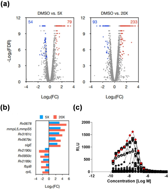
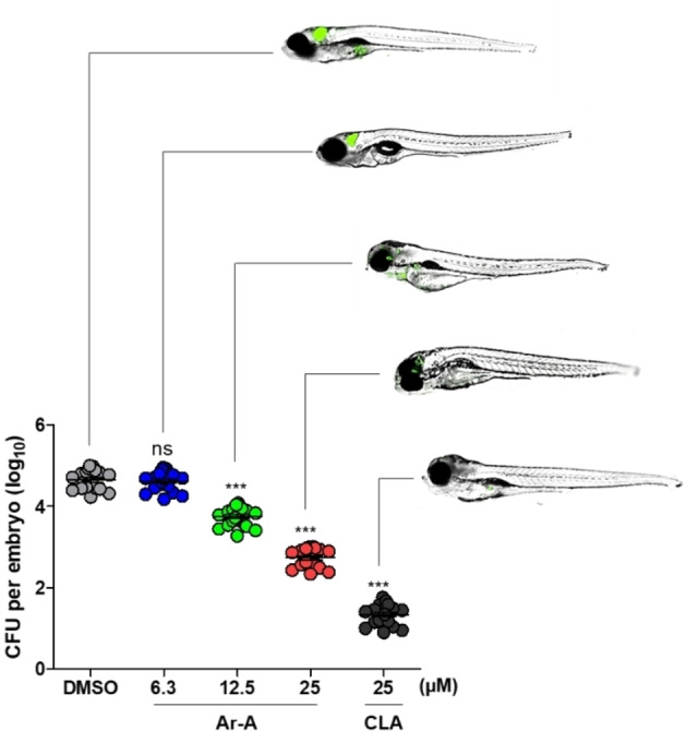
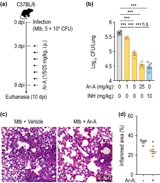
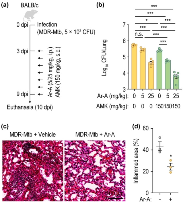
The emergence of multidrug-resistant (MDR) and extensively drug-resistant (XDR) strains of Mycobacterium tuberculosis (Mtb) poses a significant threat to health globally. During searching for new chemical entities regulating MDR- and XDR-Mtb, chemical investigation of the black oil beetle gut bacterium Micromonospora sp. GR10 led to the discovery of eight new members of arenicolides along with the identification of arenicolide A (Ar-A, 1), which was a previously reported macrolide with incomplete configuration. Genomic analysis of the bacterial strain GR10 revealed their putative biosynthetic pathway. Quantum mechanics-based computation, chemical derivatizations, and bioinformatic analysis established the absolute stereochemistry of Ar-A and arenicolides D-K (Ar-D-K, 2-9) completely for the first time. Biological studies of 1-9 revealed their antimicrobial activity against MDR and XDR strains of Mtb. Ar-A had the most potent in vitro antimicrobial efficacy against MDR- and XDR-Mtb. Mechanistically, Ar-A induced ATP depletion and destabilized Mtb cell wall, thereby inhibiting growth. Notably, Ar-A exerted a significant antimicrobial effect against Mtb in macrophages, was effective in the treatment of Mtb infections, and showed a synergistic effect with amikacin (AMK) in a mouse model of MDR-Mtb lung infection. Collectively, our findings indicate Ar-A to be a promising drug lead for drug-resistant tuberculosis.

Keywords: anti-tubercular mechanism; multidrug resistance; natural product; structure determination; tuberculosis.


Conflict of interest statement

The authors submitted patent applications for arenicolides and their biological activities.

Figures

Figure 1
Figure 1
(a) Isolation of Micromonospora sp. GR10 from the gut of the female black oil beetle, Meloe proscarabaeus. (b) Chemical structures of arenicolide A, C and eight new congeners. (c) Key nuclear magnetic resonance (NMR) correlations of Ar−D. (d) The process of conformation search and DP4 calculation to determine the unknown stereochemistry at C‐12.
Figure 2
Figure 2
(a) Organization of the putative arenicolide biosynthetic gene cluster. (b) Proposed biosynthetic pathway of arenicolides.
Figure 3
Figure 3
In vitro and ex vivo activity of Ar−A. (a) Activity of Ar−A against Mtb replicating in culture broth medium. BDQ, INH, STR, ethambutol (EMB), and LZ were used as the positive controls. Data are means±standard deviation (SD) of triplicates for each concentration. (b) Time‐kill kinetics of Ar−A for Mtb over 20 days. Bacteria were grown in Middlebrook 7H9 liquid medium in the presence of the indicated concentrations of Ar−A, BDQ, or INH (control). (c) Dose response of Ar−A in Mtb‐infected RAW264.7 macrophages. Blue bars display macrophage survival, green bars quantify intracellular Mtb‐GFP (mean±SD of 3 independent experiments). Statistical significance of quantitative analysis was determined using one‐way analysis of variance (ANOVA) comparing between treated and DMSO control. *p<0.05; ***p<0.001, ns, not significant. (d) Fluorescent microscopy of Mtb‐GFP‐infected RAW264.7 macrophages after treatment with Ar−A (10 μM) or vehicle (dimethyl sulfoxide; DMSO). Macrophage nuclei were stained with Hoechst 33342 (scale bar, 50 μm).
Figure 4
Figure 4
Ar−A induced ATP depletion in Mtb, by a different mechanism from other drugs that target ATP synthesis. (a) Ar−A induced ATP depletion in replicating Mtb. BDQ was used as the positive control and INH and STR as the negative controls. Data are means±SD of triplicates for each concentration. The Mtb and laboratory‐induced Ar−A‐resistant strains were assessed for ATP reduction for (b) Ar−A and (c) BDQ, respectively. Statistical analysis was conducted using an unpaired t test, and the results are presented as means±SD from the experiment, which was performed in duplicates for each concentration. The significance levels are denoted; ***p<0.001; ns, not significant
Figure 5
Figure 5
Transcriptional profiling and effect of Ar−A on the transcriptome and cell wall synthesis of Mtb. (a) Volcano plot of log2‐fold changes in gene expression induced by Ar−A (5‐ and 20‐fold the minimal inhibitory concentration (MIC) compared to the untreated control. Gray circles, genes with no significant changes; red and blue circles indicate up‐ and downregulated genes, respectively. FC, fold change in transcriptional level in the treatment groups (5‐ and 20‐fold the MIC of Ar−A) compared to the DMSO control. (b) Top 5 up‐ and downregulated genes according to RNA sequencing. (c) The iniBAC bioreporter assay was performed in response to Ar−A at concentrations ranging from 0.4 to 200 μM. Relative luminescence units (RLU) were measured and plotted over a 15‐day period. Red circles highlight the highest RLU peak induced by Ar−A treatment in the iniBAC reporter M. bovis strain on day 8. All symbols in the graph represent results from each different day during the 15 days following Ar−A treatment. Data are expressed as the means±standard deviation (SD) of triplicates for each concentration.
Figure 6
Figure 6
In vivo efficacy of Ar−A in a zebrafish (ZF) model of infection. ZF infected with M. marinum (Mm)–GFP were treated with Ar−A (6.3, 12.5, and 25 μM) and clarithromycin (CLA; 25 μM). The GFP signal in ZF was monitored using a fluorescence microscope. Post‐antibiotic treatment, the bacterial burden in infected ZF was quantified by enumerating CFU. Data are mean log10 CFUs per embryo (n=10 per condition) from three independent experiments. Statistical significance was determined using one‐way analysis of variance (ANOVA) comparing between treated and DMSO control. *** p<0.001.
Figure 7
Figure 7
Therapeutic effect of Ar−A in Mtb‐infected mice. (a) Schematic of the experimental design. Mice were infected with Mtb (5×10⁴ CFU/head) followed by intraperitoneal injection of vehicle, Ar−A (1, 5, or 25 mg/kg), or INH (10 mg/kg) at 3—9 dpi (n=5–6). After 10 days, mice were euthanized, and lung tissues were harvested. (b) Bacterial burden in lungs of Mtb‐infected mice. (c) Representative histopathological images of lung tissues from mice infected with Mtb for 35 days. (d) Quantitative analysis of the inflamed area (n=4). Statistical significance was determined using two‐tailed Student's t‐tests (b and d). Dpi, days post‐infection; Mtb, Mycobacterium tuberculosis; CFU, colony‐forming unit; i. p., intraperitoneal; Ar−A, arenicolide A; INH, isoniazid; n.s., not significant. Data are means±standard error of the mean (SEM) from at least three biological replications (b and d). * p<0.05; *** p<0.001.
Figure 8
Figure 8
Synergistic effect of Ar−A and AMK on MDR‐Mtb‐infected mice. (a) Schematic of the experimental design. Mice were infected with MDR‐Mtb (5×10³ CFU/head) and received intraperitoneal or subcutaneous injection with vehicle, Ar−A (5 or 25 mg/kg), or AMK (150 mg/kg) at 3—9 dpi (n=5). After 10 days, mice were euthanized, and their lungs were collected. (b) Bacterial loads in lungs of MDR‐Mtb‐infected mice. (c) Representative histopathological images of lungs from mice infected with MDR‐Mtb for 35 days. (d) Quantitative analysis of the inflamed areas (n=3). Statistical significance was determined by one‐way ANOVA with Tukey's multiple comparison test (b) or two‐tailed Student's t‐tests (d). Dpi, days post‐infection; MDR‐Mtb, multidrug‐resistant Mycobacterium tuberculosis; CFU, colony forming unit; i. p., intraperitoneal; s.c., subcutaneous; Ar−A, Arenicolide A; AMK, amikacin; n.s., not significant. Data are means±SEM from at least three biological replications (b and d). * p<0.05; *** p<0.001.

References

    1. Barberis I., Bragazzi N. L., Galluzzo L., Martini M., J. Prev. Med. Hyg. 2017, 58, E9–E12. - PMC - PubMed
    1. Moule M. G., Cirillo J. D., Front. Cell. Infect. Microbiol. 2020, 10, 65. - PMC - PubMed
    1. Falzon D., Mirzayev F., Wares F., Baena I. G., Zignol M., Linh N., Weyer K., Jaramillo E., Floyd K., Raviglione M., Eur. Respir. J. 2015, 45, 150–160. - PMC - PubMed
    1. Ou Z.-J., Yu D.-F., Liang Y.-H., He W.-Q., Li Y.-Z., Meng Y.-X., Xiong H.-S., Zhang M.-Y., He H., Gao Y.-H., Wu F., Chen Q., Infect. Dis. Poverty 2021, 10, 1–10. - PMC - PubMed
    1. Williams P. G., Miller E. D., Asolkar R. N., Jensen P. R., Fenical W., J. Org. Chem. 2007, 72, 5025–5034. - PMC - PubMed

LinkOut - more resources