. 2024 Nov 11;25(11):7078-7097.
doi: 10.1021/acs.biomac.4c00319. Epub 2024 Oct 14.

Preparation and Characterization of Photo-Cross-Linkable Methacrylated Silk Fibroin and Methacrylated Hyaluronic Acid Composite Hydrogels

Affiliations

Preparation and Characterization of Photo-Cross-Linkable Methacrylated Silk Fibroin and Methacrylated Hyaluronic Acid Composite Hydrogels

Jhaleh Amirian et al. Biomacromolecules. .

Abstract

Composite biomaterials with excellent biocompatibility and biodegradability are crucial in tissue engineering. In this work, a composite protein and polysaccharide photo-cross-linkable hydrogel was prepared using silk fibroin methacrylate (SFMA) and hyaluronic acid methacrylate (HAMA). SFMA was obtained by the methacrylation of degummed SF with glycidyl methacrylate (GMA), while HA was methacrylated by 2-aminoethyl methacrylate hydrochloride (AEMA). We investigated the effect of the addition of 1 wt % HAMA to 5, 10, and 20 wt % SFMA, which resulted in an increase in both static and cycling mechanical strengths. All composite hydrogels gelled under UV light in <30 s, allowing for rapid stabilization and stiffness increases. The biocompatibility of the hydrogels was confirmed by direct and indirect contact methods and by evaluation against the NIH3T3 and MC3T3 cell lines with a live-dead assay by confocal imaging. The range of obtained mechanical properties from developed composite and UV-cross-linkable hydrogels sets the basis as possible future biomaterials for various biomedical applications.


Conflict of interest statement

The authors declare no competing financial interest.

Figures
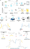
Figure 1
Figure 1
An overview of SF extraction and SFMA and HAMA synthesis, including (A) silk degumming; dissolution of SF in LiBr; and methacrylation, dialysis, and concentrating of the SFMA; (B) methacrylation of the HA by AEMA; and (C) a photo-cross-linking reaction using free radical vinyl polymerization at 365 nm to obtain the SFMA, HAMA, and SFMA–HAMA hydrogels. Figure created with Biorender.com.
Figure 2
Figure 2
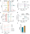
(A) 1H NMR spectra of the SF and SFMA. The presence of the methacrylate vinyl group signal with DoM of 41.6 ± 4.5% (calculated as described in methods), yellow aromatic amino acid resonances (6.7–7.5 ppm) that are not chemically modified, pink methacrylate vinyl groups that appear after the reaction (5.6–6.5 ppm), and green lysine amino acids (2.9–3.0 ppm). (B) FTIR spectra of BM cocoon, D-cocoon (degummed cocoon), SF, and SFMA, (C) 1H NMR spectra of HA before and after modification with AEMA. The presence of the methacrylate vinyl group signal with DoM of 32.5 ± 0.7% (calculated as described in methods), pink methacrylate vinyl groups that appear after the reaction amine group of AEMA with COOH groups of the HA (5.7 and 6.1 ppm), orange COOH that appear (5.2 ppm), yellow and grey methyl groups that appear (1.9 and 2.1 ppm), and purple and green methylene group that appear (3.7 and 4.2 ppm). (D) FTIR spectra of HA and HAMA. (E) FTIR spectra of SFMA and SFMA–HAMA hydrogels after UV cross-linking. (F) DoM graph of SFMA and HAMA hydrogels.
Figure 3
Figure 3
SEM images of (A) HAMA, SFMA, and SFMA–HAMA hydrogels after UV (365 nm) curation in the presence of 0.4 wt % LAP and (B) pore size distribution within the imaged hydrogels were analyzed with ImageJ and plotted. Log-normal distributions were plotted through each histogram in panel B and the averages from it are noted in the graph.
Figure 4
Figure 4
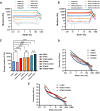
Gelation kinetics of (A) HAMA1, SFMA5, SFMA10, and SFMA20; (B) HAMA1, SFMA5–HAMA1, SFMA10–HAMA1, and SFMA20–HAMA1; (C) gelation time for HAMA1, SFMA5, SFMA10, and SFMA20; and (D) gelation time for HAMA1, SFMA5–HAMA1, SFMA10–HAMA1, and SFMA20–HAMA1 hydrogels at 37 °C (n = 3 represents three measurements taken from the same batch of samples; the bars are smaller than data marks). The in situ gelation process was measured on the quartz crystal stage by illuminating the bottom of the deposited sample with UV light. Data represented as the mean and standard deviation of n = 3 repeats (ns = not significant, *p < 0.05, **p < 0.01).
Figure 5
Figure 5
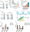
Hydrogel rheological characterization. (A) Amplitude sweep of HAMA1 and SFMA5, SFMA10, and SFMA20. (B) Amplitude sweep of HAMA1, SFMA5–HAMA1, SFMA10–HAMA1, and SFMA20–HAMA1. (C) Storage modulus (mechanical stiffness) of HAMA1, SFMA (5, 10, and 20 wt %), SFMA5–HAMA1, SFMA10–HAMA1, and SFMA20–HAMA1 hydrogels obtained from amplitude sweeps at 1 Hz and 0.2%. (D) Viscosity curves for HAMA1 and SFMA (5, 10, and 20 wt %) hydrogels. (E) HAMA and SFMA5–HAMA1, SFMA10–HAMA1, and SFMA20–HAMA1 hydrogels. Data represented at the mean and standard deviation of n = 3 repeats (ns = not significant, ****p < 0.0001).
Figure 6
Figure 6
Measurements of mechanical properties under static and dynamic conditions. (A) The illustration and working principle of the static mechanical analysis; (B) compressive strength measurement of the HAMA1, SFMA5, SFMA10, SFMA20, SFMA5–HAMA1, SFMA10–HAMA1, and SFMA20–HAMA1 hydrogels in static condition; (C) obtained Young’s modulus values from static mechanical measurements for all formulations. (D) the illustration and working principle of the dynamic mechanical analysis; (E) the representation of the schematic for calculating energy dissipation efficiency (EDE) (%); (F) measure value of EDE (%) SFMA5, SFMA10, and SFMA20; and (G) measure value of EDE (%) SFMA5–HAMA1, SFMA10–HAMA1, and SFMA20–HAMA1. Parts A and D and the scheme in panel E were created with Biorender.com. Data represented at the mean and standard deviation of n = 3 repeats. *p < 0.05, **p < 0.01, ****p < 0.0001.
Figure 7
Figure 7
Swelling ratio of (A) HAMA1 and SFMA (5, 10, and 20%) and (B) HAMA1, SFMA5–HAMA1, SFMA10–HAMA1, and SFMA20–HAMA1 hydrogels and degradation percent of the (C) HAMA1 and SFMA (5, 10, and 20%) and (D) HAMA1, SFMA5–HAMA1, SFMA10–HAMA1, and SFMA20–HAMA1 hydrogels. Data shown in the mean (n = 3) and SD are shown. The bars are smaller than data marks.
Figure 8
Figure 8
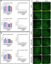
Results of live/dead assays on fibroblasts (NIH3T3) and preosteoblasts (MC3T3) and the (A) effects of the potential leachables (n = 6) and (B) direct contact with the materials (n = 3) were analyzed with one-way ANOVA followed by Dunnett’s multiple comparison test. Statistically significant differences versus control cells are indicated with p < 0.05 and *p < 0.01. (C) Representative confocal microscopy images of NIH3T3 and cells after 24 h of incubation in direct contact with the synthesized materials display the typical morphologies of NIH3T3 cells. Live cells exhibiting active metabolism were labeled with calcein-AM and appear in green, whereas the nuclei of dead cells were labeled with propidium iodide, which displays red fluorescence. The cells were maintained in direct contact with SFMA5, SFMA10, SFMA20, HAMA1, SFMA5–HAMA1, SFMA10–HAMA1, and SFMA20–HAMA1.

References

    1. Amirian J.; Zeng Y.; Shekh M. I.; Sharma G.; Stadler F. J.; Song J.; Du B.; Zhu Y. In-situ crosslinked hydrogel based on amidated pectin/oxidized chitosan as potential wound dressing for skin repairing. Carbohydr. Polym. 2021, 251, 117005.10.1016/j.carbpol.2020.117005. - DOI - PubMed
    1. Salma-Ancane K.; Sceglovs A.; Tracuma E.; Wychowaniec J. K.; Aunina K.; Ramata-Stunda A.; Nikolajeva V.; Loca D. Effect of crosslinking strategy on the biological, antibacterial and physicochemical performance of hyaluronic acid and ε-polylysine based hydrogels. Int. J. Biol. Macromol. 2022, 208, 995–1008. 10.1016/j.ijbiomac.2022.03.207. - DOI - PubMed
    1. Jongprasitkul H.; Turunen S.; Parihar V. S.; Annurakshita S.; Kellomäki M. Photocross-linkable Methacrylated Polypeptides and Polysaccharides for Casting, Injecting, and 3D Fabrication. Biomacromolecules 2021, 22 (2), 481–493. 10.1021/acs.biomac.0c01322. - DOI - PubMed
    1. Cernencu A. I.; Lungu A.; Stancu I.-C.; Serafim A.; Heggset E.; Syverud K.; Iovu H. Bioinspired 3D printable pectin-nanocellulose ink formulations. Carbohydr. Polym. 2019, 220, 12–21. 10.1016/j.carbpol.2019.05.026. - DOI - PubMed
    1. Mishbak H. H.; Cooper G.; Bartolo P. J. Development and characterization of a photocurable alginate bioink for three-dimensional bioprinting. Int. J. Bioprint. 2019, 5 (2), 189.10.18063/ijb.v5i2.189. - DOI - PMC - PubMed

LinkOut - more resources