. 2024 Oct 22;121(43):e2404892121.
doi: 10.1073/pnas.2404892121. Epub 2024 Oct 14.

The accomplices: Heparan sulfates and N-glycans foster SARS-CoV-2 spike:ACE2 receptor binding and virus priming

Affiliations

The accomplices: Heparan sulfates and N-glycans foster SARS-CoV-2 spike:ACE2 receptor binding and virus priming

Giulia Paiardi et al. Proc Natl Acad Sci U S A. .

Abstract

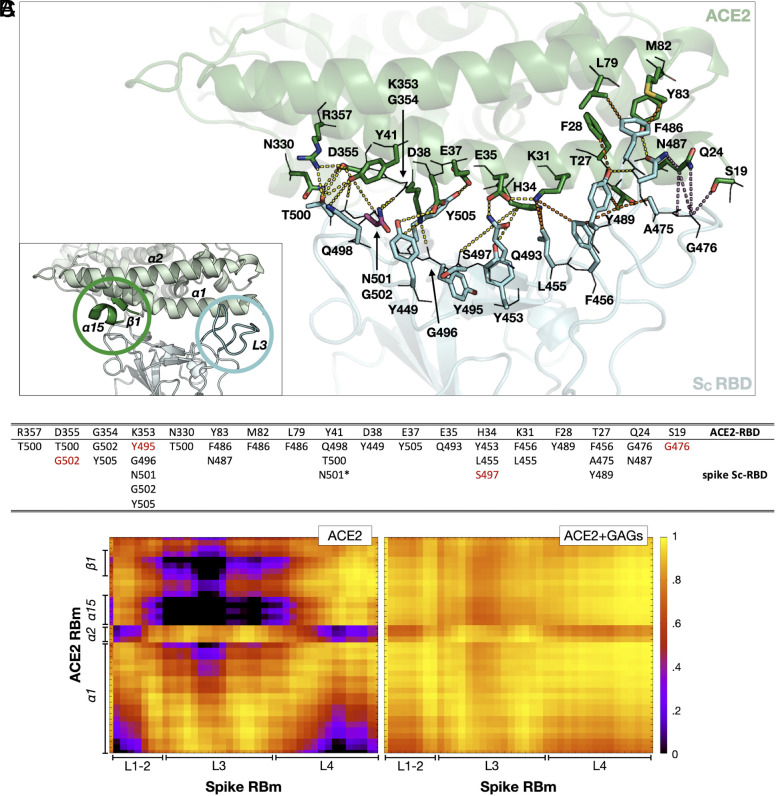
Although it is well established that the SARS-CoV-2 spike glycoprotein binds to the host cell ACE2 receptor to initiate infection, far less is known about the tissue tropism and host cell susceptibility to the virus. Differential expression across different cell types of heparan sulfate (HS) proteoglycans, with variably sulfated glycosaminoglycans (GAGs), and their synergistic interactions with host and viral N-glycans may contribute to tissue tropism and host cell susceptibility. Nevertheless, their contribution remains unclear since HS and N-glycans evade experimental characterization. We, therefore, carried out microsecond-long all-atom molecular dynamics simulations, followed by random acceleration molecular dynamics simulations, of the fully glycosylated spike:ACE2 complex with and without highly sulfated GAG chains bound. By considering the model GAGs as surrogates for the highly sulfated HS expressed in lung cells, we identified key cell entry mechanisms of spike SARS-CoV-2. We find that HS promotes structural and energetic stabilization of the active conformation of the spike receptor-binding domain (RBD) and reorientation of ACE2 toward the N-terminal domain in the same spike subunit as the RBD. Spike and ACE2 N-glycans exert synergistic effects, promoting better packing, strengthening the protein:protein interaction, and prolonging the residence time of the complex. ACE2 and HS binding trigger rearrangement of the S2' functional protease cleavage site through allosteric interdomain communication. These results thus show that HS has a multifaceted role in facilitating SARS-CoV-2 infection, and they provide a mechanistic basis for the development of GAG derivatives with anti-SARS-CoV-2 potential.

Keywords: ACE2 receptor; SARS-CoV-2; glycoprotein interactions; heparan sulfate; molecular dynamics simulation.


Conflict of interest statement

Competing interests statement:The authors declare no competing interest.

Figures

Fig. 1.
Fig. 1.
Structural model of the complex of the open, active spike homotrimer, the ACE2-RBD and three GAG chains, showing stabilizing interactions of two GAG chains and N-glycans with spike and ACE2. Two side views of a representative structure obtained from the last snapshot of one of the MD simulation replicas are shown. SA, SB, and SC subunits and ACE2 are shown as molecular surfaces in blue, teal, cyan, and green, respectively. N-glycans covalently attached to the spike and ACE2 are shown in line representation, colored according to the subunit to which they are attached. 40 frames of the N-glycan structures collected at intervals of 25 ns from the simulation are shown. The 31mer GAG chains are depicted as spheres colored by element with magenta carbons. On the Left, GAG-2 spans from the SC up-RBD to the SB S1/S2 multifunctional domain. On the Right, GAG-3 follows a similar path, simultaneously binding the SA down-RBD and the SC NTD and S1/S2.
Fig. 2.
Fig. 2.
Binding of GAGs alters the interactions at the interface between the spike SC subunit (up-RBD) and ACE2. (A) View of the SC RBD–ACE2 interface extracted from the last snapshot of the replica 1 trajectory simulated in the presence of GAGs. The SC RBD and ACE2 are shown as cartoons colored cyan and green, respectively. GAG-2 is omitted for clarity. Residues involved in the interactions are labeled and depicted as lines (main chain) and sticks (side chain) colored by element with oxygen, nitrogen, and sulfur in red, blue, and yellow, respectively. N501 is shown as magenta stick colored by element. Dashes connecting interacting residues are colored yellow, orange, and violet to represent polar, hydrophobic, and other types of contact, respectively. Contact matrices for each replica are given in SI Appendix, Fig. S4. The inset shows the location in the 3D structure of two regions of spike SC RBD and ACE2 that are dynamically coupled in the presence of GAGs (C). (B) Interactions between ACE2 (upper line) and SC RBD. Residues colored red gained the respective interactions during the simulations (both with and without GAGs) compared to the interactions reported in the literature (3, 42); SI Appendix, Table S1. The asterisk for the Y41–N501 interaction indicates that it is lost only in simulations with GAG chains. (C) Dynamic cross-correlation matrices of residues of SC RBm (x-axis) and ACE2-RBm (y-axis) for the spike:ACE2 (Left) and spike:ACE2:GAGs (Right) systems. Values range from 0 (black, uncorrelated motion) to +1 (yellow, correlated motion). The plots are shown for the replica 1 trajectory; plots for all replica trajectories are available in SI Appendix, Fig. S5.
Fig. 3.
Fig. 3.
ACE2 and the SC NTD approach each other in the presence of GAGs. (A) Side view of a representative structure of the spike head ectodomain in the open conformation (SC with up-RBD) in complex with ACE2. Spike and ACE2 are shown as cartoons colored gray and green, respectively. The SC RBD, NTD, and central helix are colored cyan. The SA and SB central helices are colored marine and teal, respectively. The spheres represent the center of mass (COM) calculated for each domain and are colored red, violet, orange, and brown for ACE2 (residues 18 to 616), SC RBD (residues 341 to 521), SC NTD (residues 16 to 271), and the three spike central helices (residues 987 to 1,034), respectively. To define the approach of ACE2 and the spike, the complex was oriented perpendicular to the xy plane. The distance d and vectors v1 and v2 were defined as follows: d (black) is the distance between the COM of ACE2 and the COM of SC NTD; v1 (magenta) points from the COM of the SC RBD to the COM of ACE2; v2 (blue) points from the COM of the three central helices to the COM of the SC NTD. The relative position of ACE2 is defined by (B) the distance d, and the angles α (C) and β (D) between the z-axis and v1 and v2, respectively. The distributions (BD) were calculated from the MD trajectories for all replicas and are colored according to system: spike:ACE2 (blue) and spike:ACE2-GAGs (orange). The motion of ACE2 and the SC RBD in the absence (E) and presence (F) of GAGs is shown by the superimposition of ten conformations extracted at equal time intervals along a representative trajectory (from gray to purple) and projected onto the first essential dynamics eigenvector. ACE2 and SC RBD are shown in cartoon representation, with GAGs omitted for ease of visualization. The corresponding plots for all the replica trajectories are similar and are shown in SI Appendix, Fig. S8.
Fig. 4.
Fig. 4.
N-glycans and GAGs contribute to stabilization of the open-spike:ACE2 complex and hinder its dissociation. (A) Interactions established by the N-glycans in the conventional MD simulations: buffering effect of spike and ACE2 N-glycans at the interface between ACE2 and SC NTD (Left); ACE2 N53 glycan:GAG-2 interactions and insertion of ACE2 N90 glycan at the interface between SC RBD and SA RBD (center) with an enlargement showing the interactions established by the N90 glycan with the spike residues (Right). (B) The dissociation pathway of ACE2 from spike in the presence of GAGs observed in the RAMD trajectories has three intermediate states. Yellow arrows show the dissociation route of ACE2 as a fan-like rotational movement in the first two intermediate states and a translating movement in the third. Blue circles (labeled AC) highlight changes in the protein:protein interactions along the dissociation pathway schematically. SA, SB, and SC subunits and ACE2 are shown as surfaces in blue, teal, cyan, and green, respectively. Mechanistically key N-glycans covalently attached to spike and ACE2 are labeled with gray circles and shown in stick representation, colored according to the subunit to which they are attached. The 31mer highly sulfated GAG-2 chain bound to the SC up-RBD is depicted as spheres colored by elements with magenta carbons. For further details of the dissociation pathway, see SI Appendix, Figs. S11–S13.
Fig. 5.
Fig. 5.
The binding of ACE2 and GAGs allosterically affects the spike protease cleavage sites, promoting virus priming. (A) Residue-based pairwise force differences in the open-spike protein when bound or not bound to ACE2 at three force thresholds (lines). The residue-based pairwise force difference upon GAG binding (not shown for clarity) follows the same distribution pattern. The spike and ACE2 glycoproteins are shown as gray cartoons with lines representing the N-glycans. Pairwise force vectors are depicted as lines colored according to the location of the initial point residue (marine, teal, cyan, and green for SA, SB, SC, and ACE2, respectively). R815 and the S1/S2 functional site residues are shown in orange and magenta, respectively. (B) Proposed force transduction from SA E773 to SA R1019 and SA E1017 (in the central helices—yellow dashed lines represent H-bonds) and (CD) the relative trajectory-averaged H-bond occupancy calculated for the last 400 ns of the MD simulations. (E) Proposed force transduction from SA R815 (cleavage site of TMPRSS2) to SA D839 and SA D843 (in the fusion peptide—yellow dashed lines represent the H-bonds) and (F) the relative trajectory-averaged H-bond occupancy calculated for the last 400 ns of the MD simulations. The solvent-accessible surface area (SASA) of SA R815 along the corresponding trajectories is reported in (G) and shows that binding of ACE2 alone or ACE2 and GAGs increase the exposure of the residue. Data are presented as mean ± SEM. *P < 0.05, two-tail ANOVA.
Fig. 6.
Fig. 6.
Schematic illustration of the proposed mechanisms by which HSPGs and N-glycans enhance SARS-CoV-2 spike:ACE2 receptor binding and virus priming. The results of the conventional and random acceleration molecular dynamics simulations reported here, along with experimental data reported in the literature, suggest the following mechanisms: HS interacts with SARS-CoV-2 spike glycoprotein, facilitating the encounter with the host cell ACE2 receptor (1). ACE2 N90 glycan drives the handshake with the spike SC up-RBD and subsequently, ACE2 N53 interacts with HS bound to the SC up-RBD, stabilizing the protein:protein complex; HS promotes the formation of residue–residue contacts, structural reordering, and the increase in coupled movement in local and distal positions at spike up-RBD:ACE2, resulting in a more energetically favorable complex (2). HS reorients the motion of ACE2 toward the spike NTD of the SC subunit to which the human receptor is bound, while the N-glycans of spike and ACE2 fill the space between the proteins, favoring a more densely packed spike S1:ACE2 complex (3). The motion upon engagement of the human receptor promotes the rearrangement of the allosteric network within spike and releases the constraints imposed on R815, the target of the TMPRSS2 human protease, making it available for cleavage. The cleavage of R815 reduces bonding cohesion and induces conformational metastability of S1-ACE2, promoting the dissociation of the S1-ACE2 component, unlocking the subsequent refolding of S2 (4). Notably, the mechanism proposed is shown for the spike SC subunit only but, based on our data, each spike subunit must undergo this process to expose R815 and be cleaved by TMPRSS2 to promote the rearrangement of S2. RBD, receptor-binding domain; NTD, N-terminal domain; S2, spike fusion domain; HSPGs, heparan sulfate proteoglycans; ACE2, angiotensin-converting enzyme 2; TMPRSS2, transmembrane protease serine subtype 2. The figure was created with BioRender (BioRender.com).

References

    1. Walls A. C., et al. , Structure, function, and antigenicity of the SARS-CoV-2 spike glycoprotein. Cell 181, 281–292.e6 (2020), 10.1016/j.cell.2020.02.058. - DOI - PMC - PubMed
    1. Wang Q., et al. , Structural and functional basis of SARS-CoV-2 entry by using human ACE2. Cell 181, 894–904.e9 (2020), 10.1016/j.cell.2020.03.045. - DOI - PMC - PubMed
    1. Lan J., et al. , Structure of the SARS-CoV-2 spike receptor-binding domain bound to the ACE2 receptor. Nature 581, 215–220 (2020), 10.1038/s41586-020-2180-5. - DOI - PubMed
    1. Yuan M., et al. , A highly conserved cryptic epitope in the receptor binding domains of SARS-CoV-2 and SARS-CoV. Science 368, 630–633 (2020), 10.1126/science.abb7269. - DOI - PMC - PubMed
    1. Watanabe Y., Allen J. D., Wrapp D., McLellan J. S., Crispin M., Site-specific glycan analysis of the SARS-CoV-2 spike. Science 369, 330–333 (2020), 10.1126/science.abb9983. - DOI - PMC - PubMed

MeSH terms

LinkOut - more resources