. 2024 Oct 22;121(43):e2406773121.
doi: 10.1073/pnas.2406773121. Epub 2024 Oct 14.

Abortive infection of bat fibroblasts with SARS-CoV-2

Affiliations

Abortive infection of bat fibroblasts with SARS-CoV-2

Punam Bisht et al. Proc Natl Acad Sci U S A. .

Abstract

Bats are tolerant to highly pathogenic viruses such as Marburg, Ebola, and Nipah, suggesting the presence of a unique immune tolerance toward viral infection. Here, we compared severe acute respiratory syndrome coronavirus 2 (SARS-CoV-2) infection of human and bat (Rhinolophus ferrumequinum) pluripotent cells and fibroblasts. Since bat cells do not express an angiotensin-converting enzyme 2 (ACE2) receptor that allows virus infection, we transduced the human ACE2 (hA) receptor into the cells and found that transduced cells can be infected with SARS-CoV-2. Compared to human embryonic stem cells-hA, infected bat induced Pluripotent Stem Cells (iPSCs)-hA produced about a 100-fold lower level of infectious virus and displayed lower toxicity. In contrast, bat embryonic fibroblast-hA produced no infectious virus while being infectable and synthesizing viral RNA and proteins, suggesting abortive infection. Indeed, electron microscopy failed to detect virus-like particles in infected bat fibroblasts in contrast to bat iPSCs or human cells, consistent with the latter producing infectious viruses. This suggests that bat somatic but not pluripotent cells have an effective mechanism to control virus replication. Consistent with previous results by others, we find that bat cells have a constitutively activated innate immune system, which might limit SARS-CoV-2 infection compared to human cells.

Keywords: SARS-CoV-2; abortive infection; bat cells; innate immune system.


Conflict of interest statement

Competing interests statement:The authors declare no competing interest.

Figures

Fig. 1.
Fig. 1.
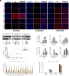
Ectopic expression of hACE2 permits replication of SARS-CoV-2 in hESCs, human fibroblast, and bat-induced pluripotent stem cells but fails to produce progeny virus in BEF. Human ACE2 was ectopically expressed in various cell types: human embryonic stem cells (hESCs-hA), bat-induced pluripotent stem cells (biPSCs-hA), human fibroblasts (IMR-90-hA), and bat fibroblasts (BEF-hA) via lentiviral transduction. These cells were subsequently infected with icSARS-CoV-2-eGFP (A). IFA at 72 hpi for SARS-CoV-2 Nucleocapsid (NC) protein (red), DAPI staining (blue) for nuclei and merged images confirmed panlocalization of NC (B). Control cells transduced but not infected with SARS-CoV-2 were included for comparison. (Scale bar, 50 µm). The mean fluorescence intensity (MFI) of eGFP and NC (red) was quantified as shown. Quantitative RT-PCR (qRT-PCR) at 72 hpi revealed the highest NC mRNA expression in IMR-90-hA cells, followed by hESCs-hA, biPSCs-hA, and BEF-hA (C). The production of infectious progeny virions was assessed using a plaque assay. Surprisingly, no virus was detected in the supernatant collected from infected BEF-hA cells (D), indicating a potential defect in viral release. A Lysate assay was conducted, and infected cells were harvested at 24-, 48-, 72-, and 96 hpi. Increased NC mRNA was observed up to 48 hpi in calu-3 and up to 72 hpi in biPSCs-hA, followed by a decrease from 72 to 96 hpi by qRT-PCR (E). Given the absence of progeny virus detection in BEF-hA cells (D), analysis of supernatant (F) and lysate (G) was performed via plaque assay. Calu-3 cells served as a positive control.
Fig. 2.
Fig. 2.
TEM of SARS-CoV-2-infected bat-induced pluripotent stem cells (biPSCs-hA), human (IMR-90-hA), and bat fibroblasts (BEF-hA). hACE2 lentivirus transduced cells were infected with SARS-CoV-2WA1 at an MOI of 0.5, 0.1, and 0.5, respectively. Mock-infected (transduced but not infected with SARS-CoV-2) or SARS-CoV-2 infected cells were fixed after 24 h and subjected to TEM. Mock-infected IMR-90-hA cells are shown in panel (A). IMR-90-hA showed SARS-CoV-2 virus particles within vesicles (B), and virus release in panel (C). Panel (D) represents an enlarged view of (C). Mock-infected biPSCs-hA cells are shown in panel (E). Panel (F and G) show complete virion inside the vesicle in infected biPSCs-hA and release in (H). Mock-infected BEF-hA cells are shown in panel (I). No such structure was seen in SARS-CoV-2 infected BEF-hA cells (JL). Red arrows indicate SARS-CoV-2 particles concentrated within vesicles in the infected cells. Mock-infected cells are shown in left panels.
Fig. 3.
Fig. 3.
Growth kinetics of SARS-CoV-2 in bat cells. Human ACE2 transduced BEF-LT-hA, or biPSCs-hA, were infected with SARS-CoV-2 with an MOI of 0.5 up to 72 h. Cells were fixed at the indicated time points for immunofluorescence analysis for Nucleocapsid (red) and DAPI (blue). Vero E6 cells were infected with an MOI of 0.1 in parallel. The merged images showing the extensive infection of the SARS-CoV-2 in the Vero E6 cells, permissive to the virus, confirmed the increase in the spread of the virus detected by NC protein expression and loss of DAPI due to cell death with progression in time of infection, scale bar, 200 μm (A). Western blot analysis of SARS-CoV-2 NC expression from the indicated cells at 8, 24, 48, and 72 hpi. GAPDH is taken as loading control. NC protein expression level was quantified using ImageJ (B). Infected cells were harvested at the indicated time points for qRT-PCR analysis of NC and ORF1ab mRNA expression. The Vero E6 and biPSCs-hA cells support viral replication with increased viral mRNA and progression in infection. In contrast, there is a decrease in the mRNA 8 hpi onward in the case of BEF-LT-hA (C). The cell-free medium was collected at 2, 8, 24, 48, and 72 hpi and subjected to progeny virus quantification by plaque assay (D). Normalized read count of SARS-CoV-2 in infected stem cells or fibroblasts of human and bat cells, in RNA seq (E). SARS-CoV-2 subgenomic RNA for membrane (M sg), envelope (E sg), and ORF3 (ORF3 sg) were detected in infected cells by qRT-PCR (F).
Fig. 4.
Fig. 4.
Differential gene expression analysis of SARS-CoV-2 infected human and bat fibroblasts: Human ACE2 expressing human fibroblasts (IMR-90-hA) and bat fibroblasts (BEF-hA) infected with SARS-CoV-2 at an MOI of 0.1 and 0.5, respectively, and subjected for RNA seq at 8 hpi and 48 hpi. (A and B) Volcano plots show differential gene expression in SARS-CoV-2 infected bat or human fibroblasts compared to mock-infected control fibroblasts at 8 hpi. Genes with an ab(log2FC) >= 0.5 are colored. Red = upregulated, blue = downregulated, yellow = SARS-CoV-2 NC. (C) Heatmap showing mean-centered expression of genes associated with innate immunity in human and bat fibroblast before and after SARS-CoV-2 exposure for 8 hpi. (D and E) Volcano plots show differential gene expression in SARS-CoV-2 infected human or bat fibroblasts compared to control fibroblasts at 48 hpi. Genes with an ab (log2FC) >= 0.5 are colored. Red = upregulated, blue = downregulated, yellow = SARS-CoV-2 NC. (F) Heatmap showing mean-centered expression of genes associated with innate immunity in human and bat fibroblast before and after SARS-CoV-2 exposure for 48 hpi. (G) Fold change of normalized gene counts from RNA seq of SARS-CoV-2 infected human and bat fibroblast to their respective control. (H) Fold change of normalized expression of selected genes in SARS-CoV-2 infected human and bat fibroblast with respect to control by qRT-PCR. Control refers to cells transduced with ACE2 lentivirus but not infected with SARS-CoV-2. Uninfected samples were neither transduced nor infected.

References

    1. Calisher C. H., et al. , Bats: Important reservoir hosts of emerging viruses. Clin. Microbiol. Rev. 9, 531–545 (2006). - PMC - PubMed
    1. Sánchez C. A., et al. , A strategy to assess spillover risk of bat SARS-related coronaviruses in Southeast Asia. Nat. Commun. 13, 4380 (2022). - PMC - PubMed
    1. Hale V. L., et al. , SARS-CoV-2 infection in free-ranging white-tailed deer. Nature 602, 481–486 (2022). - PMC - PubMed
    1. Mitchell P. K., et al. , SARS-CoV-2 B.1.1.7 variant infection in Malayan tigers, Virginia, USA. Emerg. Infect. Dis. 27, 3171–3173 (2021). - PMC - PubMed
    1. Oreshkova N., et al. , SARS-CoV-2 infection in farmed minks, the Netherlands, April and May. Euro Surveill. 25, 2001005 (2020). - PMC - PubMed

MeSH terms

Substances