Review
. 2024 Dec;11(45):e2405490.
doi: 10.1002/advs.202405490. Epub 2024 Oct 14.

Lung Immune Cell Niches and the Discovery of New Cell Subtypes

Affiliations
Review

Lung Immune Cell Niches and the Discovery of New Cell Subtypes

Qing'e Shan et al. Adv Sci (Weinh). 2024 Dec.

Abstract

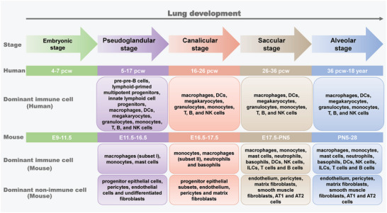
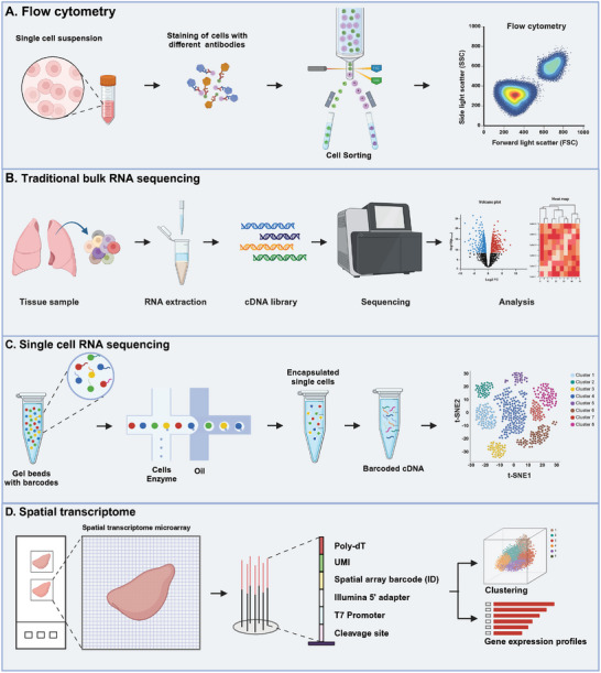
Immune cells in the lungs are important for maintaining lung function. The importance of immune cells in defending against lung diseases and infections is increasingly recognized. However, a primary knowledge gaps in current studies of lung immune cells is the understanding of their subtypes and functional heterogeneity. Increasing evidence supports the existence of novel immune cell subtypes that engage in the complex crosstalk between lung-resident immune cells, recruited immune cells, and epithelial cells. Therefore, further studies on how immune cells respond to perturbations in the pulmonary microenvironment are warranted. This review explores the processes behind the formation of the immune cell niche during lung development, and the characteristics and cell interaction modes of several major lung-resident immune cells. It indicates that distinct lung microenvironments or inflammatory niches can mediate the formation of different cell subtypes. These findings summarize and clarify paths to identify new cell subtypes that originate from resident progenitor cells and recruited peripheral cells, which are remodeled by the pulmonary microenvironment. The development of new techniques combining transcriptome analysis and location information is essential for identifying new immune cell subtypes and their relative immune niches, as well as for uncovering the molecular mechanisms of immune cell-mediated lung homeostasis.

Keywords: cell‐cell crosstalk; lung development; lung immune cells; new immune cell subtypes.


Conflict of interest statement

The authors declare no conflict of interest.

Figures

Figure 1
Figure 1
Stages of lung development and dominant composition of lung cells. pcw: post‐conception weeks; E: embryonic; PN: postnatal; DCs: dendritic cells; NK cells: natural killer cells; ILCs: innate lymphoid cells.
Figure 2
Figure 2
Self‐renewal and activation mechanisms of AMs and IMs. AMs and IMs are resident macrophages in the lungs that can not only protect the lungs from inhaled pathogens and allergens, but also prevent excessive immune response to harmless particles. The maintenance of AMs depends on the self‐renewal of resident macrophages in the alveolar lumen. AMs exert their anti‐inflammatory effects by secreting TGF‐ β, which restricts the activation of AM through the autocrine loop. AMs also provide protection against inhaled pathogens by secreting a number of cytokines and chemokines—including interleukin‐6 (IL‐6), tumor necrosis factor‐α (TNF‐α), monocyte chemoattractant protein 1, RANTES, and granulocyte colony‐stimulating factor (G‐CSF). IMs located in the lung interstitium are mainly derived from recruited circulating monocytes. They play a key role in pulmonary immunoregulation and represent an important source of steady‐state immunoregulatory cytokines such as IL‐10, TNF‐α, IL‐6, and IL‐1. In addition, IL‐10 produced by IMs may be involved in Th2 allergic inflammation and neutrophil asthma.[ 28 ] The figure was created with BioRender.com (https://biorender.com).
Figure 3
Figure 3
Interaction between alveolar macrophages and epithelial cells. Interleukin‐10 (IL‐10), plentiful in lung tissue, stimulates the production of inhibitory regulators like cytokine signal transduction inhibitor 3 (SOCS3) and the microRNA miR‐146b. This stimulation is mediated through the activation of the JAK1 signal transduction and STAT3 transcription pathways, which in turn suppresses inflammation. SOCS3 is instrumental in curbing the expression of pro‐inflammatory cytokines, while miR‐146b specifically hinders the expression and signaling of toll‐like receptor 4 (TLR4). Transforming growth factor‐β (TGF‐β) plays a role in modulating inflammatory responses via both dependent and independent signaling routes. The integrin αvβ6, predominantly found in bronchial epithelial cells but can also present in inflamed alveolar epithelial cells, binds to the latent TGF‐β. This binding induces a conformational change in TGF‐β, promoting its interaction with the TGF‐β receptor (TGFβR). The receptor TREM2, expressed in myeloid cells, is activated by the binding of DNAX‐activating protein 12 (DAP12). It helps to limit macrophage inflammation by interacting with one or more unidentified ligands. Mannose receptors serve to impede the recognition of TLR4 ligands, thereby restricting the phagocytosis of pathogens. The CD200 receptor (CD200R), upon binding to CD200 present on epithelial cells, triggers docking protein 2 (DOK2) and RAS GTPase activating protein RASA2. This interaction inhibits the inflammatory pathways, including extracellular signal‐regulated kinase (ERK), p38 mitogen‐activated protein kinase (MAPK), and Jun N‐terminal kinase (JNK). Pulmonary surfactant‐associated proteins A (SPA) and D (SPD), extensively found in the alveoli, prevent the interaction of TLR4 with its ligands as well as with TLRMD2 and CD14. This interaction inhibits the activation of nuclear factor‐κB (NF‐κB) and the initiation of binding of surfactant proteins to signal‐regulatory protein‐α (SIRPα). This process ultimately suppresses the recruitment of protein tyrosine phosphatase 1 (SHP1) containing the SH2 domain and the activation of RHOA—ultimately inhibiting phagocytosis.[ 2 , 111 ] Adapted with permission.[ 2 ] Copyright 2014, Springer Nature. The figure was created with BioRender.com (https://biorender.com).
Figure 4
Figure 4
Interaction between lung dendritic cells and lung epithelial cells. Lung dendritic cells (DCs) and epithelial cells possess pattern recognition receptors (PRRs), which allergens can activate directly. Upon allergen exposure, these epithelial cells release chemokines that draw in immature conventional DCs and inflammatory monocytes, notably through the action of CCL2 and CCL20. The activation of epithelial cells also leads to the generation of cytokines (such as IL‐1, GM‐CSF, and IL‐33) and alarm signals (like ATP and uric acid), which promote the maturation of DCs. Once activated, pulmonary DCs migrate to the nearby mediastinal lymph nodes, where they play a pivotal role in initiating Th2 and Th17 responses, critical for allergic inflammation. In addition, DCs receive help from basophils to maintain the Th2 response.[ 28 , 45 , 119 ] The figure was created with BioRender.com (https://biorender.com).
Figure 5
Figure 5
Common techniques used for identifying new lung sub‐populations include flow cytometry (A), traditional bulk RNA sequencing (B), single cell RNA sequencing (C) and spatial transcriptome (D). The figure was created with BioRender.com (https://biorender.com).

Similar articles

References

    1. Saluzzo S., Gorki A. D., Rana B. M. J., Martins R., Scanlon S., Starkl P., Lakovits K., Hladik A., Korosec A., Sharif O., Warszawska J. M., Jolin H., Mesteri I., McKenzie A. N. J., Knapp S., Cell Rep. 2017, 18, 1893. - PMC - PubMed
    1. Hussell T., Bell T. J., Nat. Rev. Immunol. 2014, 14, 81. - PubMed
    1. Domingo‐Gonzalez R., Zanini F., Che X., Liu M., Jones R. C., Swift M. A., Quake S. R., Cornfield D. N., Alvira C. M., eLife 2020, 9, 56890. - PMC - PubMed
    1. a) Kalymbetova T. V., Selvakumar B., Rodríguez‐Castillo J. A., Gunjak M., Malainou C., Heindl M. R., Moiseenko A., Chao C. M., Vadász I., Mayer K., Lohmeyer J., Bellusci S., Böttcher‐Friebertshäuser E., Seeger W., Herold S., Morty R. E., J. Pathol. 2018, 245, 153. - PubMed
    2. b) Kierdorf K., Prinz M., Geissmann F., Perdiguero E. G., Semin. Immunol. 2015, 27, 369. - PMC - PubMed
    1. Herriges M., Morrisey E. E., Development (Cambridge, England) 2014, 141, 502. - PMC - PubMed

LinkOut - more resources