. 2024 Dec:90:102046.
doi: 10.1016/j.molmet.2024.102046. Epub 2024 Oct 12.

Hypothalamic tanycytes internalize ghrelin from the cerebrospinal fluid: Molecular mechanisms and functional implications

Affiliations

Hypothalamic tanycytes internalize ghrelin from the cerebrospinal fluid: Molecular mechanisms and functional implications

Ivana M Gomez et al. Mol Metab. 2024 Dec.

Abstract

Objective: The peptide hormone ghrelin exerts potent effects in the brain, where its receptor is highly expressed. Here, we investigated the role of hypothalamic tanycytes in transporting ghrelin across the blood-cerebrospinal fluid (CSF) interface.

Methods: We investigated the internalization and transport of fluorescent ghrelin (Fr-ghrelin) in primary cultures of rat hypothalamic tanycytes, mouse hypothalamic explants, and mice. We also tested the impact of inhibiting clathrin-mediated endocytosis of ghrelin in the brain ventricular system on the orexigenic and locomotor effects of the hormone.

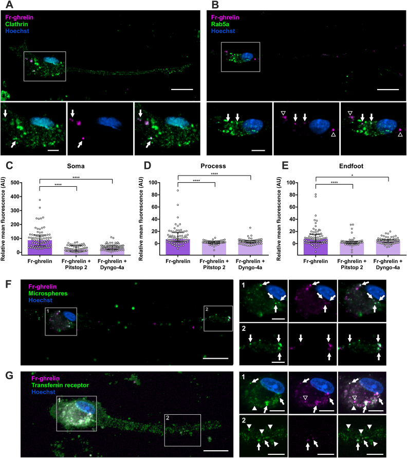
Results: In vitro, we found that Fr-ghrelin is selectively and rapidly internalized at the soma of tanycytes, via a GHSR-independent and clathrin-dependent mechanism, and then transported to the endfoot. In hypothalamic explants, we also found that Fr-ghrelin is internalized at the apical pole of tanycytes. In mice, Fr-ghrelin present in the CSF was rapidly internalized by hypothalamic β-type tanycytes in a clathrin-dependent manner, and pharmacological inhibition of clathrin-mediated endocytosis in the brain ventricular system prolonged the ghrelin-induced locomotor effects.

Conclusions: We propose that tanycyte-mediated transport of ghrelin is functionally relevant, as it may contribute to reduce the concentration of this peptide hormone in the CSF and consequently shortens the duration of its central effects.

Keywords: Blood-cerebro spinal barrier; Hypothalamus; Locomotor activity.


Conflict of interest statement

Declaration of competing interest The authors declare that the research was conducted in the absence of any commercial or financial relationships that could be construed as a potential conflict of interest.

Figures

Figure 1
Figure 1
Fr-ghrelin is selectively internalized in cultured hypothalamic tanycytes via a GHSR-independent mechanism. Panel A shows representative microphotographs of cultured tanycytes either in control conditions or after a 5 or 30-min incubation with Fr-ghrelin (magenta) imaged in fluorescence and DIC. Scale bars: 30 μm, 40 × objective. Panels B, C and D show relative mean fluorescence (AU) in soma, process and endfoot of cultured tanycytes after an incubation with vehicle alone (n = 52 cells from 3 independent experiments) or in the presence of Fr-ghrelin during 5 (n = 68, 4 independent experiments) or 30 min (n = 67, 4 independent experiments). Kruskal Wallis P < 0.0001. Panel E shows representative microphotographs of an immunocytochemistry against ghrelin (green) in a cultured tanycyte previously incubated with ghrelin for 5 min and imaged in DIC or fluorescence. Scale bars: 50 μm, 63 × objective. Panels F, G and H show relative mean fluorescence (AU) in soma, process and endfoot of cultured tanycytes after a 5-min incubation with Fr-ghrelin alone (n = 108, 5 independent experiments) or in the presence of JMV2959 (n = 76, 2 independent experiments), ghrelin (n = 34, 2 independent experiments), desacyl-ghrelin (n = 30, 2 independent experiments) or scr-F-ghrelin (n = 26, 2 independent experiments). Kruskal Wallis P < 0.0001. Panels I, J and K show relative mean fluorescence in the channel used for scr-F-ghrelin (AU) in soma, process and endfoot of cultured tanycytes after a 5-min incubation with vehicle alone (n = 17, 1 independent experiment) or in the presence of Fr-ghrelin and scr-F-ghrelin (n = 26, 2 independent experiments). Kruskal Wallis P < 0.0001. In every case, multiple comparisons were made using Dunn's post hoc test. ∗∗∗∗P < 0.0001; ∗∗∗P < 0.001; ∗∗P < 0.01; ∗P < 0.05. Panel L shows a representative microphotograph of a cultured tanycyte incubated with Fr-ghrelin (magenta) and scr-F-ghrelin (green) for 5 min. Magnified insets of the soma are presented below. Empty arrowheads indicate Fr-ghrelin and full arrowheads indicate scr-F-ghrelin. Scale bars: 50 μm for 40 × objective and 10 μm for 63 × objective. In every case, cell nuclei were labeled with Hoechst. (For interpretation of the references to colour in this figure legend, the reader is referred to the Web version of this article.)
Figure 2
Figure 2
Fr-ghrelin is internalized via a clatrhin-dependent mechanism in cultured hypothalamic tanycytes. Panel A shows an immunocytochemistry against clathrin (green) in a cultured tanycyte previously incubated with Fr-ghrelin for 5 min (magenta). Magnified insets are presented below, showing the soma of the cell. Arrows indicate clatrhin-IR signal that colocalize with Fr-ghrelin. Scale bars: 20 μm, 63 × objective. Panel B shows an immunocytochemistry against Rab5a (green) in a cultured tanycyte previously incubated with Fr-ghrelin for 5 min (magenta). Magnified insets are presented below, showing the soma of the cell. Arrows indicate Rab5a-IR signal that colocalize with Fr-ghrelin and arrowheads indicate Fr-ghrelin that does not colocalize with Rab5a-IR signal. Scale bars: 20 μm, 63 × objective. Panels C, D and E show relative mean fluorescence in soma, process and endfoot of cultured tanycytes after a 5-min incubation with Fr-ghrelin alone (n = 74, 2 independent experiments) or in the presence of Pitstop 2 (n = 73, 2 independent experiments) or Dyngo-4a (n = 61, 2 independent experiments). Kruskal Wallis P < 0.0001. Multiple comparisons were made using Dunn's post hoc test. ∗∗∗∗P < 0.0001; ∗P < 0.05. Panel F shows a representative microphotograph of a cultured tanycyte previously incubated with Fr-ghrelin (magenta) and fluorescent microspheres (green) for 5 min. Arrows indicate fluorescent microspheres colocalizing with Fr-ghrelin signal. Scale bars: 30 μm for low or 10 μm for high magnification, 63 × objective. Panel G shows an immunocytochemistry against Transferrin Receptor (TfR, green) in a cultured tanycyte previously incubated with Fr-ghrelin for 5 min (magenta). Magnified insets showing the soma and endfoot of the cell are presented. Arrows indicate Fr-ghrelin colocalizing with TfR-IR signal, empty arrowheads indicate Fr-ghrelin that does not colocalize with TfR and full arrowheads indicate TfR-IR signal that does not colocalize with Fr-ghrelin. Scale bars: 30 μm for low or 10 μm for high magnification, 63 × objective. In every case, cell nuclei were labeled with Hoechst. (For interpretation of the references to colour in this figure legend, the reader is referred to the Web version of this article.)
Figure 3
Figure 3
Fr-ghrelin is mainly internalized at the soma of hypothalamic tanycytes in vitro. Panels A, B and C show relative mean fluorescence (AU) in soma, processes and endfeet of cultured tanycytes after a 5-min incubation either with vehicle (n = 23, 2 independent experiments) or Fr-ghrelin alone (n = 25, 2 independent experiments) or in presence of colchicine (n = 25, 2 independent experiments). Kruskal Wallis P < 0.0001 for soma, P = 0.0054 for process and P = 0.0093 for endfoot. Panel D shows the experimental outline used in the pulse-chase experiments. After a 5-min incubation with Fr-ghrelin, cells were washed an incubated in fresh medium for 10 or 25 min previous to fixation. Panel E shows the total fluorescence intensity per cell (AU) of cultured tanycytes at 0 (n = 83, 3 independent experiments), 10 (n = 101, 3 independent experiments), and 25 min (n = 79, 3 independent experiments) post incubation with Fr-ghrelin. Kruskal Wallis P < 0.0001. Bar graph in panel f shows the mean of the fluorescence intensity (AU) for soma, process and endfoot of the cultured tanycytes mentioned in panel e. Panel G, H and I show fluorescence intensity (AU) in soma, process and endfoot of cultured tanycytes at 0, 10 and 25 min post incubation with Fr-ghrelin. Kruskal Wallis P < 0.0001. In every case, multiple comparisons were made using the Dunn's post hoc test. ∗∗∗∗P < 0.0001; ∗∗P < 0.01; ∗P < 0.05.
Figure 4
Figure 4
Fr-ghrelin is mainly internalized at the apical side of the hypothalamic tanycytes ex vivo. Panel a shows the experimental outline used in ex vivo assays. Hypothalamus explants were obtained and incubated with Fluorogold or Fr-ghrelin, either within the ventricle or in contact with its external region. Panels B and C show representative micrographs of coronal sections of explants incubated with Fluorogold (green) inside the ventricle or on their external side, respectively. Magnified insets are presented below. Arrows indicate fluorescent signal corresponding to Fluorogold in the somas of tanycytes. Panels D and E show representative micrographs of coronal sections of explants incubated with Fr-ghrelin (magenta) inside the ventricle or on their external region, respectively. Magnified insets are presented below. Arrows indicate fluorescent signal corresponding to Fr-ghrelin in the somas of tanycytes in d. In both cases, cell nuclei were labeled with Hoechst. Scale bars: 100 μm for low magnification (10 × objective) or 30 μm for high magnification (40 × objective). (For interpretation of the references to colour in this figure legend, the reader is referred to the Web version of this article.)
Figure 5
Figure 5
Fr-ghrelin present in the CSF is rapidly internalized in hypothalamic tanycytes in vivo, in a similar fashion as seen in vitro. Panel A shows representative microphotographs of the median eminence of mice centrally injected either with vehicle (control) or Fr-ghrelin and perfused after 15, 30, 60 or 90 min. Scale bar: 200 μm (10 × objective). Panels B and C show the mean fluorescence intensity (AU) either in the floor of the 3V or the external ME of mice belonging to the experimental groups mentioned above (control conditions: n = 4; Fr-ghrelin 15 min: n = 4, 30 min: n = 4, 60 min: n = 4, 90 min: n = 3). One-way ANOVA of the logarithm of mean fluorescence intensity (Ffloor of the 3V (4, 14) = 34.53; P < 0.0001; Fexternal ME (4, 15) = 14.33; P < 0.0001). In every case, multiple comparisons were made using Sidak's post hoc test. For the floor of the 3V: ∗∗∗∗P < 0.0001 vs vehicle; ∗P < 0.05 vs vehicle; ####P < 0.0001 vs Fr-ghrelin 15 min. For external ME: ∗∗∗∗P < 0.0001 vs vehicle; ###P < 0.001 vs Fr-ghrelin 15 min; ##P < 0.01 vs Fr-ghrelin 15 min. Panel D and E show the mean fluorescence intensity (AU) in the floor of the 3V or the external ME of mice ICV injected with F-ghrelin alone (n = 5) or in the presence of Pitstop 2 (n = 7). Unpaired t-test of the logarithm of mean fluorescence intensity Pfloor of the 3V = 0.0048; Pexternal ME = 0.0037. Panel F shows representative microphotographs of the 3V wall of mice centrally injected with Scr-F-ghrelin (green) and Fr-ghrelin (magenta). Magnified insets are presented in f, showing Scr-F-ghrelin and Fr-ghrelin signal colocalizing in β-tanycytes. Panel G shows representative microphotographs of the 3V wall of mice centrally injected with fluorescent microspheres (green) and Fr-ghrelin (magenta). Magnified insets are presented in g, showing microspheres and Fr-ghrelin signal colocalizing in the soma of β-tanycytes. Scale bars: 200 μm for low magnification (10 × objective) and 30 μm for high magnification (63 × objective). In every case, cell nuclei were labeled with Hoechst. (For interpretation of the references to colour in this figure legend, the reader is referred to the Web version of this article.)
Figure 6
Figure 6
The internalization of ghrelin from the CSF has functional implications. Panel A shows the experimental outline used in the behavioral assessments. Mice were centrally injected with vehicle or Pitstop 2 and after 5 min injected with vehicle or ghrelin, as appropriate. Food intake was monitored for 120 min or locomotor activity was registered for 60 min after the injections. Panel B shows the food intake (mg) over time (min) of mice subjected to the experimental scheme described in A (vehicle + vehicle: n = 8; vehicle + ghrelin: n = 10; Pitstop 2 + vehicle: n = 8; Pitstop 2 + ghrelin: n = 13). Panel C shows the quantitative analysis of 30 min-food intake for the mice mentioned in panel B. Two-way ANOVA (Fpre-treatment (1,35) = 0.2546; P = 0.6170; Fpost-treatment (1,35) = 44.69; P < 0.0001; Finteraction (1,35) = 1.191; P = 0.2826). Panel D show the distance traveled (cm) over time (min) of mice subjected to the experimental scheme described above (n = 8 for every condition). Panel E shows the quantitative analysis of locomotor activity in the 51–60 min period for the mice mentioned in panel d. Two-way ANOVA (Fpre-treatment (1,7) = 40.23; P = 0.0004; Fpost-treatment (1,7) = 8.533; P = 0.0223; Finteraction (1,7) = 8.836; P = 0.0207). In every case, multiple comparisons were made using Sidak's post hoc test. ∗∗∗∗P < 0.0001; ∗∗P < 0.01; ∗P < 0.05.

Similar articles

References

    1. Banks W.A. Viktor Mutt lecture: peptides can cross the blood-brain barrier. Peptides. 2023;169 doi: 10.1016/j.peptides.2023.171079. - DOI - PubMed
    1. Banks W.A. From blood-brain barrier to blood-brain interface: new opportunities for CNS drug delivery. Nat Rev Drug Discov. 2016;15(4):275–292. doi: 10.1038/nrd.2015.21. - DOI - PubMed
    1. Cui H., López M., Rahmouni K. The cellular and molecular bases of leptin and ghrelin resistance in obesity. Nat Rev Endocrinol. 2017;13(6):338–351. doi: 10.1038/nrendo.2016.222. - DOI - PMC - PubMed
    1. Edwards A., Abizaid A. Clarifying the ghrelin system's ability to regulate feeding behaviours despite enigmatic spatial separation of the GHSR and its endogenous ligand. Int J Mol Sci. 2017;18(4):859. doi: 10.3390/ijms18040859. - DOI - PMC - PubMed
    1. Perello M., Cabral A., Cornejo M.P., De Francesco P.N., Fernandez G., Uriarte M. Brain accessibility delineates the central effects of circulating ghrelin. J Neuroendocrinol. 2019;31(7) doi: 10.1111/jne.12677. - DOI - PubMed

LinkOut - more resources