. 2024 Oct 15;10(1):97.
doi: 10.1038/s41526-024-00435-y.

Surface tension enables induced pluripotent stem cell culture in commercially available hardware during spaceflight

Affiliations

Surface tension enables induced pluripotent stem cell culture in commercially available hardware during spaceflight

Maedeh Mozneb et al. NPJ Microgravity. .

Abstract

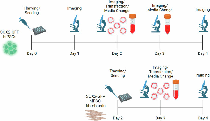
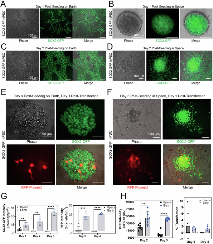
Low Earth Orbit (LEO) has emerged as a unique environment for evaluating altered stem cell properties in microgravity. LEO has become increasingly accessible for research and development due to progress in private spaceflight. Axiom Mission 2 (Ax-2) was launched as the second all-private astronaut mission to the International Space Station (ISS). Frozen human induced pluripotent stem cells (hiPSCs) expressing green fluorescent protein (GFP) under the SOX2 promoter, as well as fibroblasts differentiated from SOX2-GFP hiPSCs, were sent to the ISS. Astronauts then thawed and seeded both cell types into commercially available 96-well plates, which provided surface tension that reduced fluid movement out of individual wells and showed that hiPSCs or hiPSC-derived fibroblasts could survive either in suspension or attached to a Matrigel substrate. Furthermore, both cell types could be transfected with red fluorescent protein (RFP)-expressing plasmid. We demonstrate that hiPSCs and hiPSC-fibroblasts can be thawed in microgravity in off-the-shelf, commercially-available cell culture hardware, can associate into 3D spheroids or grow adherently in Matrigel, and can be transfected with DNA. This lays the groundwork for future biomanufacturing experiments in space.


Conflict of interest statement

M.M. (none). M.A. (none). P.M. (employee of Axiom Space, Inc.). D.M.N.M. (employee of BioServe Space Technologies). S.P. (none). George Lawless (none). S.D. (employee of BioServe Space Technologies). S.A.N. (none). R.B. (none). A.A.Q. (none). P.W. (employee of Axiom Space, Inc.). J.S. (none). Jana Stoudemire (employee of Axiom Space, Inc.). S.C. (employee of BioServe Space Technologies). C.S. (none). Arun Sharma (none).

Figures

Fig. 1
Fig. 1. Experimental workflow of hiPSC and hiPSC-fibroblast culture and transfection experiment for Axiom Space Mission 2 (Ax-2).
Workflow of hiPSC and hiPSC-fibroblast transfection experiment during Ax-2. Figures were created with BioRender software.
Fig. 2
Fig. 2. hiPSC culture conducted aboard the ISS.
Astronauts (A) Sultan Al Neyadi from the United Arab Emirates, (B) Peggy Whitson from the United States and Axiom Space, and (C) Rayyanah Barnawi from Saudi Arabia and Axiom Space conduct experimental operations on 96-well plates containing SOX2-GFP hiPSCs and SOX2-GFP hiPSC-fibroblasts in the Life Sciences Glovebox (LSG) aboard the ISS. Images courtesy of NASA and Sultan Al Neyadi.
Fig. 3
Fig. 3. hiPSCs spontaneously aggregate into spheroids, adhere to extracellular matrix, and are amenable to transfection in space microgravity.
A Fluorescence and phase contrast images of SOX2-GFP hiPSCs cultured in 2D on Earth. B Fluorescence and phase contrast images of hiPSCs aggregated into a spheroid 1 day after seeding hiPSCs in a 96-well plate aboard the ISS. C Fluorescence and phase contrast images of adherent hiPSCs 2 days after seeding hiPSCs in a 96-well plate on Earth. D Fluorescence and phase contrast images of adherent hiPSCs 2 days after seeding hiPSCs in a 96-well plate aboard the ISS. E Fluorescence and phase contrast images of adherent hiPSCs 3 days after seeding hiPSCs and 1 day after lipofectamine-based transfection with an RFP-expressing plasmid in a 96-well plate on Earth. F Fluorescence and phase contrast images of adherent hiPSCs 3 days after seeding hiPSCs and 1 day after lipofectamine-based transfection with an RFP-expressing plasmid in a 96-well plate aboard the ISS. The seeding day is considered as Day 0. G Quantification of SOX2-GFP signal and RFP plasmid signal in SOX2-GFP hiPSCs on Earth and in space. GFP (left) and RFP (right) signal intensity per unit area from different days of operation for SOX2-GFP hiPSCs cultured in space vs. on Earth is shown. Each dot represents an independent well of a 96-well plate. Data (n = 6) is shown as scatter dots ± std. 2-way ANOVA, α < 0.05, 95% CI. Tukey’s multiple comparisons test showed the level of significance for SOX2 signal intensity per well (left) to be P = 0.0032 (**) on Day 2, P = 0.0041 (**) on Day 3, and P < 0.0001 (****) on Day 4. For the RFP signal intensity per well (right) the level of significance was P < 0.0001 (****) for both Days 3 and 4. The seeding day is considered as Day 0. H Quantification of SOX2-GFP intensity per single cell (left) and transfection efficiency difference (right) from different days of operation for SOX2-GFP hiPSCs cultured in space and on Earth (n = 6). Single datapoints are reported as scatter dots ± std. 2-way ANOVA, 95% CI. Tukey’s multiple comparisons test showed the level of significance for pluripotency (left) to be p = 0.0011 (**) for Day 2 and P < 0.0001(****) for Day 3. The difference in percentage of cells transfected in space and on Earth (right) was non-significant.
Fig. 4
Fig. 4. hiPSC-fibroblasts spontaneously aggregate into spheroids, adhere to extracellular matrix, and are susceptible to transfection in space microgravity.
Representative fluorescence images of SOX2-GFP hiPSC-fibroblasts at day 1 post-seeding (A) on Earth and (B) in space. In space, fluorescence and phase contrast images of hiPSC-fibroblasts aggregated into a spheroid 1 day after seeding in a 96-well plate aboard the ISS. (C) Representative fluorescence and phase contrast images of adherent hiPSC-fibroblasts 2 days after seeding and 1 day after lipofectamine-based transfection with an RFP-expressing plasmid in a 96-well plate on Earth and (D) in space. (E) Quantification of SOX2-GFP hiPSC-fibroblasts RFP signal on Earth and in space at day 1 post-transfection (n = 6). Intensity of RFP signal per unit area is shown. Two-tailed paired t-test showed the level of significance between Earth and space samples to be P = 0.033 (*). (F) Quantification of the difference in transfection efficacy between hiPSC-fibroblasts in space and on Earth at day 1 post-transfection (n = 6). Single datapoints are reported as scatter dots ± std. Welch’s two tailed t-test showed the significant of difference between Earth and space sample to be p = 0.0004 (***), 95% CI.

References

    1. Sharma, A. et al. Biomanufacturing in low earth orbit for regenerative medicine. Stem Cell Rep.17, 1–13 (2022). - PMC - PubMed
    1. Arzt, M., Mozneb, M., Escopete, S., Moses, J. & Sharma, A. The benefits of stem cell biology and tissue engineering in low-earth orbit. Stem Cells Dev.33, 143-147 (2024) - PubMed
    1. Chua, C. Y. X. et al. Advanced material technologies for space and terrestrial medicine. Nat. Rev. Mater.10.1038/s41578-024-00691-0. (2024)
    1. Baio, J. et al. Spaceflight activates proein kinase C alpha signaling and modifies the developmental stage of human neonatal cardiovascular progenitor cells. Stem Cells Dev.27, 805–818 (2018). - PubMed
    1. Jha, R. et al. Simulated microgravity and 3D culture enhance induction, viability, proliferation and differentiation of cardiac progenitors from human pluripotent stem cells. Sci. Rep.6, 30956 (2016). - PMC - PubMed

LinkOut - more resources