. 2024 Oct 14;14(1):24072.
doi: 10.1038/s41598-024-75637-1.

Proteomic analysis of CD29+ Müller cells reveals metabolic reprogramming in rabbit myopia model

Affiliations

Proteomic analysis of CD29+ Müller cells reveals metabolic reprogramming in rabbit myopia model

Chae-Eun Moon et al. Sci Rep. .

Abstract

The prevalence of myopia is rapidly increasing, significantly impacting the quality of life of affected individuals. Prior research by our group revealed reactive gliosis in Müller cells within myopic retina, prompting further investigation of their role in myopia, which remains unclear. In this study, we analyzed protein expression changes in CD29+ Müller cells isolated from a form deprivation-induced rabbit model of myopia using magnetic activated cell sorting to investigate the role of these cells in myopia. As the principal glial cells in the retina, Müller cells exhibited significant alterations in the components of metabolic pathways, particularly glycolysis and angiogenesis, including the upregulation of glycolytic enzymes, such as lactate dehydrogenase A and pyruvate kinase, implicated in the adaptation to increased metabolic demands under myopic stress. Additionally, a decrease in the expression of proteins associated with oxygen transport suggested enhanced vulnerability to oxidative stress. These findings highlight the proactive role of CD29+ Müller cells in modifying the retinal environment in response to myopic stress and provide valuable insights into mechanisms that could help mitigate myopia progression.

Keywords: Glycolysis; Myopia; Müller cells; Oxidative stress; Proteomics.


Conflict of interest statement

The authors declare no competing interests.

Figures

Fig. 1
Fig. 1
Representation of proteomic analysis of Müller cells derived from the retina of myopia-induced rabbits. (a) Establishment of a myopia model in rabbits through form deprivation. The retina was segmented into central and peripheral regions based on the distance from the visual streak to the limbus. The central retina was defined as the area within 4 mm from the visual streak, while the peripheral retina included areas beyond this boundary. (b) Isolation of Müller cells from both the central and peripheral regions of the retina using magnetic-activated cell sorting (MACS) with CD29 antibody selection. (c) Subsequent proteomic analysis performed on the isolated Müller cells.
Fig. 2
Fig. 2
Change in the refractive error and axial length after form deprivation myopia induction. Compared to the control eye, the myopic eye showed significant reduction in the refractive error and axial elongation.
Fig. 3
Fig. 3
Identification of reactive gliosis in Müller cells via glial fibrillary acidic protein (GFAP) immunohistochemistry of the myopic retina. (a) GFAP expression increased in Müller cells of the myopic retina compared to that of control retina. (b) Quantitative analysis showed a significant increase in mean GFAP immunofluorescence intensity in the retina of myopic rabbits compared to controls.
Fig. 4
Fig. 4
Validation of CD29+ cell identity as Müller cells. (a) Comparison of CD29+ and CD29- cells revealed that CD29+ cells exhibited larger and more elongated morphology, characteristic of Müller cells, as opposed to CD29- cells. The cytoplasm of CD29+ cells showed a significantly stronger CD29 immunofluorescence signal, indicating effective isolation. (b) Quantitative analysis showed a significant increase in mean CD29 immunofluorescence intensity in the retina of myopic rabbits compared to controls. (c) Immunofluorescence staining of CD29+ cells from both the central and peripheral retinal regions demonstrated co-expression of glial fibrillary acidic protein (GFAP) and glutamine synthetase (GLUL), confirming their identity as Müller cells.
Fig. 5
Fig. 5
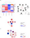
Proteomics analysis of CD29+ Müller cells in the myopic retina of rabbits. (a) Heatmap depicting unsupervised hierarchical clustering of protein expression profiles in CD29+ Müller cells, highlighting significant differences between the myopic and control groups. The analysis includes a Gene Ontology biological process breakdown using ClueGO to identify key signaling pathways. (b) Protein-protein interaction (PPI) network constructed using differentially expressed proteins (p-value < 0.5), illustrating the interactions and potential regulatory mechanisms differentially regulated in the myopic retina. (c) Comprehensive protein-protein interaction (PPI) network incorporating proteins universally expressed in CD29+ Müller cells across both the central and peripheral regions of the retina.

Similar articles

Cited by

References

    1. Holden, B. A. et al. Global prevalence of myopia and high myopia and temporal trends from 2000 through 2050. Ophthalmology. 123, 1036–1042. 10.1016/j.ophtha.2016.01.006 (2016). - PubMed
    1. Frick, K. D. What the comprehensive economics of blindness and visual impairment can help us understand. Indian J. Ophthalmol. 60, 406–410. 10.4103/0301-4738.100535 (2012). - PMC - PubMed
    1. Holden, B. et al. Myopia, an underrated global challenge to vision: Where the current data takes us on myopia control. Eye (London England). 28, 142–146. 10.1038/eye.2013.256 (2014). - PMC - PubMed
    1. Mérida, S. et al. Glutathione and a pool of metabolites partly related to oxidative stress are associated with low and high myopia in an altered bioenergetic environment. Antioxid. (Basel Switzerland). 13. 10.3390/antiox13050539 (2024). - PMC - PubMed
    1. Yu, Q. et al. Association between inflammatory cytokines and oxidative stress levels in aqueous humor with axial length in human myopia. Exp. Eye Res. 237, 109670. 10.1016/j.exer.2023.109670 (2023). - PubMed

LinkOut - more resources