. 2024 Oct 14;7(1):1322.
doi: 10.1038/s42003-024-06899-8.

Golgi-localized Ring Finger Protein 121 is necessary for MYCN-driven neuroblastoma tumorigenesis

Affiliations

Golgi-localized Ring Finger Protein 121 is necessary for MYCN-driven neuroblastoma tumorigenesis

Belamy B Cheung et al. Commun Biol. .

Abstract

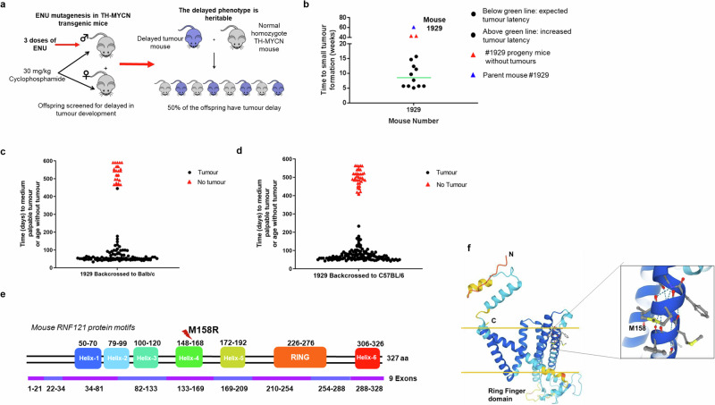
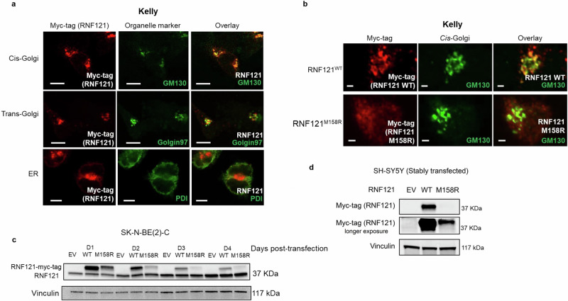
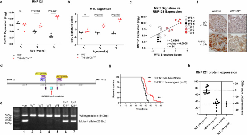
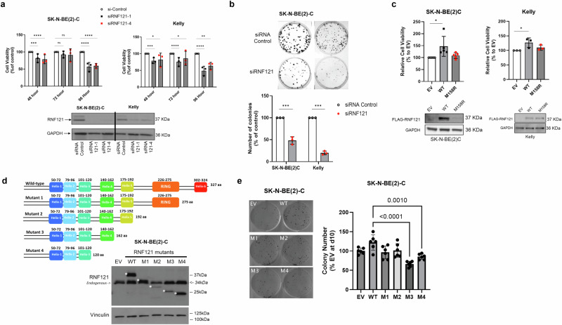
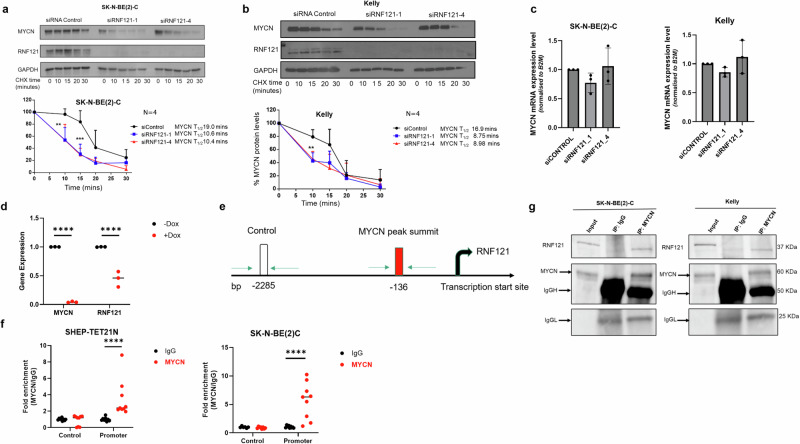
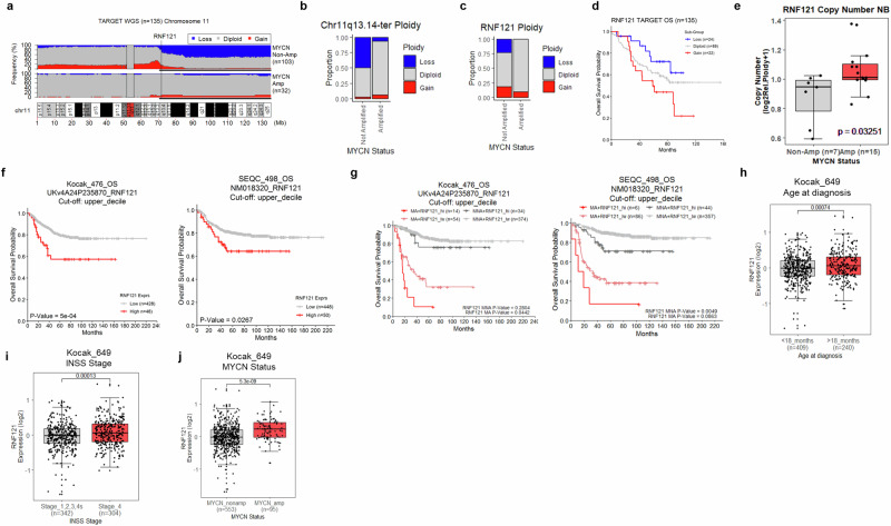
MYCN amplification predicts poor prognosis in childhood neuroblastoma. To identify MYCN oncogenic signal dependencies we performed N-ethyl-N-nitrosourea (ENU) mutagenesis on the germline of neuroblastoma-prone TH-MYCN transgenic mice to generate founders which had lost tumorigenesis. Sequencing of the mutant mouse genomes identified the Ring Finger Protein 121 (RNF121WT) gene mutated to RNFM158R associated with heritable loss of tumorigenicity. While the RNF121WT protein localised predominantly to the cis-Golgi Complex, the RNF121M158R mutation in Helix 4 of its transmembrane domain caused reduced RNF121 protein stability and absent Golgi localisation. RNF121WT expression markedly increased during TH-MYCN tumorigenesis, whereas hemizygous RNF121WT gene deletion reduced TH-MYCN tumorigenicity. The RNF121WT-enhanced growth of MYCN-amplified neuroblastoma cells depended on RNF121WT transmembrane Helix 5. RNF121WT directly bound MYCN protein and enhanced its stability. High RNF121 mRNA expression associated with poor prognosis in human neuroblastoma tissues and another MYC-driven malignancy, laryngeal cancer. RNF121 is thus an essential oncogenic cofactor for MYCN and a target for drug development.


Conflict of interest statement

The authors declare no competing interests.

Figures

Fig. 1
Fig. 1. RNF121M158R mutation blocks neuroblastoma development in TH-MYCN mice.
a Homozygous TH-MYCN (TH-MYCN+/+) mice were screened for delay in tumour development after ENU chemical treatment. Homozygous TH-MYCN (TH-MYCN+/+) breeder mice were treated for 2 weeks with cyclophosphamide to prevent tumour progression, then subjected to N-ethyl-N-nitrosourea (ENU) chemical mutagenesis of their germline at 6 weeks of age to induce random mutations in their sperm genome. b Tumour latency in 13 offspring of mouse 1929. Approximately half of the progeny developed tumours by the expected 7 weeks postnatal, whereas the remainder exhibited increased tumour latency. A subset of these mice did not develop tumours. Parent mouse 1929, that did not develop a tumour, is shown as a comparison. c, d Mouse 1929 or non-ENU-treated TH-MYCN+/+ mice were crossed with BALB/c (c) and C57BL/6 (d) wild-type mice before backcrossing with Cyclophosphamide-treated TH-MYCN+/+ mice. The mice represented by red triangles were selected for exome sequencing. e An RNF121M158R mutation was identified by exome sequencing of tumours from all 14 offspring with the tumour-suppressed phenotype. f A 3D model of RNF121M158R showing the location of the mutation. The secondary structures with the light and darker blue colours are predicted with the most confidence, whereas the yellow and red colours signify the least confident predictions. The yellow horizontal bars signify the approximate locations of the membrane surfaces. A zoomed in view of the Met 158 microenvironment is shown in the box. Met 158, located in transmembrane helix 4, and surrounding residues are shown in ball-and-stick format.
Fig. 2
Fig. 2. The RNF121M158R mutation reduces RNF121 protein levels, stability and localization to the cis-Golgi.
a Immunofluorescent staining using a: an anti-Myc-tag antibody to identify RNF121 co-expressed with the Myc tag; GM130 antibody as a cis-Golgi marker; Golgin-97 antibody as a trans-Golgi marker; and, PDI antibody as an endoplasmic reticulum (ER) marker, in a human MYCN amplified neuroblastoma cell line, Kelly. Scale bar: 100 μm. b Kelly cells transiently transfected with Myc-tagged wild-type RNF121 (RNF121WT) or mutant RNF121M158R for 48 h. Shown here are representative images of immunofluorescent staining using an anti-Myc-tag antibody for the RNF121 protein and a GM130 antibody for the cis-Golgi. Scale bar: 20 μm. c Immunoblot analysis of RNF121 expression in human MYCN amplified neuroblastoma cell line, SK-N-BE(2)-C, across the 4 days following transfection with either RNF121WT or mutant RNF121M158R. d Immunoblot analysis of RNF121 expression in human MYCN non-amplified neuroblastoma cell line, SH-SY5Y, following transfection with either RNF121WT wild type or RNF121M158R.
Fig. 3
Fig. 3. RNF121 is necessary for neuroblastoma development in TH-MYCN mice.
mRNA expression level of a RNF121WT, and, b a MYC gene signature in ganglia tissue and tumours from wild-type (WT) and TH-MYCN+/+ mice, obtained at different postnatal ages (1, 2, and 6 weeks of age; n  =  4 per group). Gene expression values are represented as log2-transformed microarray probe intensities of RNA levels. c The Pearson correlation coefficient (r) and p value for RNF121WT and the MYC signature expression scores for the tissue samples in (a) and (b). d Schematic of targeted RNF121 conditional allele: Two sgRNAs (yellow) were complexed with Cas9 nuclease to generate double stranded breaks in the RNF121 locus. A targeting vector featuring exon 3 (blue) of the RNF121 gene flanked by loxP sites was used as a template for homologous recombination. The resulting RNF121 conditional mice was crossed to Cre deleter mice to delete exon 3, leading to the generation of RNF121 knockout mice. e PCR products of mouse tail genomic DNA using primers which distinguished RNF121WT (540 bp in lanes 2–5) and the successfully floxed RNF121+/ allele (269 bp in lanes 6–7). Positive control for RNF121 hemizygous mutant allele (+ ve): DNA from RNF121 targeted embryonic stem cell clone. f Immunohistochemical staining for RNF121WT protein in coeliac ganglia from 20-week-old RNF121WT or RNF121+/− mice using an anti-mouse RNF121 polyclonal antibody and a control IgG antibody. g Tumour-free survival assessed using Kaplan–Meier analyses of TH-MYCN+/+ mice back-crossed with either RNF121WT (n = 25, black line) or floxed RNF121+/− knockout (n = 21, red line) mice (**p < 0.01). h A graphical representation showing RNF121 protein expression levels in tumour tissues from TH-MYCN+/+ mice crossed with either RNF121WT or floxed RNF121+/− knockout mice, across 5 independent western Blots (each with 3 different tumour lysates per group).
Fig. 4
Fig. 4. The RNF121 transmembrane domain 5 is required for growth and clonogenicity in vitro.
a Top panel: Cell viability analyses (n = 3) of SK-N-BE(2)-C and Kelly human neuroblastoma cells transfected with RNF121 siRNA-1, RNF121 siRNA-4 or Control siRNA for 96 h. Two-sided unpaired Student’s t-tests were performed to derive p-values. Differences in cell growth were compared to Control siRNA transfected cells. ***p < 0.001 and ****p < 0.0001. Bottom panel: Immunoblot analysis of RNF121 expression in SK-N-BE(2)-C and Kelly cells following siRNA-mediated RNF121 knockdown for 48 h. b SK-N-BE(2)-C and Kelly cells transfected with RNF121 siRNA-1 or Control siRNA were grown for 10 days in vitro and then assessed for colony formation. c Top panel: Cell viability analysis of SK-N-BE(2)-C and Kelly cells transfected with either Flag-RNF121WT, Flag-RNF121M158R or Empty vector control for 72 h. Two-sided unpaired Student’s t-tests were performed to derive p-values. *p < 0.05. Differences in cell growth were compared to Empty vector control expressing cells. Bottom panel: Immunoblot analysis of Flag-RNF121 expression in SK-N-BE(2)-C and Kelly cells following either overexpression of Flag-RNF121WT, Flag-RNF121M158R or Empty vector control for 24 h. d Top panel: Schematic representation of the Myc-tag-RNF121 deletion mutants. Bottom Panel: Representative immunoblot analysis for SK-N-BE(2)-C cells either overexpressing for 48 h empty Vector (EV), Myc-tag-RNF121WT full-length or each of the 4 Deletion Mutants of RNF121WT, probed with an anti-Myc-tag antibody. *predicated molecular weights of ectopically overexpressed Myc-RNF121 full-length and 4 Mutants with Myc-tag. e Left panel: SK-N-BE(2)C cells transfected with empty vector (EV), RNF121WT or each of the four RNF121 Deletion Mutants grown for 10 days and then assessed for colony formation. Right panel: Histogram representing the number of colonies 10 days after transfection as a percentage of control EV. **p < 0.05 for the comparison of RNF121 Mutant 3 and 4 with RNF121WT.
Fig. 5
Fig. 5. RNF121 directly binds MYCN protein and increases its stability.
a, b Representative immunoblots from cycloheximide (CHX) chase experiments 48 h after RNF121 knockdown using control siRNA or RNF121-specific siRNAs #1 or #4 in SK-N-BE(2)-C (a) and Kelly (b) cells. Cells were then treated with 100 µg/ml CHX for up to 30 min followed by immunoblotting of total cell lysates with anti-RNF121 or anti-MYCN antibodies. The ratio of MYCN protein/Actin protein was artificially set at 1.0 for control samples and then the half-life of MYCN was calculated from the line graph. c RT-PCR of MYCN mRNA expression levels following 48 h of siRNA knockdown of RNF121. d RT-PCR of MYCN and RNF121 mRNA expression levels following 24 h of Doxycyline (Dox) treatment of Dox-inducible MYCN knockdown neuroblastoma cell line, SHEP-Tet21N. e Schematic of PCR primer sites in the RNF121 gene promoter used for ChIP-PCR, detailing the MYCN peak binding summit (−136 bp) and an upstream negative control site (−2285 bp). f ChIP-PCR assays assessing fold enrichment of MYCN protein binding at two sites within the RNF121 gene promoter: the MYCN protein binding summit (−136 bp [Promoter]) or the negative control region (−2285 bp [Control]) using an anti-IgG (control) or anti-MYCN antibody in total cell lysates from either SHEP-TET21N or SK-N-BE(2)-C neuroblastoma cells, both with basal high MYCN expression. g Representative Western blot analysis for endogenous RNF121 after immunoprecipitation of endogenous MYCN in total cell lysates from MYCN amplified Kelly and SK-N-BE(2)-C cells. 5% of the cell lysate was loaded for input.
Fig. 6
Fig. 6. The RNF121WT gene is preferentially conserved in MYCN amplified human neuroblastoma tumours and associates with poor patient outcome.
a Frequency of segmental copy number variation (loss [blue], gain [red]) across Chromosome 11 (10 kilobase genomic bins), from whole-genome sequencing of somatic tumour DNA (WGS) from the TARGET neuroblastoma patient cohort (n  =  135). The 11q13.4-ter region is emphasised by a thin black bar underline. Genomic coordinates are provided in megabase measurements (Mb) below the plot. Centromeric regions were excluded from analyses (11p11.11/q11). b The proportion of patients with chromosome 11q13.4-ter segmental copy number variation (loss [blue], gain [red]) in MYCN non-amplified and amplified patient subgroups. c The proportion of patients with RNF121WT copy number variation (loss [blue], gain [red]) in MYCN non-amplified and amplified patient subgroups. d Kaplan–Meier curve of overall neuroblastoma patient survival in the TARGET cohort (n  =  135) subdivided by RNF121WT ploidy. e RNF121WT copy number for 22 neuroblastoma cell lines divided into MYCN-non-amplified and amplified subgroups. Data is derived from the Dependency Map portal (DepMap; 21Q2 release). The p-value is calculated using a two-sided t-test between each subgroup. f Kaplan–Meier curves of Overall Survival probability of patients from the Kocak (n = 476) or SEQC neuroblastoma cohorts (n = 498) subgrouped above or below the upper-decile of RNF121 mRNA expression for the overall group. The p-values are calculated using a log-rank test. g Kaplan–Meier curves of Overall Survival probability for patients from the Kocak (n = 476) or SEQC neuroblastoma cohort (n = 498) subdivided by RNF121 mRNA expression and MYCN amplification status (Amplified; MA, Non-amplified; MNA). Patients were subgrouped above or below the upper decile of RNF121 mRNA expression (High; RNF121_hi, Low; RNF121_lw). The p-values were calculated using two-sided log-rank tests between each RNF121 (lw, hi) expression group after subdividing cohorts by MYCN amplification status. p values were also adjusted using the Benjamini–Hochberg method to account for multiple comparisons. hj Comparison of RNF121 mRNA expression levels for patients subdivided by either the age at diagnosis (<18 months or >18 months), disease stage (INSS stage 1,2,3,4s vs stage 4) or MYCN amplification status (MYCN non-amplified vs amplified) in the Kocak dataset (n = 649).

Similar articles

References

    1. Darom, A., Bening-Abu-Shach, U. & Broday, L. RNF-121 is an endoplasmic reticulum-membrane E3 ubiquitin ligase involved in the regulation of beta-integrin. Mol. Biol. Cell21, 1788–1798 (2010). - PMC - PubMed
    1. Zhao, Y., Hongdu, B., Ma, D. & Chen, Y. Really interesting new gene finger protein 121 is a novel Golgi-localized membrane protein that regulates apoptosis. Acta Biochim. Biophys. Sin.46, 668–674 (2014). - PubMed
    1. Ogino, K. et al. RING finger protein 121 facilitates the degradation and membrane localization of voltage-gated sodium channels. Proc. Natl Acad. Sci. USA112, 2859–2864 (2015). - PMC - PubMed
    1. Zemirli, N., Pourcelot, M., Dogan, N., Vazquez, A. & Arnoult, D. The E3 ubiquitin ligase RNF121 is a positive regulator of NF-κB activation. Cell Commun. Signal.12, 72 (2014). - PMC - PubMed
    1. Zeng, Y. et al. Ring finger protein 6 promotes breast cancer cell proliferation by stabilizing estrogen receptor alpha. Oncotarget8, 20103–20112 (2017). - PMC - PubMed

Substances

LinkOut - more resources