. 2024 Oct 15;31(1):92.
doi: 10.1186/s12929-024-01084-9.

CD81-guided heterologous EVs present heterogeneous interactions with breast cancer cells

Affiliations

CD81-guided heterologous EVs present heterogeneous interactions with breast cancer cells

Elena Gurrieri et al. J Biomed Sci. .

Abstract

Background: Extracellular vesicles (EVs) are cell-secreted particles conceived as natural vehicles for intercellular communication. The capacity to entrap heterogeneous molecular cargoes and target specific cell populations through EV functionalization promises advancements in biomedical applications. However, the efficiency of the obtained EVs, the contribution of cell-exposed receptors to EV interactions, and the predictability of functional cargo release with potential sharing of high molecular weight recombinant mRNAs are crucial for advancing heterologous EVs in targeted therapy applications.

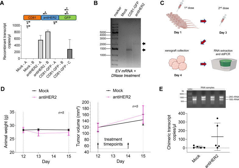
Methods: In this work, we selected the popular EV marker CD81 as a transmembrane guide for fusion proteins with a C-terminal GFP reporter encompassing or not Trastuzumab light chains targeting the HER2 receptor. We performed high-content imaging analyses to track EV-cell interactions, including isogenic breast cancer cells with manipulated HER2 expression. We validated the functional cargo delivery of recombinant EVs carrying doxorubicin upon EV-donor cell treatment. Then, we performed an in vivo study using JIMT-1 cells commonly used as HER2-refractory, trastuzumab-resistant model to detect a more than 2000 nt length recombinant mRNA in engrafted tumors.

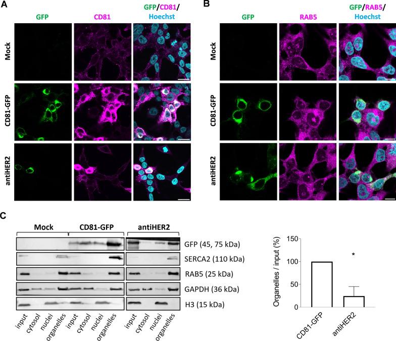
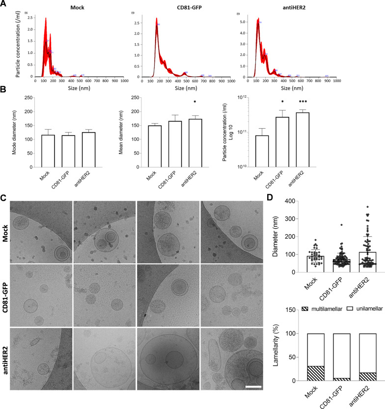
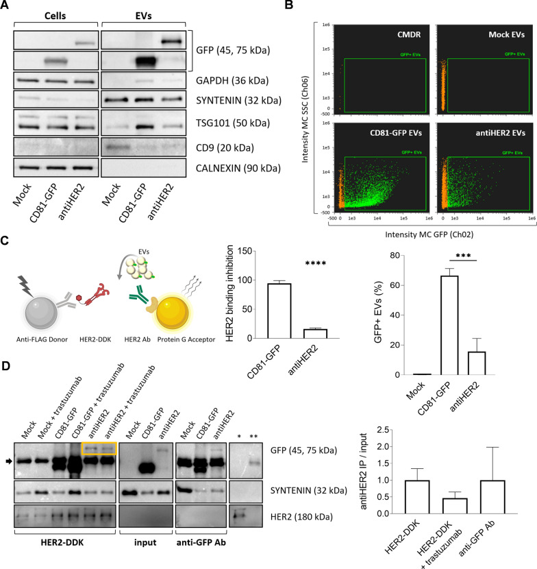
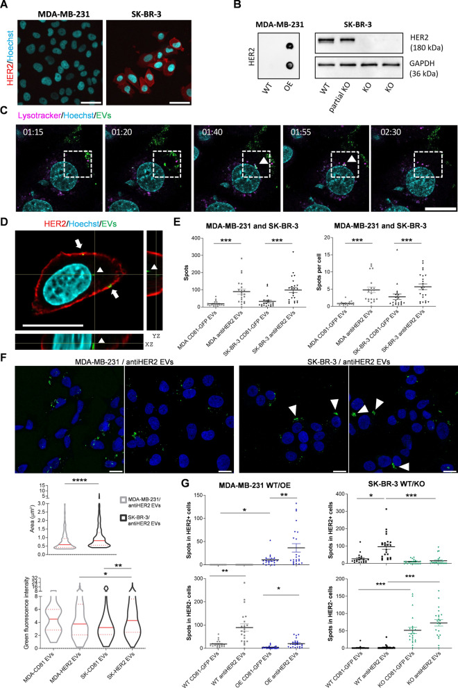
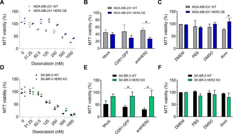
Results: Fusion proteins participated in vesicular trafficking dynamics and accumulated on secreted EVs according to their expression levels in HEK293T cells. Despite the presence of GFP, secreted EV populations retained a HER2 receptor-binding capacity and were used to track EV-cell interactions. In time-frames where the global EV distribution did not change between HER2-positive (SK-BR-3) or -negative (MDA-MB-231) breast cancer cell lines, the HER2 exposure in isogenic cells remarkably affected the tropism of heterologous EVs, demonstrating the specificity of antiHER2 EVs representing about 20% of secreted bulk vesicles. The specific interaction strongly correlated with improved cell-killing activity of doxorubicin-EVs in MDA-MB-231 ectopically expressing HER2 and reduced toxicity in SK-BR-3 with a knocked-out HER2 receptor, overcoming the effects of the free drug. Interestingly, the fusion protein-corresponding transcripts present as full-length mRNAs in recombinant EVs could reach orthotopic breast tumors in JIMT-1-xenografted mice, improving our sensitivity in detecting penetrant cargoes in tissue biopsies.

Conclusions: This study highlights the quantitative aspects underlying the creation of a platform for secreted heterologous EVs and shows the limits of single receptor-ligand interactions behind EV-cell engagement mechanisms, which now become the pivotal step to predict functional tropism and design new generations of EV-based nanovehicles.

Keywords: Drug-loading; Extracellular vesicles; Nanovehicles; RNA cargo; Receptor-binding; Tetraspanin.


Conflict of interest statement

The authors declare that they have no competing interests.

Figures

Fig. 1
Fig. 1
CD81 fusion proteins are expressed in HEK293T upon transient transfection and co-sediment with organelle-enriched sub-cellular fractions. A, B GFP detection and immunofluorescence staining of endogenous CD81 and RAB5 proteins in transfected HEK293T cells. Cell were subjected to confocal microscopy after 48 h of transfection with CD81-GFP and antiHER2 plasmids. Recombinant proteins are visualized in green (GFP), endogenous CD81 or RAB5 in magenta (Alexa Fluor 568), and cell nuclei in cyan (Hoechst). Scale bar is 20 μm in A and 10 μm in B. C Immunoblotting of sub-cellular fractions obtained through a sequential lysis buffer-centrifugation protocol. Separation of subcellular fractions was confirmed by the enrichment of corresponding protein markers: Cytosol (GAPDH), nuclei (histone H3), and organelles (SERCA2 for endoplasmic reticulum, RAB5 for early-endosomes). GFP-positive chimeric proteins were detected at the expected molecular weight (45 for CD81-GFP and 75 kDa for antiHER2). The histogram reports the densitometric quantification normalized over CD81-GFP condition, with mean and SD of two independent experiments. Significance is *P < 0.05
Fig. 2
Fig. 2
Profiling of particles recovered from transfected HEK293T cells. A, B Nanoparticle Tracking Analysis (NTA) of particles secreted by transfected HEK293T cells. Representative size distribution profiles of Mock, CD81-GFP, anti-HER2 samples. The black curve indicates the mean of three measurements, with SE in red. Mode and Mean diameters, and particle concentration are plotted. Error bars include at least three biological replicates. Significance * is P < 0.05, ***P < 0.001, vs Mock condition. C Representative Cryo-EM images of Mock, CD81-GFP and antiHER2 EV samples confirming the vesicular structure and size heterogeneity of recovered vesicles. The indicated scale bar is 100 nm. D Plot of the observed diameter of vesicles in Cryo-EM images (n = 35 for Mock, n = 99 for CD81-GFP, n = 74 for antiHER2) and lamellarity, expressed as percentage of unilamellar and multilamellar vesicles over the observed bulk EV populations
Fig. 3
Fig. 3
CD81-guided fusion proteins are cargo of secreted EV populations. A Representative immunoblotting of cell and EV lysates (1 μg proteins/well). EVs are positive to transmembrane (CD9) and cytosolic proteins (SYNTENIN, TSG101), while negative to CALNEXIN, and with low detectable levels of GAPDH compared to cell lysates. B Dot plots of imaging flow cytometry to detect GFP-positive EVs. The green fluorescent signal (Ch02, 488 nm laser) was detected as sub-gating of EVs labeled with Cell Mask Deep Red (CMDR, in orange, Ch11, 635 nm) to side-scatter (Ch06). Non-fluorescent, calibrator SpeedBeads, Amnis (1 µm) were continuously run during acquisitions. The graph shows the quantification of double-positive particles. Mean and error bars derive from three independent experiments. Significance antiHER2 vs CD81-GFP EVs is ***P < 0.001. C Sandwich designed for the AlphaLISA competitive assay. CD81-GFP and antiHER2 EVs were tested for competition with HER2-DDK. The graph shows the measured alpha counts normalized to the GFP-positive EV population as calculated by NTA and imaging flow cytometry. Mean and SD derive from three independent experiments (significance is ****P < 0.0001). D Representative western blotting of recombinant EVs immunoprecipitation with HER2-DDK or anti-GFP antibody in serum-free DMEM. AntiHER2 GFP-positive fusion proteins are enclosed in the yellow box above the antibody heavy chains (black arrow). Controls of beads flow through with HER2-DDK (*) or anti-GFP antibody (**) are shown on the right, indicating saturation of the beads’ surface to avoid non-specific binding. The graph shows the densitometric quantification of antiHER2 EVs captured by both HER2-DDK and anti-GFP Ab, with a competition effect of Trastuzumab. Mean and SD refer to two independent experiments
Fig. 4
Fig. 4
Recombinant EVs show heterogeneous interactions with breast cancer cells. A Immunofluorescence of MDA-MB-231 and SK-BR-3 breast cancer cell lines as HER2 negative or positive cells, respectively. HER2 receptor is in red (Alexa Fluor 633), nuclei are shown in cyan (Hoechst). The indicated scale bar is 50 μm. B Left: HER2 protein detection by Dot blot in lysates from wild-type or transfected (OE) MDA-MB-231 cells. Right: Immunoblot for checking the selection of SK-BR-3 cells with HER abrogation (ERBB2-KO, or KO). C Representative confocal time lapse of recombinant EVs incubated with live cells (time points are indicated). GFP-EVs are shown in green, lysosomes are shown in magenta (Lysotracker red), and nuclei in cyan (Hoechst). The white squares highlight the co-localization between EVs and lysosomes (white arrowhead). The indicated scale bar is 20 μm and the time points expressed as hh:mm. D Representative confocal image of fixed SK-BR-3 cells recognizing HER2 (Alexa Fluor 633) and nuclei (Hoechst) after 4 h incubation with CD81-GFP EVs (green spots). XY, YZ and XZ orthogonal views are reported. White arrows or the arrowhead indicate different localization of EVs upon cell interaction. Scale bar is 20 μm. E–G Quantification of recombinant EVs in recipient breast cancer cell lines. Cells were incubated with EVs for 4 h, then washed with PBS before fixation and HER2 immunofluorescence. Fourteen Z-stacks were acquired within around 11 μm of total Z-size and the Maximum Intensity Projections have been analyzed with an automated pipeline (using CellProfilerTM 4.0.7). Graphs report Mean and SD of the spot distribution from three independent experiments (*P < 0.05, **P < 0.01, ***P < 0.001, ****P < 0.0001). E) GFP + spots per field (left) or per cell (right) are quantified for CD81-GFP or antiHER2 EVs on MDA-MB-231 and SK-BR-3 WT. F) Representative images of the analyzed confocal acquisitions. Accumulation of GFP + spots in SK-BR-3 cells is indicated with arrowheads. Nuclei are in blue and the scale bar is 20 μm. Bottom graphs report GFP + spots’ area and fluorescence intensity. G) GFP + spots are quantified for CD81-GFP or antiHER2 EVs on MDA-MB-231 (WT and HER2 OE) and SK-BR-3 (WT and KO), with distinction of HER2 + (top) and HER2- (bottom) cells based on HER2 IF
Fig. 5
Fig. 5
Functional interactions of recombinant doxo-EVs with breast cancer cells. A, D Cell viability following doxorubicin treatment of MDA-MB-231 WT and HER2 OE (A) and SK-BR-3 WT and KO (D) for 72 h. B, E MTT assay on MDA-MB-231 WT and HER2 OE (B) and SK-BR-3 WT and KO (E) after 72 h of incubation with recombinant doxo-EVs (60 nM of secreted doxorubicin). Effect of doxo-EVs was normalized on recombinant EVs without the drug and doxorubicin alone at the corresponding concentration (60 nM). C, F MTT controls including doxorubicin alone are reported for the same cells. Graphs show Mean and SD of three biological replicates (*P < 0.05)
Fig. 6
Fig. 6
Recombinant EVs reach breast orthotopic tumors and share their full-length mRNA cargo. A Schematic representation of primers tested in ddPCR to detect the recombinant transcripts in EV-RNAs. The histogram below shows the observed transcript copy number per microliter in ddPCR experiments. Mean and SD refer to two independent experiments. B cDNA synthetized from EV-RNA was amplified for detecting the recombinant transcripts in the full-length form (indicated by arrows) on agarose gel. C Schematic representation of the in vivo study, from mice treatment (5 per condition) to recombinant EV-RNA detection by ddPCR in tumor xenografts. Mock doxo-EVs and antiHER2 doxo-EVs were tested. Subcutaneously injection was performed with a doxorubicin concentration of 0.5 µg/kg at tumor implantation site on day 0 and day 2. D Animal weight measured during the in vivo study at day 12, 13 and 15 (left) and tumor volume measured before and after mice treatment, at day 12 and 15, respectively (right). Mean and SEM are reported in the graphs. E Representative agarose gel of the RNA quality (18S and 28S rRNAs, 2 μg RNA/well) obtained from tumor xenografts (top) and copies/µl of recombinant transcripts detected in tumor xenografts (bottom). Each dot corresponds to one animal

Similar articles

Cited by

References

    1. Théry C, Witwer KW, Aikawa E, Alcaraz MJ, Anderson JD, Andriantsitohaina R, et al. Minimal information for studies of extracellular vesicles 2018 (MISEV2018): a position statement of the International Society for Extracellular Vesicles and update of the MISEV2014 guidelines. J Extracell vesicles [Internet]. 2018 [cited 2019 Feb 12];7:1535750. Available from: https://www.tandfonline.com/doi/full/10.1080/20013078.2018.1535750 - PMC - PubMed
    1. Liu Y-J, Wang C. A review of the regulatory mechanisms of extracellular vesicles-mediated intercellular communication. Cell Commun Signal. 2023;21:77. - PMC - PubMed
    1. Anand S, Samuel M, Mathivanan S. Exomeres: a new member of extracellular vesicles family. Subcell Biochem. 2021;97:89–97. - PubMed
    1. Welsh JA, Goberdhan DCI, O’Driscoll L, Buzas EI, Blenkiron C, Bussolati B, et al. Minimal information for studies of extracellular vesicles (MISEV2023): from basic to advanced approaches. J Extracell vesicles. 2024;13: e12404. - PMC - PubMed
    1. Van Niel G, D’Angelo G, Raposo G. Shedding light on the cell biology of extracellular vesicles. Nat Rev Mol Cell Biol. 2018;2:213–28. - PubMed