. 2024 Jan-Dec;16(1):2410316.
doi: 10.1080/19420862.2024.2410316. Epub 2024 Oct 14.

CD200R1 immune checkpoint blockade by the first-in-human anti-CD200R1 antibody 23ME-00610: molecular mechanism and engineering of a surrogate antibody

Affiliations

CD200R1 immune checkpoint blockade by the first-in-human anti-CD200R1 antibody 23ME-00610: molecular mechanism and engineering of a surrogate antibody

Cristina Melero et al. MAbs. 2024 Jan-Dec.

Abstract

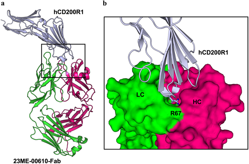
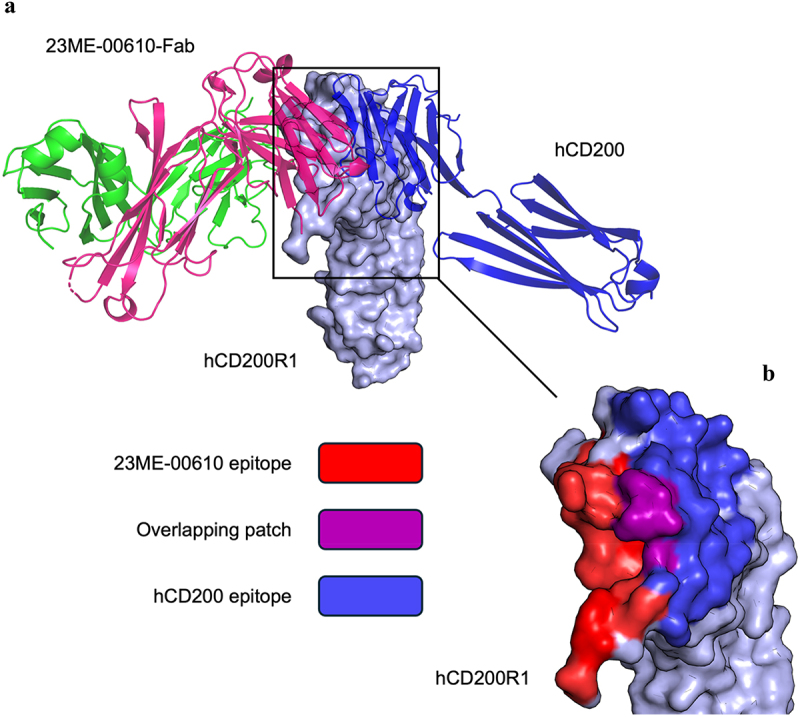
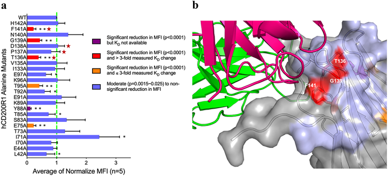
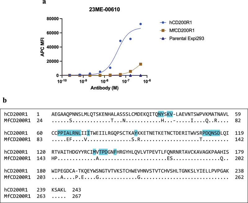
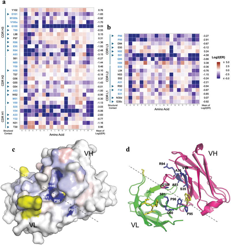
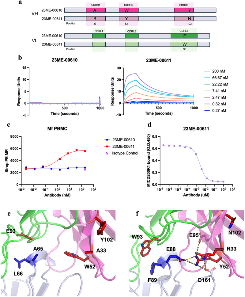
Human CD200R1 (hCD200R1), an immune inhibitory receptor expressed predominantly on T cells and myeloid cells, was identified as a promising immuno-oncology target by the 23andMe database. Blockade of CD200R1-dependent signaling enhances T cell-mediated antitumor activity in vitro and in vivo. 23ME-00610 is a potential first-in-class, humanized IgG1 investigational antibody that binds hCD200R1 with high affinity. We have previously shown that 23ME-00610 inhibits the hCD200R1 immune checkpoint function. Herein, we dissect the molecular mechanism of 23ME-00610 blockade of hCD200R1 by solving the crystal structure of 23ME-00610 Fab in complex with hCD200R1 and performing mutational studies, which show 23ME-00610 blocks the interaction between hCD200 and hCD200R1 through steric hindrance. However, 23ME-00610 does not bind CD200R1 of preclinical species such as cynomolgus monkey MfCD200R1. To enable preclinical toxicology studies of CD200R1 blockade in a pharmacologically relevant non-clinical species, we engineered a surrogate antibody with high affinity toward MfCD200R1. We used phage display libraries of 23ME-00610 variants with individual CDR residues randomized to all 20 amino acids, from which we identified mutations that switched on MfCD200R1 binding. Structural analysis suggests how the surrogate, named 23ME-00611, acquires the ortholog binding ability at the equivalent epitope of 23ME-00610. This engineering approach does not require a priori knowledge of structural and functional mapping of antibody-antigen interaction and thus is generally applicable for therapeutic antibody development when desired ortholog binding is lacking. These findings provide foundational insights as 23ME-00610 advances in clinical studies to gain understanding of the hCD200R1 immune checkpoint as a target in immuno-oncology.

Keywords: 23ME-00610; CD200; CD200R1; cancer immunotherapy; deep mutational scan; immune checkpoint.


Conflict of interest statement

AD, LL, GF, YH: Employees of 23andMe. OG: Employees of Proteros Biostructures. BC, CM, AJH, and SJB were employees of 23andMe at the time this work was performed.

Figures

Figure 1.
Figure 1.
Crystal structure of 23ME-00610-Fab bound to hCD200R1-iso4-ref reveals binding interface. (a) Structure of 23ME-00610-Fab:hCD200R1 complex shown in ribbon. hCD200R1 is in light blue color. 23ME-00610 heavy chain is in magenta, and light chain in green. (b) Zoom-in view of the binding interface with 23ME-00610 is represented as a surface. R67 in hCD200R1, shown as a stick, protrudes into a pocket cavity created at the intersection of the heavy (HC) and light (LC) chains of 23ME-00610.
Figure 2.
Figure 2.
23ME-00610 epitope overlaps with the hCD200 binding site. (a) Structure of 23ME-00610-Fab:hCD200R1 complex is superposed with hCd200:hcd200r1 complex model. 23ME-00610-Fab is shown as ribbons (HC in magenta; LC in green). Surface representation of hCD200R1 is shown in light blue; hCD200 depicted in blue ribbons. Homology model of the hCd200:hcd200r1 complex was generated by aligning AlphaFold predicted structures of hCD200 and hCD200R1 to the known mouse structure (PDB ID 4BFI). (b) Surface representation of hCD200R1 shows structural epitope residues (<5 Å) for 23ME-00610 (red) and hCD200 (blue) and the overlap between the two epitopes (purple).
Figure 3.
Figure 3.
Structural overlaps of hCD200 and 23ME-00610 CD200R1 epitopes are functionally important for 23ME-00610 binding. (a) Relative binding potency of 23ME-00610-Fab to Expi293 cells expressing either wild type (WT) or alanine mutants of hCD200R1-iso4-ref was evaluated by flow cytometry and SPR. Binding MFI (mean fluorescence intensity) was first normalized by the variant expression levels (see methods) and then the ratios to normalized MFI of WT (as 1, green dotted line) were plotted to represent relative binding potency for 23ME-00610. Attenuated binding with p = 0.0015-0.025 (*) or p < 0.0001 (**) relative to WT are shown. Coloring of each bar accords to relative binding potency of variants expressed as full-length protein on Expi293 cells and SPR KD measured as purified ECD protein as labeled. Positions highlighted with a red star are those that overlap between structural epitope of 23ME-00610 and hCD200 as in Figure 2b. (b) With hCD200R1 as surface representation, positions are colored according to the relative binding potency of alanine mutation variants shown in (A) where a significant decrease (**p < 0.0001) in 23ME-00610 binding potency verified by SPR are colored red. 23ME-00610-Fab is shown as ribbons (HC in magenta; LC in green).
Figure 4.
Figure 4.
23ME-00610 exhibits minimal cross-reactivity to cynomolgus CD200R1 ortholog (MfCD200R1). (a) Binding of 23ME-00610 to MfCD200R1 or hCD200R1 over-expressed on Expi293 cells was evaluated by flow cytometry. (b) Sequence alignment of the extracellular domains of hCD200R1-iso4-ref and MfCD200R1 (UniProt: G7NZT0) with dots indicating identical sequence. hCD200R1 residues with atoms within 5Å of 23ME-00610 in the solved structure are shaded in cyan.
Figure 5.
Figure 5.
Deep mutational scan of 23ME-00610 for mapping the high affinity interaction with hCD200R1. Single mutation enrichment of all randomized CDR positions of 23ME-00610 VH (a) or VL (b) from hCD200R1 panning is shown with Log2(ER) colored as labeled. Positions with low solvent accessibility are in blue, which is defined as < 25% of the whole residue area being solvent accessible as unbound 23ME-00610 fab using FreeSASA. CDR positions in the scan that are part of the structural contact sites with hCD200R1 are pointed with arrowheads. For each position, the mean of the Log2(ER) is calculated as the average of Log2(ER) of all mutation except cysteine. (c) The mean of the Log2(ER) is mapped on the structure of 23ME-00610 fab in a top-down view using the same color scale as A and B. Approximate border between VH and VL is denoted with a dotted line. Residues in yellow are part of the structural contact but not included in the mutational scan. (d) Same view of fab as (C) is plotted in ribbon with residues with Log2(ER) < −2 in blue sticks.
Figure 6.
Figure 6.
Mutations in 23ME-00611 surrogate antibody confers high affinity binding to MfCD200R1. (a) Schematics that show the VH or VL CDR positions mutated from 23ME-00610 to 23ME-00611; (b) biacore sensorgrams that show lack of 23ME-00610 binding to MfCD200R1 with concentration up to 200 nM (left) and 23ME-00611’s binding to MfCD200R1 at KD = 2 nM at 37°C (right); (c) cynomolgus PBMC FACS study confirms lack of 23ME-00610 binding to MfCD200R1 while 23ME-00611showed good binding potency. (d) 23ME-00611’s potent inhibition of MfCD200R1:MfCD200 interaction as shown by ELISA; (e) zoom-in views of 23ME-00610:hCD200R1 crystal structure and (f) 23ME-00611:MfCD200R1AlphaFold model show the two structurally equivalent residues (blue sticks) in hCD200R1 or MfCD200R1 that can explain the lack of 23ME-0610 binding and the four fab mutations in red sticks that confer 23ME-00611 high affinity binding to MfCD200R1. 23ME-00611 A33R mutation permits a network of charge interaction (dotted line) to accommodate E88 in MfCD200R1. Heavy and light chains indexed using kabat and chothia numbering, respectively.

References

    1. Wright GJ, Cherwinski H, Foster-Cuevas M, Brooke G, Puklavec MJ, Bigler M, Song Y, Jenmalm M, Gorman D, McClanahan T, et al. Characterization of the CD200 receptor family in mice and humans and their interactions with CD200. J Immunol. 2003;171(6):3034–12. doi:10.4049/jimmunol.171.6.3034. - DOI - PubMed
    1. Hatherley D, Lea SM, Johnson S, Barclay AN.. Structures of CD200/CD200 receptor family and implications for topology, regulation, and evolution. Structure. 2013;21(5):820–832. doi:10.1016/j.str.2013.03.008. - DOI - PMC - PubMed
    1. Foster-Cuevas M, Wright GJ, Puklavec MJ, Brown MH, Barclay AN. Human herpesvirus 8 K14 protein mimics CD200 in down-regulating macrophage activation through CD200 receptor. J Virol. 2004;78(14):7667–7676. doi:10.1128/JVI.78.14.7667-7676.2004. - DOI - PMC - PubMed
    1. Zhang S, Cherwinski H, Sedgwick JD, Phillips JH. Molecular mechanisms of CD200 inhibition of mast cell activation. J Immunol. 2004;173(11):6786–6793. doi:10.4049/jimmunol.173.11.6786. - DOI - PubMed
    1. McWhirter JR, Kretz-Rommel A, Saven A, Maruyama T, Potter KN, Mockridge CI, Ravey EP, Qin F, Bowdish KS. Antibodies selected from combinatorial libraries block a tumor antigen that plays a key role in immunomodulation. Proc Natl Acad Sci USA. 2006;103(4):1041–1046. doi:10.1073/pnas.0510081103. - DOI - PMC - PubMed

MeSH terms

Substances

LinkOut - more resources