. 2024 Oct 15;84(20):3419-3434.
doi: 10.1158/0008-5472.CAN-24-0293.

The USP1 Inhibitor KSQ-4279 Overcomes PARP Inhibitor Resistance in Homologous Recombination-Deficient Tumors

Affiliations

The USP1 Inhibitor KSQ-4279 Overcomes PARP Inhibitor Resistance in Homologous Recombination-Deficient Tumors

Louise Cadzow et al. Cancer Res. .

Abstract

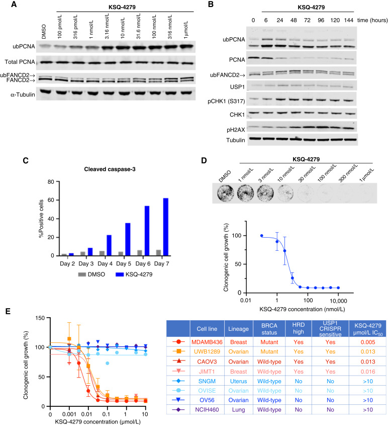
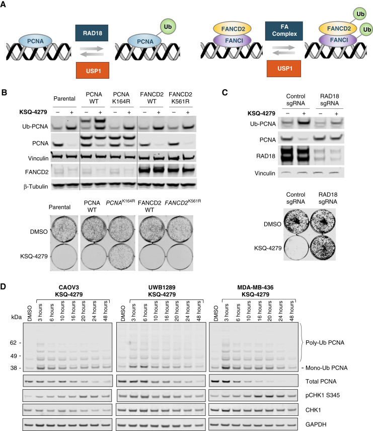
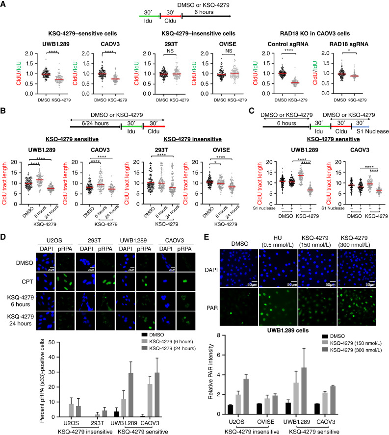
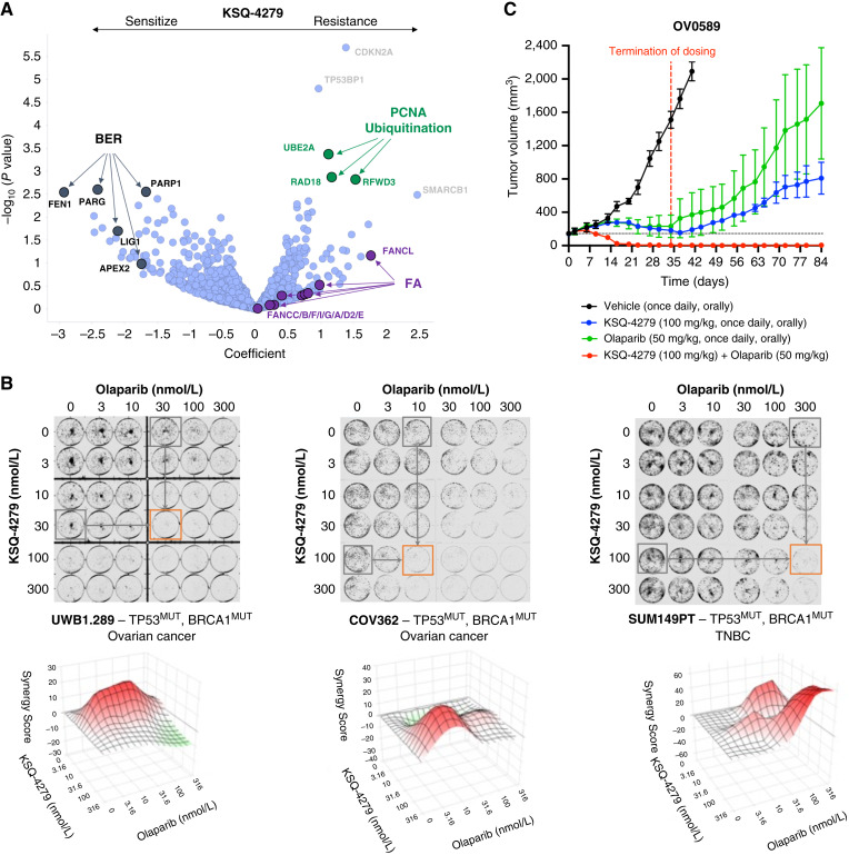
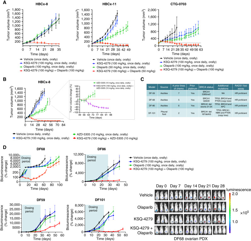
Defects in DNA repair pathways play a pivotal role in tumor evolution and resistance to therapy. At the same time, they create vulnerabilities that render tumors dependent on the remaining DNA repair processes. This phenomenon is exemplified by the clinical activity of PARP inhibitors in tumors with homologous recombination (HR) repair defects, such as tumors with inactivating mutations in BRCA1 or BRCA2. However, the development of resistance to PARP inhibitors in BRCA-mutant tumors represents a high unmet clinical need. In this study, we identified deubiquitinase ubiquitin-specific peptidase-1 (USP1) as a critical dependency in tumors with BRCA mutations or other forms of HR deficiency and developed KSQ-4279, the first potent and selective USP1 inhibitor to enter clinical testing. The combination of KSQ-4279 with a PARP inhibitor was well tolerated and induced durable tumor regression across several patient-derived PARP-resistant models. These findings indicate that USP1 inhibitors represent a promising therapeutic strategy for overcoming PARP inhibitor resistance in patients with BRCA-mutant/HR-deficient tumors and support continued testing in clinical trials. Significance: KSQ-4279 is a potent and selective inhibitor of USP1 that induces regression of PARP inhibitor-resistant tumors when dosed in combination with PARP inhibitors, addressing an unmet clinical need for BRCA-mutant tumors.


Conflict of interest statement

E. Krall reports personal fees from Merck Sharp and Dohme LLC outside the submitted work, as well as a patent 11485736 issued. E. Chipumuro reports other support from KSQ Therapeutics outside the submitted work. K. Sinkevicius reports employment with KSQ Therapeutics and receiving KSQ Therapeutics stock options. P.C. Gokhale reports grants from KSQ Therapeutics, Inc. during the conduct of the study, as well as grants from Kymera Therapeutics, Pfizer Inc., Treeline Biosciences, and Arvinas outside the submitted work. U.A. Matulonis reports personal fees from Tango Therapeutics, ProfoundBio, NextCure, Eisai, ImmunoGen, AbbVie, Alkermes, Novartis, and GSK outside the submitted work. J.F. Liu reports personal fees from AstraZeneca, Bristol Myers Squibb, Clovis Oncology, Daiichi Sankyo, Eisai, Genentech/Roche, GlaxoSmithKline, Regeneron Therapeutics, and Zentalis Pharmaceuticals outside the submitted work. M. Schlabach reports a patent for US 2021/0115049 A1 pending. F. Stegmeier reports full-time employment with KSQ Therapeutics during the time of this work, as well as being on the Board of Directors and ownership of stocks in KSQ Therapeutics. A.A. Wylie reports personal fees from KSQ Therapeutics outside the submitted work, as well as a patent for WO2021163530A1 issued, WO20230203046A1 issued, and WO2023147311A1 issued. J. Brenneman, J.J. Ali, S. Shenker, M. McGuire, P. Grasberger, Y. Mishina, E. Krall, J. Hixon, E. Chipumuro, K. Sinkevicius, A. Olaharski, H. Liu, J. Wilt, M. Schlabach, and F. Stegmeier conducted this research while employed by KSQ Therapeutics. No disclosures were reported by the other authors.

Figures

Figure 1.
Figure 1.
A, USP1 dependency profile using a CRISPR knockout screen across >500 cell lines. B, Chemical structure of KSQ-4279. C, KSQ-4279 binding affinity was determined by measuring the initial rate dependence of USP1 with Ub-Rho substrate as a function of fixed increasing concentrations of KSQ-4279. Solid lines are simulated from the mixed linear inhibition model global fit parameters: Vmax 1.24 (±0.04) nmol/L minutes−1 (kcat = 0.82 seconds−1), Km = 1,038 (±90) nmol/L, Kic = 6.9 (±3) nmol/L, and Kiu = 2.3 (±0.3) nmol/L. Points averaged from N = 4 determinations. Error bars, SD. D, Selectivity assessment of KSQ-4279 was established by testing a panel of deubiquitinases using a ubiquitin–rhodamine biochemical assay using 1-µmol/L KSQ-4279. E, Cryo-EM structure of the KSQ-4279 inhibited USP1–UAF1–ubiquitin complex. USP1, UAF1, and ubiquitin are rendered as cyan, blue, and green ribbons, respectively. KSQ-4279 is depicted as a yellow stick. F, KSQ-4279 shown inside a semitransparent molecular surface as it is cradled by the palm and thumb subdomains of USP1. G, USP1-apo (brown) is rendered as a ribbon superposed on the USP1/KSQ-4279 ribbon structure (light blue). KSQ-4279 is depicted as a blue molecular surface.
Figure 2.
Figure 2.
A, Immunoblot analysis of MDA-MB-436 cells treated with a dose titration of KSQ-4279 for 6 hours. B, MDA-MB-436 cells treated with 300-nmol/L KSQ-4279 across a time course to assess pharmacodynamic response and DNA damage induction. C, Cleaved caspase-3 expression in MDA-MB-436 cells treated with either 300-nmol/L KSQ-4279 or DMSO over a 7-day time course. D, KSQ-4279 induced growth inhibition of MDA-MB-436 cells as assessed via clonogenic assays. Data are representative of three independent experiments, with one representative example shown. E, KSQ-4279 induced growth effects across a panel of cell lines as assessed via clonogenic assays.
Figure 3.
Figure 3.
A, KSQ-4279 plasma and tumor exposure was determined at 4, 8, and 24 hours following a single oral dose administered at 10, 30, 100, or 300 mg/kg. B, Tumors were collected at 4, 8, or 24 hours after single dose of control or KSQ-4279, formalin-fixed, paraffin-embedded, sectioned, and stained for Ub-PCNA, and representative images are shown. Scale bar, 100 µm. IHC H-score was established based on percentage of positive cells and intensity of Ub-PCNA staining. C, Tumor growth curve for OV0589 human ovarian PDX model treated with per oral once daily 10 mL/kg control, 10-, 30-, 100-, or 300-mg/kg KSQ-4279. Mice (n = 3/group) were randomized, and treatment administered once the mean tumor volume reached approximately 200 mm3, denoted as day 0. The dotted black line represents the mean tumor volume in the combination-treated group on day 0, prior to treatment initiation. qd, once daily; po, orally.
Figure 4.
Figure 4.
A, Schematic overview of the ubiquitination and deubiquitination of the USP1 substrates, PCNA, and FANCD2. B, Top, immunoblot analysis of CAOV3 cells engineered to overexpress PCNA and FANCD2 constructs after treatment with 300 nmol/L KSQ-4279. Note that the PCNA constructs contained a Flag-tag, which runs as the upper band in the PCNA blots. Bottom, 14-day clonogenic assays to determine the impact of the point mutations on the sensitivity to KSQ-4279. C, Top, immunoblot analysis of CAOV3 cells containing CRISPR knockout of RAD18 after treatment with KSQ-4279. Bottom, 14-day clonogenic assay results showing differential responses to 300 nmol/L KSQ-4279. D, Immunoblot analysis of cells treated with 300 nmol/L KSQ-4279 across a range of time points showing the impact of USP1 inhibition on PCNA and CHK1 levels.
Figure 5.
Figure 5.
A–C, DNA fiber assays were performed to study the impact of KSQ-4279 on replication fork stability, replication fork progression, and the presence of ssDNA gaps in nascent DNA fibers. For each experiment, the drug treatment and CldU/IdU incorporation schedules are illustrated. Each dot represents one fiber and between 100 and 200 fibers are quantified from two independent experiments. Bars, means ± SD. Statistical analysis was performed according to the two-tailed Mann–Whitney test. D, Representative images of cells stained for DAPI and phospho-RPA(Ser33) upon treatment with DMSO, 10-µmol/L camptothecin (CPT) for 24 hours, and 300 nmol/L KSQ-4279 for 6 and 24 hours. Images were taken at 60× oil immersion and foci quantified. Data were collected by running replicates in two independent experiments. E, Representative images of UWB1.289 cells stained for DAPI and PAR upon treatment with DMSO, 0.5 mmol/L hydroxyurea (HU), and different doses of KSQ-4279 in the presence of 10 µmol/L PARGi for 30 minutes. Images were taken at 60× oil immersion and PAR intensity quantified. Data were collected by running replicates in two independent experiments. *, P < 0.05; ****, P < 0.0001.
Figure 6.
Figure 6.
A, A DDR-enriched CRISPR library was used to run a CRISPR/Cas9 functional genomics screen in UWB1.289 cells in the presence or absence of 300 nmol/L KSQ-4279. Cells were harvested, genomic DNA isolated, and deep sequencing performed to identify which guides were enriched or depleted from the cells. B, Clonogenic assay results in a panel of BRCA-mutant cell lines after 14-day combination treatments of KSQ-4279 and olaparib. C, Tumor growth curves for the PDX model, OV0589, treated once daily via oral gavage, with vehicle control, 100 mg/kg KSQ-4279, 50-mg/kg olaparib or the combination of 100 mg/kg KSQ-4279, and 50 mg/kg olaparib. Mice (n = 3/group) were randomized and treatment administered once the mean tumor volume reached approximately 200 mm3, denoted as day 0. The horizontal dotted black line represents the mean tumor volume in the combination-treated group on day 0, prior to treatment initiation. Compounds were administered once daily for 34 days, at which point, dosing was terminated (dotted red line) and tumor size monitored.
Figure 7.
Figure 7.
A, Tumor growth curves for the PDX models HBCx-8, HBCx-11, and CTG-0703, treated once daily via oral gavage, with vehicle control, KSQ-4279, olaparib or the combination of KSQ-4279 and olaparib at the indicated doses. Mice (n = 3–10/group) were randomized, and treatment administered once the mean tumor volume reached approximately 200 mm3, denoted as day 0. The horizontal dotted black line represents the mean tumor volume in the combination-treated group on day 0, prior to treatment initiation. B, Tumor growth curves for the HBCx-8 model, treated with KSQ-4279, AZD5305, or the combination of KSQ-4279 and AZD5305 at the indicated doses. Around day 50, tumors that were progressing on single-agent treatment with AZD5305 were randomized and allocated into either receiving AZD5305 alone or a combination of KSQ-4279 and AZD5305. C, Clinical history and mutational profiles of the four PARPi-resistant BRCA1-mutant orthotopic ovarian cancer ascites models evaluated in the study. D, Following initiation of dosing, each orthotopic model was monitored by bioluminescence, and percent of change was plotted graphically, with representative images of luciferase bioluminescence signal for the ovarian PDX DF68 shown. Each orthotopic model was treated once daily for 28 days via oral gavage with vehicle control, 100 mg/kg olaparib, 100 mg/kg KSQ-4279, or the combination of 100 mg/kg olaparib and 100 mg/kg KSQ-4279. Mice were randomized, and treatment was administered 7 days after transfer of luciferase-tagged human ovarian cancer cells, denoted as day 0.

Similar articles

Cited by

References

    1. Negrini S, Gorgoulis VG, Halazonetis TD. Genomic instability-an evolving hallmark of cancer. Nat Rev Mol Cell Biol 2010;11:220–8. - PubMed
    1. Hanahan D, Weinberg RA. Hallmarks of cancer: the next generation. Cell 2011;144:646–74. - PubMed
    1. Shaheen FS, Znojek P, Fisher A, Webster M, Plummer R, Gaughan L, et al. . Targeting the dna double strand break repair machinery in prostate cancer. PLoS One 2011;6:e20311. - PMC - PubMed
    1. Gudmundsdottir K, Ashworth A. The roles of BRCA1 and BRCA2 and associated proteins in the maintenance of genomic stability. Oncogene 2006;25:5864–74. - PubMed
    1. Lomonosov M, Anand S, Sangrithi M, Davies R, Venkitaraman AR. Stabilization of stalled DNA replication forks by the BRCA2 breast cancer susceptibility protein. Genes Dev 2003;17:3017–22. - PMC - PubMed

MeSH terms

LinkOut - more resources