. 2024 Sep 30:11:1432665.
doi: 10.3389/fvets.2024.1432665. eCollection 2024.

Comparisons of longitudinal radiographic measures of keel bones, tibiotarsal bones, and pelvic bones versus post-mortem measures of keel bone damage in Bovans Brown laying hens housed in an aviary system

Affiliations

Comparisons of longitudinal radiographic measures of keel bones, tibiotarsal bones, and pelvic bones versus post-mortem measures of keel bone damage in Bovans Brown laying hens housed in an aviary system

Moh Sallam et al. Front Vet Sci. .

Abstract

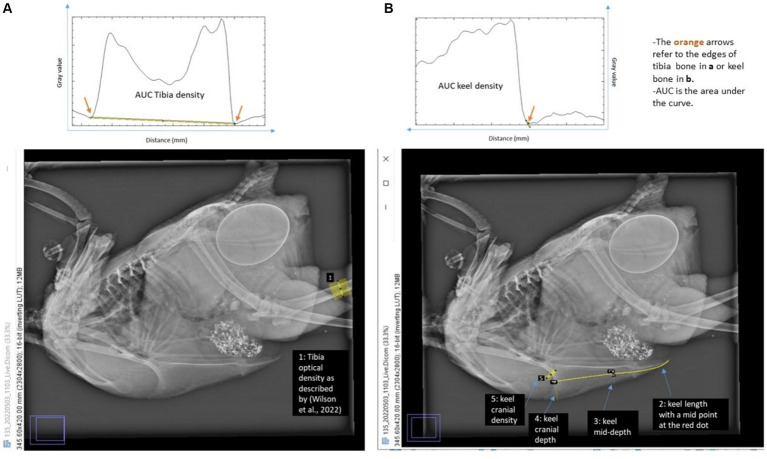
Keel bone damage, include deviations and fractures, is common in both white and brown laying hens, regardless of the housing system. Radiography for assessing birds' keel bones is was proposed by previous studies. However, radiographs show only 2 out of 3 dimensions of the dissected keel bones. The current study aimed to (1) investigate the association of radiographic optical density (keel and tibiotarsal) and geometry (keel) with dissected keel bone pathology. Previous studies suggested that keel bone fractures may result from internal pressure exerted by pelvic cavity contents. The current study also aimed to (2) investigate the potential associations between pelvic dimensions and measures of keel bone damage. A sample of 200 laying hens on a commercial farm were radiographed at 16, 29, 42, 55, and 68 weeks, and culled at the end of the laying period (week 74). The birds were examined post-mortem for pelvic dimensions and underwent whole-body radiography, followed by keel and tibiotarsal bone dissection and radiography, and keel bone scoring. The radiographs were used to estimate radiographic optical density (keel and tibiotarsal bone) and keel bone geometry (ratio of keel bone length to mid-depth). The method for on-farm radiography of laying hens, including live bird restraint, positioning for live keel imaging, and post-imaging measurements, was developed, tested, and found to be reproducible. The radiographs (1,116 images of 168 birds) and the respective measurements and post-mortem scores of keel bones are also provided for further development of radiographic metrics relevant to keel bone damage. Some longitudinal radiographic measurements of keel geometry (ratio of length to mid-depth) and optical density (keel and tibiotarsal) showed associations with the damage (deviations/fractures) observed on the dissected keel bones. The associations of keel damage were clearer with the radiographic keel geometry than with keel and tibiotarsal optical density, also clearer for the keel deviations than for keel fractures. The higher radiography ratio of keel length to mid-depth at weeks 42, 55 and 68 of age, the larger deviations size observed on the dissected keels at age of 74 weeks. The higher the tibiotarsal radiographic optical density at week 55 of age, the lower deviations size and fractures count observed on the dissected keels at age of 74 weeks. Pelvic dimensions showed a positive correlation with body weight, but a larger pelvic cavity was associated with increased keel bone damage. These findings lay the foundations for future use of on-farm radiography in identifying appropriate phenotypes for genetic selection for keel bone health.

Keywords: animal welfare; bone radiodensity; fractures; on-farm; pelvic cavity; poultry.


Conflict of interest statement

The authors declare that the research was conducted in the absence of any commercial or financial relationships that could be construed as a potential conflict of interest.

Figures

Figure 1
Figure 1
(Left) Set-up used for on-farm radiographic examination and (right) a live bird restrained and positioned for radiographic examination. The distance between radiography source and the flat panel detector was 100 cm.
Figure 2
Figure 2
Examples of radiographic images of the same live bird (at different ages), and of the whole body. Whole-body radiograph orientations: for the body and keel bone (cranial to the left, caudal to the right, dorsal at the top of image, ventral at the bottom of image) and for the tibiotarsal bone (cranial to the bottom of image, caudal to the top of image, proximal to the left of image, distal to the right of image). Dissected bones radiograph orientations: for the keel bone (caudal to the bottom of image, cranial to the top of image, ventral margin to the left of image, dorsal margin to the right of image) and for the tibiotarsal bone (cranial to the right of image, caudal to the left of image, proximal at the top of image, distal at the bottom of image).
Figure 3
Figure 3
Protocol used in scoring dissected keel bones. Keel bone orientation (cranial to the left, caudal to the right, dorsal margin of keel bone on bottom of image, and ventral margin of keel bone on top of image).
Figure 4
Figure 4
Measurement locations on radiography using the ImageJ program and estimated density of (A) tibiotarsal and (B) keel bone. Orientations: for the body and keel bone (cranial to the left, caudal to the right, dorsal at the top of image, ventral at the bottom of image) and for tibiotarsal bone (cranial to the bottom of image, caudal to the top of image, proximal at the left of image, distal at the right of image).
Figure 5
Figure 5
Mean of ratio of keel length to mid-depth across the levels of keel bone deviation size (A), fracture count (B), and callus size (C). Different letters on score group boxes indicate significantly different mean value (Tukey statistics, p < 0.05).
Figure 6
Figure 6
Linear model of extent of keel deviation, with body weight, keel, tibiotarsal optical density, and ratio of keel length to mid-depth as predictors, at different radiographic measurement points (age 16, 29, 42, 55, 68 weeks), post-mortem (PM), and post-mortem dissection (PMD).
Figure 7
Figure 7
Linear model of fracture count, with body weight, keel, tibiotarsal optical density, and ratio of keel bone length to mid-depth as predictors, at different radiographic measurement points (age 16, 29, 42, 55, 68 weeks), post-mortem (PM), and post-mortem dissection (PMD).
Figure 8
Figure 8
Linear model of keel density, with pelvic capacity, tibiotarsal density, and their interaction as predictors.
Figure 9
Figure 9
Ventral (top row) and lateral (bottom row) views of the same three keel bones showing ventral and dorsal deviations, respectively. Keel bone orientation at the top row (cranial to the left, caudal to the right, left side of keel bone on top of image, right side of keel bone on bottom of image) and at the bottom row (cranial to the left, caudal to the right, ventral margin of keel bone on top of image, dorsal margin of keel on bottom of image).

Similar articles

References

    1. Heerkens JLT, Delezie E, Ampe B, Rodenburg TB, Tuyttens FAM. Ramps and hybrid effects on keel bone and foot pad disorders in modified aviaries for laying hens. Poult Sci. (2016) 95:2479–88. doi: 10.3382/ps/pew157, PMID: - DOI - PubMed
    1. Petrik MT, Guerin MT, Widowski TM. On-farm comparison of keel fracture prevalence and other welfare indicators in conventional cage and floor-housed laying hens in Ontario, Canada. Poult Sci. (2015) 94:579–85. doi: 10.3382/ps/pev039, PMID: - DOI - PubMed
    1. Rodenburg T, Tuyttens F, De Reu K, Herman L, Zoons J, Sonck B. Welfare assessment of laying hens in furnished cages and non-cage systems: assimilating expert opinion. Anim Welf. (2008) 17:355–61. doi: 10.1017/S0962728600027858 - DOI
    1. Sandilands V. The laying hen and bone fractures. Vet Rec. (2011) 169:411–2. doi: 10.1136/vr.d6564, PMID: - DOI - PubMed
    1. Stratmann A, Fröhlich EKF, Harlander-Matauschek A, Schrader L, Toscano MJ, Würbel H, et al. . Soft perches in an aviary system reduce incidence of keel bone damage in laying hens. PLoS One. (2015) 10:e0122568. doi: 10.1371/journal.pone.0122568, PMID: - DOI - PMC - PubMed

LinkOut - more resources