. 2024 Sep 28:29:101279.
doi: 10.1016/j.mtbio.2024.101279. eCollection 2024 Dec.

Enhancing retention and permeation of rapamycin for osteoarthritis therapy using a two-stage drug delivery system

Affiliations

Enhancing retention and permeation of rapamycin for osteoarthritis therapy using a two-stage drug delivery system

Guangyong Lin et al. Mater Today Bio. .

Abstract

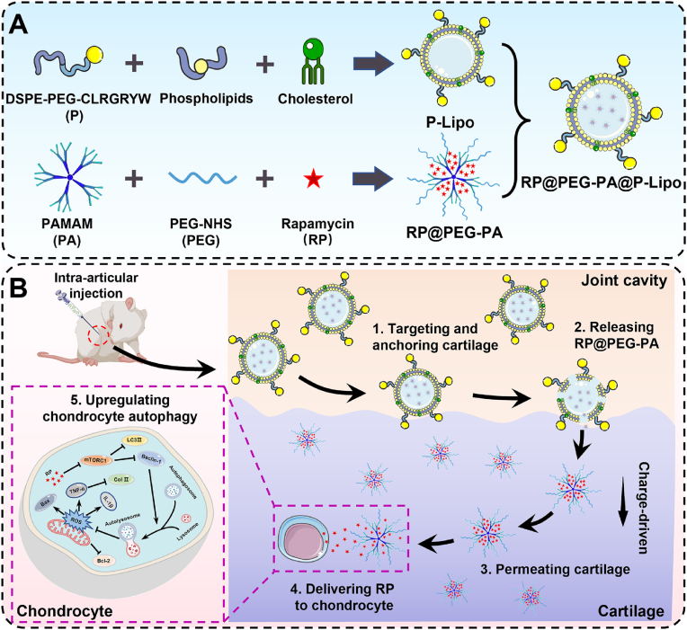
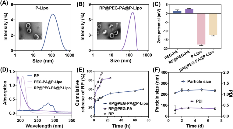
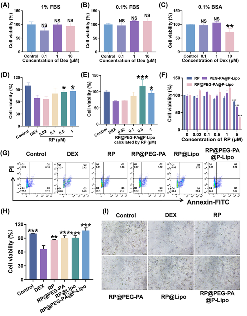
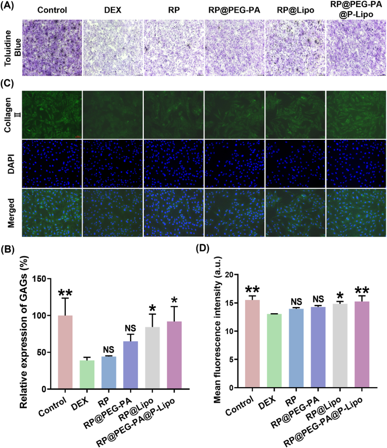
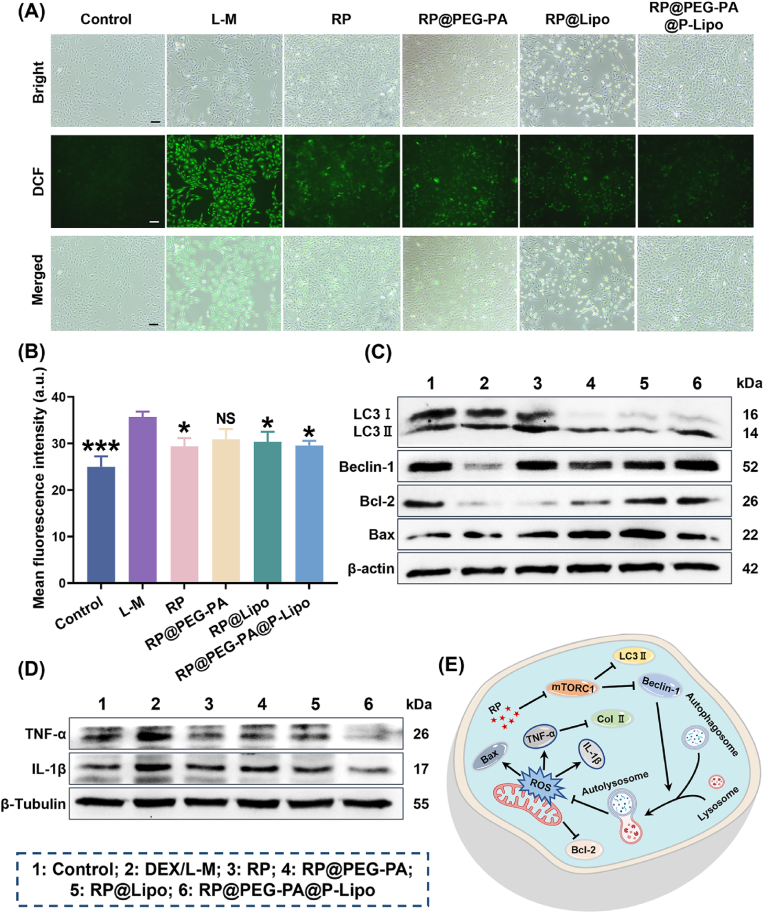
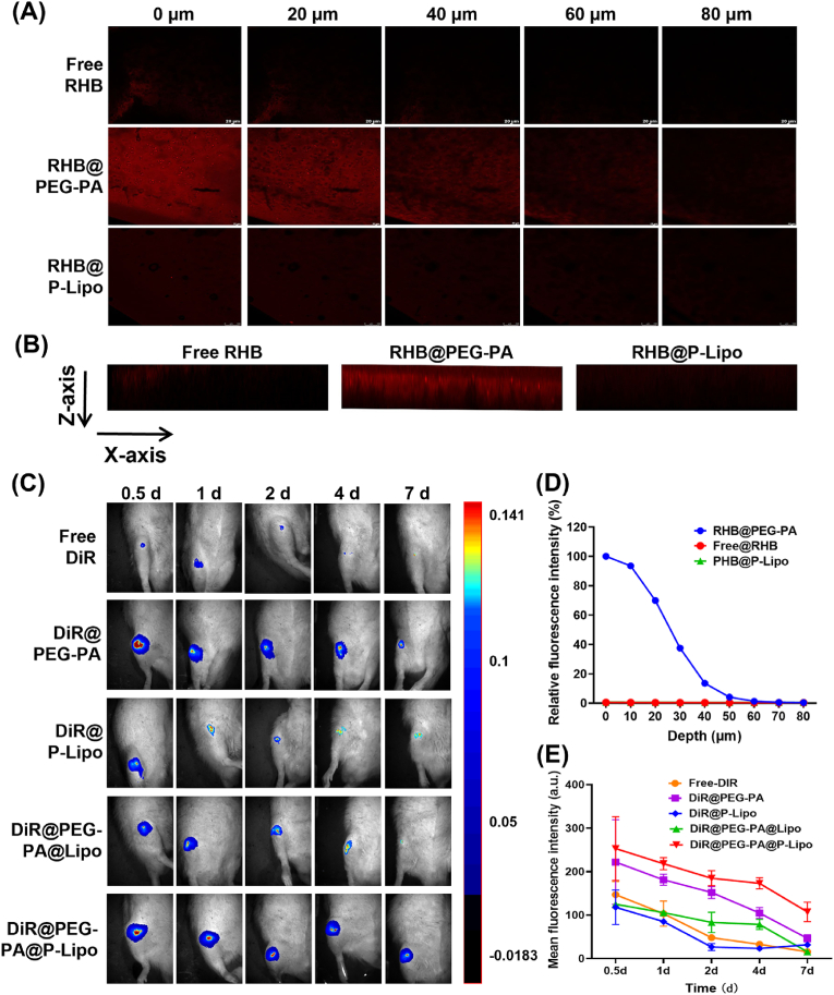
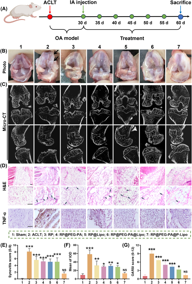
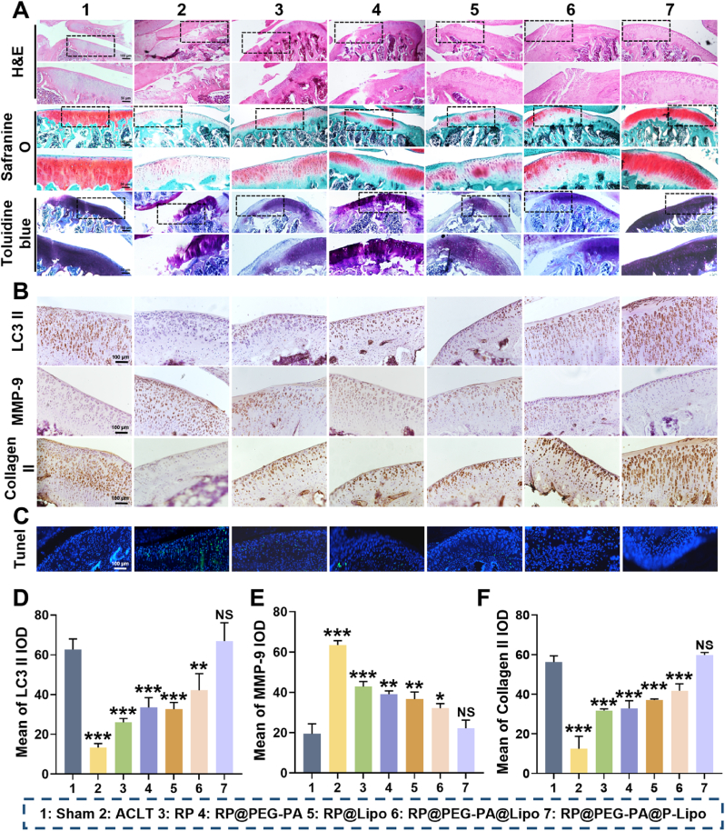
Osteoarthritis (OA) remains a challenging degenerative joint disease, largely associated with chondrocyte apoptosis during its development. Preserving chondrocytes stands as a promising strategy for OA treatment. Rapamycin (RP) exhibits chondrocyte protection by fostering autophagy. Nevertheless, the swift clearance of intra-articular injections and the dense cartilage extracellular matrix (ECM) hinder RP from effectively reaching chondrocytes. Herein, we developed a "two-stage" drug delivery system (RP@PEG-PA@P-Lipo). This system comprises primary nanoparticles (P-Lipo), liposomes modified with a collagen II targeting peptide (WYRGRLC), and secondary nanoparticles (RP@PEG-PA), PEG-modified PAMAM encapsulating rapamycin (RP). RP@PEG-PA@P-Lipo demonstrates adherence to the cartilage surface with WYRGRLC, substantially prolonging retention within the joint cavity. Subsequently, released RP@PEG-PA can effectively penetrate the cartilage and deliver RP to chondrocytes through small size and charge-driven forces. In vitro and in vivo experiments corroborate its notable therapeutic effects on OA. This study holds promise in offering a novel approach for clinical drug delivery and OA treatment.

Keywords: Autophagy; Chondrocyte; Osteoarthritis; Rapamycin; Two-stage nanoparticle.


Conflict of interest statement

The authors declare that they have no known competing financial interests or personal relationships that could have appeared to influence the work reported in this paper.

Figures

Image 1
Graphical abstract
Scheme 1
Scheme 1
Schematic illustration (A) the construction and (B) working mechanism of “two-stage” nanoparticles RP@PEG-PA@P-Lipo for OA treatment.
Fig. 1
Fig. 1
Characterization of “two-stage” nanoparticles. TEM images and DLS characterization of (A) P-Lipo and (B) RP@PEG-PA@P-Lipo, scale bar = 100 nm. (C) Zeta potential of PEG-PA, RP@PEG-PA, P-Lipo and RP@PEG-PA@P-Lipo. (D) UV–vis–NIR spectra of free RP, PEG-PA@P-Lipo and RP@PEG-PA@P-Lipo. (E) In vitro release of RP from free RP, RP@PEG-PA and RP@PEG-PA@P-Lipo in pH7.4 PBS containing 0.1 % SDS. (F) The stability analysis of RP@PEG-PA@P-Lipo in pH 7.4 PBS at 4 °C. Data in C, E, and F are given as mean ± SD, n = 3.
Fig. 2
Fig. 2
Anti-apoptotic effects of RP@PEG-PA@P-Lipo on RCCs. The optimization concentration of DEX in (A) 1 % FBS, (B) 0.1 % FBS, and (C) 0.1 % BSA mediums, respectively. Cell viability of RCCs treated with different concentration of (D) RP and (E) RP@PEG-PA@P-Lipo. (F) Cytotoxicity of different concentration of RP, PEG-PA@P-Lipo and RP@PEG-PA@P-Lipo. (G) Effect of RP@PEG-PA@P-Lipo on the rate of chondrocyte apoptosis induced by DEX. Representative scatter plots showed apoptosis levels determined by flow cytometry. (H) Effect of RP@PEG-PA@P-Lipo on the survival rate of chondrocytes by MTT. (I) Effect of RP@PEG-PA@P-Lipo on morphology and number of chondrocytes, scale bar = 200 μm. Data are given as mean ± SD, n = 3. NS p > 0.05, ∗p < 0.05, ∗∗p < 0.01, ∗∗∗p < 0.001, data in A – C, F are compared to control group, data in D - E, H are compared to DEX group.
Fig. 3
Fig. 3
The protection effect of RP@PEG-PA@P-Lipo on the chondrocytes. (A) Evaluation of GAG content in chondrocytes by Toluidine Blue staining, scale bar = 100 μm. (C) IF was used to evaluate the expression of collagen II (green). DAPI (blue) was used to stain the nucleus, scale bar = 20 μm. The quantitative analysis of (B) the Toluidine Blue staining and (D) the IF results. Data are given as mean ± SD, n = 3. NS p > 0.05, ∗p < 0.05, ∗∗p < 0.01, compared to DEX group.
Fig. 4
Fig. 4
The underlying anti-apoptosis and anti-inflammation mechanisms of RP@PEG-PA@P-Lipo in RCCs cells. (A) ROS levels was analyzed by DCFH-DA probe showing the preventive effect of RP@PEG-PA@P-Lipo against ROS generation in RCCs, scale bar = 20 μm. (B) Mean fluorescence intensity was calculated using Image J software. (C–D) Western blot analyses for protein expression in RCCs stimulated with DEX or L-M and treated with different formulations. β-actin and β-Tubulin were used as an internal control. (E) The mechanism of anti-apoptosis and anti-inflammation of RP@PEG-PA@P-Lipo. Data in (B) are shown as mean ± SD, n = 3. NS p > 0.05, ∗p < 0.05, ∗∗∗p < 0.001, compared to L-M group.
Fig. 5
Fig. 5
Enhanced permeation and retention of RP@PEG-PA@P-Lipo in cartilage tissue. (A) Representative fluorescence confocal images of porcine cartilage tissues treated with free RHB, RHB@PEG-PA and RHB@P-Lipo. Images were obtained at 20 μm intervals below the lateral porcine cartilage, scale bar = 20 μm. (B) Representative x-z orthogonal images of porcine cartilage tissues treated with free RHB, RHB@PEG-PA and RHB@P-Lipo. Horizontal image length, 160 μm; vertical image length, 80 μm. (C) The distribution and retention of free DiR, DiR@PEG-PA, DiR@P-Lipo, DiR@PEG-PA@Lipo and DiR@PEG-PA@P-Lipo in knee joints of OA rat model after intra-articular injection. DiR was used as probe to track the in vivo retention of nanoparticles, and the whole process was monitored by a in vivo imaging system at 0.5 d, 1 d, 2 d, 4 d,7 d after administration. (D) The relative fluorescence intensity of porcine cartilage tissues at different depths. (E) The mean fluorescence intensity of OA joints at different times. Data in (E) are shown as mean ± SD, n = 3.
Fig. 6
Fig. 6
The in vivo therapeutic effect of RP@PEG-PA@P-Lipo in OA rat models. (A) In vivo experimental schedule. (B) Knee joint closeup photo. (C) Micro-CT images of the knees in OA rat after treatment. (D) H&E staining and TNF-α evaluation of synovium sections, scale bar = 100 μm. The black arrows represent inflammatory cells. (E) Synovitis score. (F) IHC analysis of TNF-α in synovium, scale bar = 100 μm. (G) OARIS score. NS p > 0.05, ∗p < 0.05, ∗∗p < 0.01, ∗∗∗p < 0.001, compared to sham group.
Fig. 7
Fig. 7
The repaired effect of RP@PEG-PA@P-Lipo on cartilage in OA model rats. (A) H&E, Safranine O, and Toluidine Blue staining of cartilage sections of treatment with different agents, scale bar = 100 μm. (B) IHC analysis of cartilage after treatment with different agents, scale bar = 100 μm. (C) Tunel staining of cartilage from different groups. Quantitative analysis of (D) LC3Ⅱ, (E) MMP-9 and (F) Collagen II positive area of IHC analysis. Data are given as mean ± SD, n = 3. NS p > 0.05, ∗p < 0.05, ∗∗p < 0.01, ∗∗∗p < 0.001, compared to sham group.

Similar articles

Cited by

References

    1. Leifer V.P., Katz J.N., Losina E. The burden of OA-health services and economics. Osteoarthritis Cartilage. 2022;30(1):10–16. - PMC - PubMed
    1. Xia B., Di C., Zhang J., Hu S., Jin H., Tong P. Osteoarthritis pathogenesis: a review of molecular mechanisms. Calcif. Tissue Int. 2014;95(6):495–505. - PMC - PubMed
    1. Rodriguez-Merchan E.C. Topical therapies for knee osteoarthritis. Postgrad. Med. 2018;130(7):607–612. - PubMed
    1. Arden N.K., Perry T.A., Bannuru R.R., Bruyere O., Cooper C., Haugen I.K., Hochberg M.C., McAlindon T.E., Mobasheri A., Reginster J.Y. Non-surgical management of knee osteoarthritis: comparison of ESCEO and OARSI 2019 guidelines. Nat. Rev. Rheumatol. 2021;17(1):59–66. - PubMed
    1. Fraenkel L., Buta E., Suter L., Dubreuil M., Levy C., Najem C., Brennan M., Corn B., Kerns R., Goulet J. Nonsteroidal anti-inflammatory drugs vs cognitive behavioral therapy for arthritis pain: a randomized withdrawal trial. JAMA Intern. Med. 2020;180(9):1194–1202. - PMC - PubMed

LinkOut - more resources