. 2024 Sep 28:23:3445-3453.
doi: 10.1016/j.csbj.2024.09.014. eCollection 2024 Dec.

Uncovering conserved networks and global conformational changes in G protein-coupled receptor kinases

Affiliations

Uncovering conserved networks and global conformational changes in G protein-coupled receptor kinases

Min Jae Seo et al. Comput Struct Biotechnol J. .

Abstract

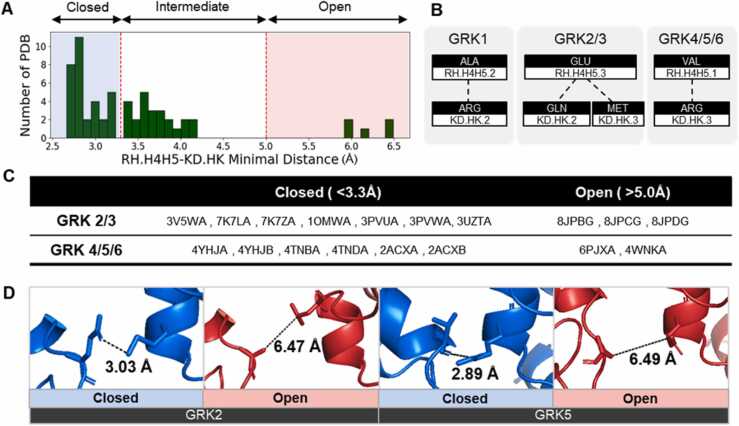
G protein-coupled receptor kinases (GRKs) are essential regulators of signaling pathways mediated by G protein-coupled receptors. Recent research suggests that GRK-mediated phosphorylation patterns dictate functional selectivity, leading to biased cellular responses. However, a comprehensive understanding of the structural mechanisms at the single-residue level remains elusive. This study aims to define the general conformational dynamics of GRKs with a particular focus on quantifying the transitions between the closed and open states. Specifically, we examined these transitions, classified based on the ionic lock between the regulatory G protein signaling homology domain and kinase domain. To facilitate a precise structural comparison, we assigned common labels to topologically identical positions across the 47 GRK structures retrieved from the Protein Data Bank. Our analysis identified both general and subfamily-specific dynamic movements within the networks and measured the conformational change scores between the two states. Elucidating these structural dynamics could provide significant insights into the regulatory mechanisms of GRK.

Keywords: Conformational change; G protein-coupled receptor kinase; GPCR; GRK; Phosphorylation.


Conflict of interest statement

The authors declare no conflicts of interest..

Figures

ga1
Graphical abstract
Fig. 1
Fig. 1
(A) Simple mechanism of G protein-coupled receptor kinase (GRK). The bottom box shows a schematic presentation of GRK in the closed and open states. (B) Simple workflow from common labeling with Alphafold2 structure to contact score networks. (C) Secondary structure element labels according to the GRK Common Label (GCL) system.
Fig. 2
Fig. 2
(A) Histogram of the minimal distance between residues in RH.H4H5 and KD.HK. The Y-axis represents the number of Protein Data Bank (PDB) files, and the X-axis represents the distance. These data also include the G protein-coupled receptor kinase (GRK)1 structure. (B) Amino acid information of the ionic lock between RH.H4H5 and KD.HK according to each GRK subfamily. (C) Filtered PDB files according to the selection criteria. The last letter of the PDB file names is the chain identifier. (D) Structures of the closed and open states. From left to right: PDB codes 3PVW, 8JPB, 4TND, and 4WNK.
Fig. 3
Fig. 3
Flow of contact score calculation. G protein-coupled receptor kinase (GRK) Common Label (GCL) Contact Score (upper left box): long-range heavy atom contacts at GCL positions in GRK structures. Average Contact Score (ACS) and Secondary Structure Element (SSE)-ACS (lower left boxes): contact scores of residues and secondary structure elements, respectively. The right box shows the conformational change (CC) score at the residue level and secondary structure level. A score of zero indicates no contact change between the closed and open structures.
Fig. 4
Fig. 4
(A) Conformational change of each G protein-coupled receptor kinase (GRK)2/3 and GRK4/5/6 subfamily. The X-axis represents the secondary structure element-conformational change (SSE-CC) score. The Y-axis represents secondary structure pairs showing common conformational movements. (B) Network showing the sum of GRK2/3 and GRK4/5/6 SSE-CC scores. Thicker edges indicate a higher tendency for contact in either the closed or open state (Fig. S7B). Each node represents a secondary structure element. (C) Example of conformational differences between closed and open states. Only common movements are shown in the network. In the left box, RH.H10- KD.HCS4 interactions are more prominent in the closed state. In the right box, KD.HKHL-RH.HN interactions show closer contact in the open state.
Fig. 5
Fig. 5
(A) Conformational changes at the residue level. Each square represents a residue at a specific G protein-coupled receptor kinase (GRK) Common Label (GCL) position in the GRK structure. Only residues showing common conformational movements are presented. (B) Conservation scores of all positions and those shown in (A), calculated by Jalview software. Statistical analysis was performed using the Mann–Whitney U test (*: p-value < 0.1, **:p-value < 0.01, ***:p-value<0.001). (C) Conservation scores of each of the 78 GCL positions in (A), categorized by GRK subfamily. The X-axis labels for GCL positions are provided in the attached files.

References

    1. Zhai R., Snyder J., Montgomery S., Sato P.Y. Double life: how GRK2 and β-arrestin signaling participate in diseases. Cell Signal. 2022;94 - PMC - PubMed
    1. Lymperopoulos A. GRK2 and β-arrestins in cardiovascular disease: something old, something new. Am J Cardiovasc Dis. 2011;1:126. - PMC - PubMed
    1. Choi M., et al. G protein–coupled receptor kinases (GRKs) orchestrate biased agonism at the β2-adrenergic receptor. Sci Signal. 2018;11 - PubMed
    1. Claing A., Laporte S.A., Caron M.G., Lefkowitz R.J. Endocytosis of G protein-coupled receptors: roles of G protein-coupled receptor kinases and ß-arrestin proteins. Prog Neurobiol. 2002;66:61–79. - PubMed
    1. Krupnick J.G., Benovic J.L. The role of receptor kinases and arrestins in G protein–coupled receptor regulation. Annu Rev Pharmacol Toxicol. 1998;38:289–319. - PubMed

LinkOut - more resources