Review
. 2024 Sep 10:7:32.
doi: 10.20517/cdr.2024.55. eCollection 2024.

Cancer-associated fibroblast cell surface markers as potential biomarkers or therapeutic targets in lung cancer

Affiliations
Review

Cancer-associated fibroblast cell surface markers as potential biomarkers or therapeutic targets in lung cancer

Samaneh Tokhanbigli et al. Cancer Drug Resist. .

Abstract

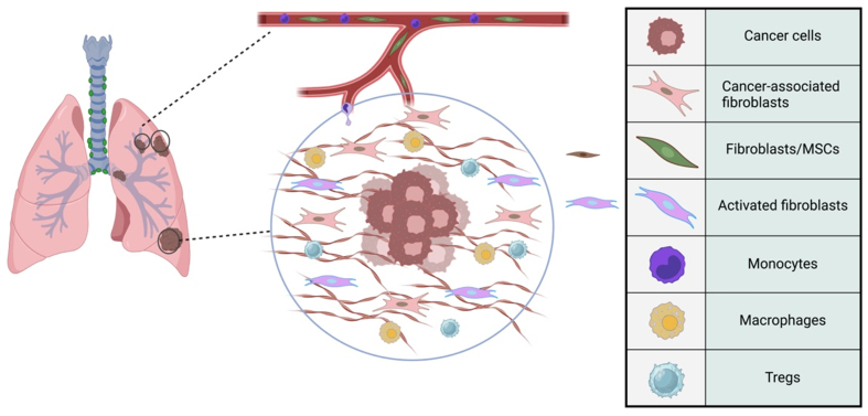
Cancer-associated fibroblasts (CAFs) are the vital constituent of the tumor microenvironment, and in communication with other cells, they contribute to tumor progression and metastasis. Fibroblasts are the proposed origin of CAFs, which are mediated by pro-inflammatory cytokines and the recruitment of immune cells akin to wound healing. Although various studies have identified different subpopulations of CAFs in lung cancer, the heterogeneity of CAFs, particularly in lung cancer, and their potential as a therapeutic target remain largely unknown. Notwithstanding CAFs were previously thought to have predominantly tumor-promoting features, their pro- or anti-tumorigenic properties may depend on various conditions and cell origins. The absence of distinct markers to identify CAF subpopulations presents obstacles to the successful therapeutic targeting and treatment of CAFs in cancer. Human clinical and animal studies targeting CAFs have shown that targeting CAFs exacerbates the disease progression, suggesting that subpopulations of CAFs may exert opposing functions in cancer progression. Therefore, it is essential to pinpoint specific markers capable of characterizing these subpopulations and revealing their mechanisms of function. The cell-specific surface markers of CAFs will serve as an initial step in investigating precise CAF subpopulations and their role in diagnosing and targeting therapy against cancer-promoting CAF subsets in lung cancer.

Keywords: Fibroblasts; inflammation; lung cancer; targeted therapy; therapeutic markers.


Conflict of interest statement

All authors declared that there are no conflicts of interest.

Figures

Figure 1
Figure 1
Schematic Crosstalk of CAFs and immune cells in the tumor microenvironment of lung cancer. CAFs orchestrate the tumor microenvironment, interacting with various cell types within the tumor microenvironment of lung cancer. An example of this immune component is cancer cells, MSCs, activated fibroblasts, monocytes, macrophages, and Tregs. MSCs could differentiate into CAFs and other cell types, supporting the tumor. Activated fibroblasts are involved in remodeling the ECM and promoting tumor invasion. Monocytes can differentiate into macrophages within the tumor microenvironment. Macrophages can have pro-tumorigenic roles, promoting tumor growth and suppressing the immune response. Tregs contribute to the immunosuppressive environment within the tumor, aiding in tumor evasion from the immune system. CAFs: Cancer-associated fibroblasts; MSCs: mesenchymal stem cells, Tregs: regulatory T cells; ECM: extracellular matrix.
Figure 2
Figure 2
The origin of CAFs and their crosstalk signaling pathways regulating tumor microenvironment. (A) CAFs in lung cancer are potentially derived from tissue-resident fibroblasts, mesenchymal stem cells, epithelial cells, and pericytes; (B) These CAFs modulate the tumor microenvironment by activating a variety of signaling pathways, including TGF-β, Wnt/β-catenin, MAPK, IL6, EGFR, JAK/STAT, and NF-κB, resulting in the orchestration of processes such as proliferation and ECM remodeling, immunosuppression, EMT, drug resistance, and angiogenesis. CAFs: Cancer-associated fibroblasts; TGF-β: transforming growth factor beta; MAPK: mitogen-activated protein kinases; IL: interleukin; EGFR: epidermal growth factor receptor; JAK/STAT: Janus kinases/signal transducers and activators of transcription; NF-κB: nuclear factor kappa-light-chain-enhancer of activated B cells; ECM: extracellular matrix, EMT: epithelial-mesenchymal transition.
Figure 3
Figure 3
A schematic presentation of different branches/subtypes of CAFs in lung cancer CAFs. Single-cell RNAseq analysis revealed heterogeneity of CAFs in NSCLC. Kim et al. identified different branches of CAFs[150], including immunosuppressive, ApCAFs, myCAFs, and proliferative CAFs. Hu et al. introduced three subtypes with various HGF and FGF expression levels[151]. Five unique CAFs were found in Lambrechts’s investigation; one was categorized as non-malignant fibroblasts[152]. CAFs: Cancer-associated fibroblasts; NSCLC: non-small cell lung cancer; ApCAFs: antigen-presenting CAFs; myCAFs: myofibroblast-like CAFs; HGF: hepatocyte growth factor; FGF: fibroblast growth factor.

Similar articles

Cited by

References

    1. Sung H, Ferlay J, Siegel RL, et al. Global Cancer Statistics 2020: GLOBOCAN estimates of incidence and mortality worldwide for 36 cancers in 185 countries. CA Cancer J Clin. 2021;71:209–49. doi: 10.3322/caac.21660. - DOI - PubMed
    1. Ferone G, Lee MC, Sage J, Berns A. Cells of origin of lung cancers: lessons from mouse studies. Genes Dev. 2020;34:1017–32. doi: 10.1101/gad.338228.120. - DOI - PMC - PubMed
    1. Fidler-Benaoudia MM, Torre LA, Bray F, Ferlay J, Jemal A. Lung cancer incidence in young women vs. young men: a systematic analysis in 40 countries. Int J Cancer. 2020;147:811–9. doi: 10.1002/ijc.32809. - DOI - PubMed
    1. Miller KD, Nogueira L, Mariotto AB, et al. Cancer treatment and survivorship statistics, 2019. CA Cancer J Clin. 2019;69:363–85. doi: 10.3322/caac.21565. - DOI - PubMed
    1. Hanahan D, Coussens LM. Accessories to the crime: functions of cells recruited to the tumor microenvironment. Cancer Cell. 2012;21:309–22. doi: 10.1016/j.ccr.2012.02.022. - DOI - PubMed

LinkOut - more resources