

Clinical outcomes of using operating microscope for alveolar ridge preservation: A randomized controlled trial
- PMID: 39403776
- PMCID: PMC11951953
- DOI: 10.1002/JPER.24-0081
Clinical outcomes of using operating microscope for alveolar ridge preservation: A randomized controlled trial
Abstract
Background: The use of the operating microscope (OM) for extraction and alveolar ridge augmentation (ARP) is increasing due to enhanced magnification and illumination. The primary objective was to compare the wound healing and crestal bone quality after the use of OM and dental loupes (DL) for ARP.
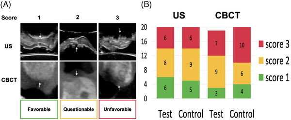
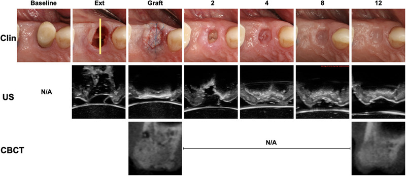
Methods: Forty non-molar teeth with periapical lesions in need of extraction and ARP from 33 patients were randomly assigned to 2 groups: DL (control) or OM (test). All procedures were performed by 1 surgeon and assessments done by masked examiners. ARP was performed with an allograft and a resorbable collagen membrane. The presence of granulomatous tissue remnants after debridement was recorded. Cone-beam computed tomography (CBCT) and ultrasound (US) scans were taken during the healing phase up to 16-week visits. Bone cores were retrieved from implant osteotomies for histologic analysis. Patient-reported outcome measurements (PROMs) were assessed.
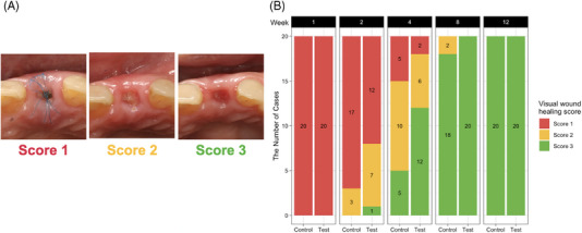
Results: All patients completed all study visits except 1 who dropped out before the last visit. After socket debridement, the test group exhibited significantly fewer sites with tissue remnants (p = 0.01) and a better healing score at 2-week (p = 0.04) and 4-week (p = 0.01) time points. There were no significant differences in 12-week crestal bone healing by histology (p = 0.1), US (p = 0.85), and CBCT healing (p = 0.64) at 12 weeks, as well as PROMs (p > 0.1).
Conclusion: Within the limitation of the study, the use of OM for ARP resulted in significantly fewer tissue remnants and favorable early visual wound healing. CBCT and US-derived-crestal bone quality did not show a difference between the 2 groups.
Plain language summary: Alveolar ridge preservation (ARP) by placing bone particulates in the extraction socket, covered by a wound dressing material, is commonly applied immediately after tooth extraction to reduce jawbone shrinkage in preparation for implant placement later. The jawbone healing varies, depending largely on the ability to remove the etiology, socket features, extent of surgical trauma, and wound stability. Healed jawbone with good quality is favorable for easiness of implant placement and could be related to maintenance of long-term implant health. The surgical microscope with high magnification (up to ∼25×) and co-axial illumination is ideal for assistance in the removal of granulomatous tissue that is believed to interfere with healing, performance of minimally invasive extraction, and stabilization of the wound with meticulous tissue management and fine sutures. This study compared the use of the surgical microscope to dental loupes for ARP in a randomized controlled design. The microscope-assisted ARP is associated with a significantly higher chance of removing granulomatous tissue, favorable early healing, and similar crestal bone quality. Removal of granulomatous tissue is significant for immediate implant placement. This study serves as a model for testing the benefits of the surgical microscope for encouraging early healing in more challenging intraoral surgical procedures.
Keywords: alveolar bone; alveolar ridge augmentation; microscope; microsurgery; ultrasonography; wound healing.
© 2024 The Author(s). Journal of Periodontology published by Wiley Periodicals LLC on behalf of American Academy of Periodontology.
Conflict of interest statement
The authors declare no conflicts of interest.
Figures

References
-
- Avila‐Ortiz G, Chambrone L, Vignoletti F. Effect of alveolar ridge preservation interventions following tooth extraction: a systematic review and meta‐analysis. J Clin Periodontol. 2019;46(Suppl 21):195‐223. - PubMed
-
- MacBeth N, Trullenque‐Eriksson A, Donos N, Mardas N. Hard and soft tissue changes following alveolar ridge preservation: a systematic review. Clin Oral Implants Res. 2017;28(8):982‐1004. - PubMed
-
- Leblebicioglu B, Tatakis DN. Complications following alveolar ridge augmentation procedures. Periodontol 2000. 2023;93(1):221‐235. - PubMed
-
- Chan HL, Lin GH, Fu JH, Wang HL. Alterations in bone quality after socket preservation with grafting materials: a systematic review. Int J Oral Maxillofac Implants. 2013;28(3):710‐720. - PubMed
Publication types
MeSH terms
Substances
Grants and funding
LinkOut - more resources
Full Text Sources
Miscellaneous
