

Accuracy of the LaserSAFE technique for detecting positive surgical margins during robot-assisted radical prostatectomy: blind assessment and inter-rater agreement analysis
- PMID: 39403832
- PMCID: PMC11707496
- DOI: 10.1111/his.15336
Accuracy of the LaserSAFE technique for detecting positive surgical margins during robot-assisted radical prostatectomy: blind assessment and inter-rater agreement analysis
Abstract
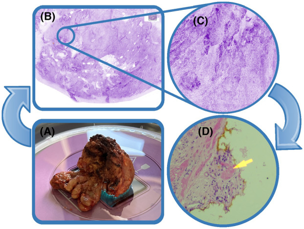
Introduction and objectives: Fluorescence confocal microscopy (FCM) is a new imaging modality capable of generating digital microscopic resolution scans of fresh surgical specimens, and holds potential as an alternative to frozen section (FS) analysis for intra-operative assessment of surgical margins. Previously, we described the LaserSAFE technique as an application of FCM for margin assessment in robot-assisted radical prostatectomy (RARP) using the Histolog® scanner. This study describes the accuracy and inter-rater agreement of FCM imaging compared to corresponding paraffin-embedded analysis (PA) among four blinded pathologists for the presence of positive surgical margins (PSM).
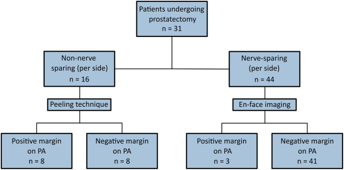
Materials and methods: RARP specimens from patients enrolled in the control arm of the NeuroSAFE PROOF study (NCT03317990) were analysed from April 2022 to February 2023. Prostate specimens were imaged using the Histolog® scanner before formalin fixation and PA. Four trained assessors, blinded to PA, reviewed and analysed FCM images of the posterolateral prostatic surface.
Results: A total of 31 prostate specimens were included in the study. PA per lateral side of the prostate identified 11 instances of positive margins. Among the four histopathologists included in our study, FCM achieved a sensitivity of 73-91 and specificity of 94-100% for the presence of PSM. Fleiss' Kappa for inter-rater agreement on PSM was 0.78 (95% confidence interval = 0.64-0.92), indicating substantial agreement.
Conclusion: This blinded analysis of FCM versus PA among histopathologists with different experience levels demonstrated high accuracy and substantial inter-rater agreement for diagnosing PSM. This supports the role of the FCM as an alternative to FS.
Keywords: fluorescence confocal microscopy; frozen section analysis; margin assessment; prostate cancer.
© 2024 The Author(s). Histopathology published by John Wiley & Sons Ltd.
Conflict of interest statement
R.A.M. receives a salary from NIHR RFPB PB‐PG‐1216‐20013 and PCUK MA‐CT20‐11. G.S. receives consulting fees from Angle plc and has received travel expenses from Janssen and Johnson and Johnson (not related to the present manuscript). M.A., T.Al‐H., K.D, L.S.T.M., E.D., W.V., A.F. and A.H. report no conflicts of interest related to this work. SamanTree Medical SA (Lausanne, Switzerland) provided the Histolog® scanner, training and consumables but was not involved in the study design, execution, results or writing of the manuscript.
Figures
References
-
- Averbeck MA, Marcelissen T, Anding R, Rahnama'I MS, Sahai A, Tubaro A. How can we prevent postprostatectomy urinary incontinence by patient selection, and by preoperative, peroperative, and postoperative measures? International Consultation on Incontinence‐Research Society 2018. Neurourol. Urodyn. 2019; 38(Suppl 5); S119–S126. - PubMed
-
- Nguyen LN, Head L, Witiuk K et al. The risks and benefits of cavernous neurovascular bundle sparing during radical prostatectomy: a systematic review and meta‐analysis. J. Urol. 2017; 198; 760–769. - PubMed
-
- Walsh PC, Lepor H, Eggleston JC. Radical prostatectomy with preservation of sexual function: anatomical and pathological considerations. Prostate 1983; 4; 473–485. - PubMed
Publication types
MeSH terms
Grants and funding
LinkOut - more resources
Full Text Sources
Medical
Miscellaneous
