An 8-Year 5-Month-Old Boy with a Basal Ganglia Lesion with Triphasic Waves on Electroencephalogram
- PMID: 39403907
- PMCID: PMC11474962
- DOI: 10.62641/aep.v52i5.1741
An 8-Year 5-Month-Old Boy with a Basal Ganglia Lesion with Triphasic Waves on Electroencephalogram
Abstract
Background: Triphasic waves (TWs) on electroencephalograms (EEGs) have predominantly been observed in adults, often associated with Creutzfeldt‒Jakob disease and metabolic encephalopathy. However, TWs have also been linked to various nonmetabolic and structural abnormalities. Additionally, reports of TWs in children are rare.
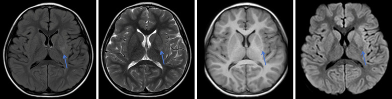
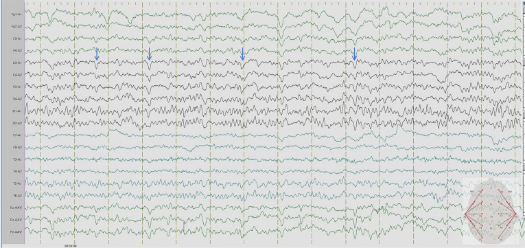
Case presentation: We present the case of an 8-year and 5-month-old boy with basal ganglia lesion who exhibited TWs in the local C3 lead on electroencephalography. Subsequent EEGs revealed no additional abnormalities. During the follow-up at 1 year and 8 months, there was no significant change in the patient's condition.
Conclusion: Triphasic waves can occur in children with basal ganglia lesions, but their underlying causes may differ from those previously reported. Further research is needed to elucidate the mechanisms and clinical significance of TWs in pediatric patients.
Conflict of interest statement
The authors declare no conflict of interest.
Figures


References
-
- Fernández-Torre JL, Kaplan PW. Triphasic Waves: Historical Overview of an Unresolved Mystery. Journal of Clinical Neurophysiology: Official Publication of the American Electroencephalographic Society . 2021;38:399–409. - PubMed
-
- Kwon OY, Jung KY, Park KJ, Kang JK, Shon YM, Lee IK, et al. Source localization of triphasic waves: implications for the pathophysiological mechanism. Clinical EEG and Neuroscience . 2007;38:161–167. - PubMed
-
- Sundaram MB, Blume WT. Triphasic waves: clinical correlates and morphology. The Canadian Journal of Neurological Sciences. Le Journal Canadien des Sciences Neurologiques . 1987;14:136–140. - PubMed
-
- Foley JM, Watson CW, Adams RD. Significance of the electroencephalographic changes in hepatic coma. Transactions of the American Neurological Association . 1950;51:161–165. - PubMed
Publication types
MeSH terms
LinkOut - more resources
Full Text Sources
Miscellaneous