. 2024 Oct 15;134(20):e181604.
doi: 10.1172/JCI181604.

Autoinflammation in patients with leukocytic CBL loss of heterozygosity is caused by constitutive ERK-mediated monocyte activation

Jonathan Bohlen  1   2 Ivan Bagarić  1   2   3 Taja Vatovec  1   2   3 Masato Ogishi  4 Syed F Ahmed  5 Axel Cederholm  6 Lori Buetow  5 Steicy Sobrino  2   7   8 Corentin Le Floc'h  1   2 Carlos A Arango-Franco  1   2   9 Luis Seabra  2 Marine Michelet  10 Federica Barzaghi  11 Davide Leardini  12 Francesco Saettini  13 Francesca Vendemini  14 Francesco Baccelli  12 Albert Catala  15 Eleonora Gambineri  16   17 Marinella Veltroni  17 Yurena Aguilar de la Red  18 Gillian I Rice  19 Filippo Consonni  17   20 Laureline Berteloot  21   22 Laetitia Largeaud  23 Francesca Conti  24   25 Cécile Roullion  2   26 Cécile Masson  2   27 Boris Bessot  2 Yoann Seeleuthner  1   2 Tom Le Voyer  1   2   4   28 Darawan Rinchai  4 Jérémie Rosain  1   2   4   29 Anna-Lena Neehus  1   2 Lucia Erazo-Borrás  1   2   9 Hailun Li  1   2 Zarah Janda  1   3 En-Jui Cho  1   3 Edoardo Muratore  12 Camille Soudée  1   2 Candice Lainé  1   2 Eric Delabesse  30 Claire Goulvestre  31 Cindy S Ma  32   33 Anne Puel  1   2   4 Stuart G Tangye  32   33 Isabelle André  2 Christine Bole-Feysot  2   26 Laurent Abel  1   2   4 Miriam Erlacher  34   35 Shen-Ying Zhang  1   2   4 Vivien Béziat  1   2   4 Chantal Lagresle-Peyrou  2   36 Emmanuelle Six  2   8 Marlène Pasquet  37 Laia Alsina  38 Alessandro Aiuti  11   39 Peng Zhang  4 Yanick J Crow  2   40 Nils Landegren  6   41 Riccardo Masetti  10 Danny T Huang  5   42 Jean-Laurent Casanova  1   2   4   43   44 Jacinta Bustamante  1   2   4   29
Affiliations

Autoinflammation in patients with leukocytic CBL loss of heterozygosity is caused by constitutive ERK-mediated monocyte activation

Jonathan Bohlen et al. J Clin Invest. .

Abstract

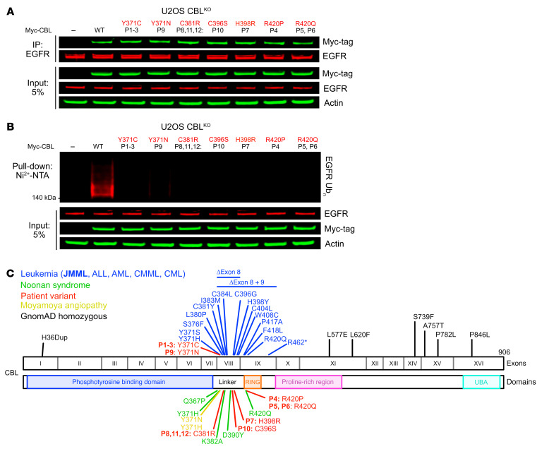
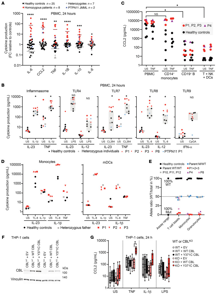
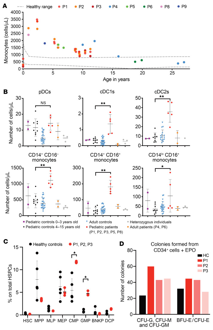
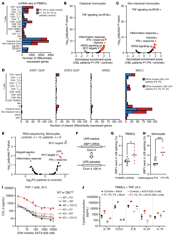
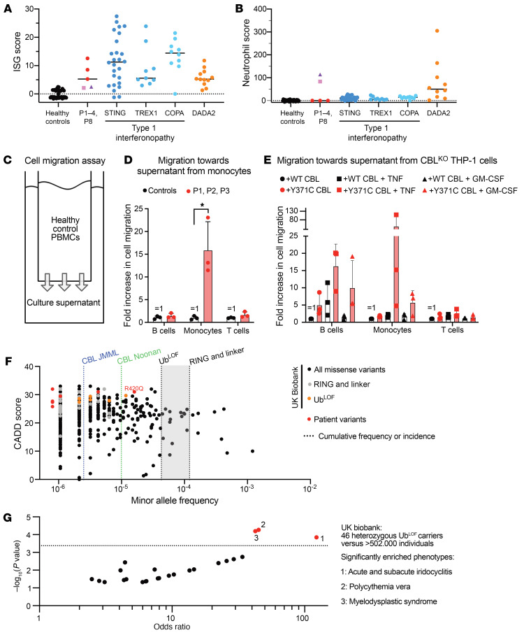
Patients heterozygous for germline CBL loss-of-function (LOF) variants can develop myeloid malignancy, autoinflammation, or both, if some or all of their leukocytes become homozygous for these variants through somatic loss of heterozygosity (LOH) via uniparental isodisomy. We observed an upregulation of the inflammatory gene expression signature in whole blood from these patients, mimicking monogenic inborn errors underlying autoinflammation. Remarkably, these patients had constitutively activated monocytes that secreted 10 to 100 times more inflammatory cytokines than those of healthy individuals and CBL LOF heterozygotes without LOH. CBL-LOH hematopoietic stem and progenitor cells (HSPCs) outgrew the other cells, accounting for the persistence of peripheral monocytes homozygous for the CBL LOF variant. ERK pathway activation was required for the excessive production of cytokines by both resting and stimulated CBL-LOF monocytes, as shown in monocytic cell lines. Finally, we found that about 1 in 10,000 individuals in the UK Biobank were heterozygous for CBL LOF variants and that these carriers were at high risk of hematological and inflammatory conditions.

Keywords: Autoimmunity; Immunology; Innate immunity; Monocytes.


Figures

Figure 1
Figure 1. The patients’ CBL variants are UbLOF but retain substrate-binding activity.
(A) Intact substrate binding by the CBL variants from the patients. Coimmunoprecipitation of EGFR and overexpressed Myc-tagged patient CBL variants or empty vector (EV) in CBLKO U2OS cells. Anti-EGFR antibody immunoprecipitates and cell lysates were analyzed by immunoblotting. (B) Defective substrate ubiquitination by the CBL variants from the patients. CBLKO U2OS cell lysates with overexpressed Myc-tagged patient CBL variants or EV, as indicated, along with His-Ub. (C) Summary of the CBL variants reported in patients with leukemia, NS, and moyamoya angiopathy and of the CBL variants present in the homozygous state in gnomAD v2.1 and the variants of the patients studied here.
Figure 2
Figure 2. High levels of cytokine secretion by the monocytes of patients with CBL LOH.
(A and B) The PBMCs of patients with CBL LOH produce excessively large amounts of inflammatory cytokines. Cytokine levels in the supernatants of PBMCs from the indicated individuals after 24 hours of culture ex vivo without stimulation (A) or with the indicated stimuli (B). Heterozygous individuals are: patients P10, P11, and P12, the father of patients P1–P3, and the grandmother, uncle, and brother of P7. Cytokine levels were assessed by bead-based ELISA. The statistical significance of differences between healthy controls and homozygous patients was assessed in multiple Mann-Whitney tests, corrected for multiple testing. *P < 0.05, **P < 0.005, ***P < 0.0005, ****P < 0.00005. FC, fold change; US, unstimulated. (C) PBMCs or the indicated leukocyte subsets from the indicated individuals, purified by magnetic sorting, were stimulated as indicated for 24 hours, and CCL2 levels were assessed in the supernatant. The statistical significance of differences was assessed in multiple Mann-Whitney tests, corrected for multiple testing. *P < 0.05. (D) Monocytes or mDCs from the indicated individuals were obtained by magnetic sorting and stimulated with the indicated agonists for 24 hours. (E) Variant allele frequencies of CBL variants in the indicated leukocyte subsets of the indicated individuals, as determined by amplicon sequencing. (F) Engineered THP-1 cell lines. Western blot of wild-type (CBLWT) and CBL-knockout (CBLKO) THP-1 cells generated by CRISPR/Cas9 genome editing and stably transduced with constructs: empty vector (EV), wild-type CBL (WT CBL), or Y371C-mutated CBL (Y371C CBL). (G) Cytokine levels in the supernatant were assessed by bead-based ELISA on the THP-1 cells shown after stimulation, as indicated, for 24 hours. *P < 0.05 by Mann-Whitney test, with correction for multiple testing.
Figure 3
Figure 3. Monocytosis during childhood in CBL-LOH patients.
(A) Monocyte counts per microliter of blood, as determined from clinical and research blood counts for the patients at the ages indicated. (B) Immunophenotyping of myeloid leukocyte subsets by mass cytometry, for the individuals indicated. *P < 0.05, **P < 0.005 by Mann-Whitney test, corrected for multiple testing. (C) Bone marrow phenotyping for 5 healthy controls, P1, P2, and P3. The statistical significance of differences was assessed in multiple Mann-Whitney tests, with correction for multiple testing. *P < 0.05. CMP, common myeloid progenitor; GMP, granulocytic myeloid progenitor; HSC, hematopoietic stem cell; MPP, multipotent progenitor; MLP, multi-lymphoid progenitor; MEP, megakaryocyte-erythroid progenitor; BNKP, B/NK cell progenitor; DCP, dendritic cell progenitor. (D) Number of colony-forming units (CFU), including CFU-GM, CFU-G, and CFU-M, and of erythroid burst-forming units (BFU-E) and erythroid colony-forming units (CFU-E) for the CD34+ cells of a healthy control (HC), P1, P2, and P3.
Figure 4
Figure 4. Characterization of activated monocytes in CBL-LOH patients.
(A) Number of differentially expressed genes in the detected leukocyte subsets on scRNA-Seq on cryopreserved PBMCs from healthy adult (n = 11) and pediatric (n = 6) controls, and adults (n = 2) and children (n = 4) with CBL-LOH. (B and C) Gene set enrichment analysis of genes differentially expressed in classical (B) and non-classical (C) monocytes of CBL-LOH patients relative to healthy controls. (D) Numbers of differentially expressed genes in the detected leukocyte subsets common to CBL-LOH patients and patients with heterozygous gain-of-function (GOF) variants of STAT1, STAT3, and PIK3CD (n = 1, 1, and 2, respectively), and an MIS-C patient with RNase L deficiency. (E) Bulk RNA-Seq on healthy control (n = 10) and CBL-LOH patient monocytes after 24 hours of culture without stimulation ex vivo. Gene set enrichment analysis was performed, and the pathways for which significant enrichment was detected are shown in blue and red. (F) Diagram of UPR stress-induced XBP1 splicing. (G and H) Quantification of stress-dependent XBP1 splicing in PBMCs (G) and monocytes (H) from healthy controls (n = 10) and CBL-LOH patients (n = 6). *P < 0.05, ***P < 0.0005 by Mann-Whitney test. (I) Cytokine production by the indicated THP-1 cell lines following TNF stimulation with or without ASTX-029 at the indicated concentrations, including pretreatment with the inhibitor for 1 hour. Supernatants were collected after 24 hours. Dose-response curves were plotted. (J) Cytokine production by monocytes from patients and healthy controls stimulated with TNF with or without 1 μM ASTX-029, including pretreatment with the inhibitor for 1 hour. Supernatants were collected after 24 hours.
Figure 5
Figure 5. Inflammation in vivo, ex vivo, and in vitro in CBL-LOH patients and CBL-mutated THP-1 cells.
(A) Relative expression levels for the 24 interferon-stimulated genes (ISGs) comprising the ISG score in patients P1, P2, P3, P4, and P8 relative to 27 controls. P1–P3, red; P4, purple; P8, pink. (B) Neutrophil scores for 27 healthy controls, the patients, previously reported individuals with type 1 interferonopathy with mutations of the indicated genes, and individuals with DADA2. P1–P3, red; P4, purple; P8, pink. (C) Experimental setup for PBMC Transwell migration assays. (D) Transwell migration of healthy control PBMCs toward cell culture supernatants from the monocytes of healthy controls or patients (P1, P2, and P3). The statistical significance of differences was assessed in a Mann-Whitney test. *P < 0.05. (E) Transwell migration of healthy control PBMCs toward supernatants from cultures of the indicated THP-1 cell lines after pretreatment with TNF. (F) Population genetics of CBL and CBL-driven disease. Combined annotation-dependent depletion (CADD)–minor allele frequency (MAF) plot of non-synonymous variants of CBL found in the UK Biobank genetic database (black). Patient variants, red; known UbLOF variants, orange; missense variants in the RING and linker domains, gray. The cumulative frequencies of these groups of variants are indicated. The incidence of CBL-driven NS and JMML is shown. (G) Phenotypes for which significant enrichment was detected in 46 carriers of UbLOF CBL variants in the UK Biobank relative to the 502,365 individuals of the database.

Similar articles

Cited by

References

    1. Afonina IS, et al. Limiting inflammation—the negative regulation of NF-κB and the NLRP3 inflammasome. Nat Immunol. 2017;18(8):861–869. doi: 10.1038/ni.3772. - DOI - PubMed
    1. O’Shea JJ, et al. Cytokines and autoimmunity. Nat Rev Immunol. 2002;2(1):37–45. doi: 10.1038/nri702. - DOI - PubMed
    1. Van Etten RA. Aberrant cytokine signaling in leukemia. Oncogene. 2007;26(47):6738–6749. doi: 10.1038/sj.onc.1210758. - DOI - PMC - PubMed
    1. Swaminathan G, Tsygankov AY. The Cbl family proteins: ring leaders in regulation of cell signaling. J Cell Physiol. 2006;209(1):21–43. doi: 10.1002/jcp.20694. - DOI - PubMed
    1. Zheng N, et al. Structure of a c-Cbl-UbcH7 complex: RING domain function in ubiquitin-protein ligases. Cell. 2000;102(4):533–539. doi: 10.1016/S0092-8674(00)00057-X. - DOI - PubMed