. 2024 Nov 20;31(12):e240061.
doi: 10.1530/ERC-24-0061. Print 2024 Dec 1.

AGO2 protein: a key enzyme in the miRNA pathway as a novel biomarker in adrenocortical carcinoma

Affiliations

AGO2 protein: a key enzyme in the miRNA pathway as a novel biomarker in adrenocortical carcinoma

Anila Hashmi et al. Endocr Relat Cancer. .

Abstract

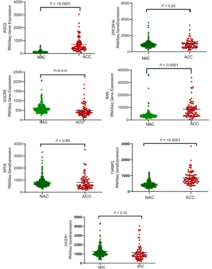
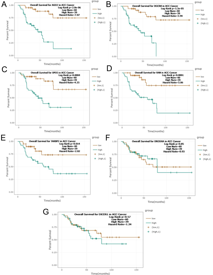
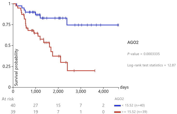
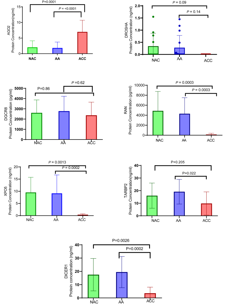
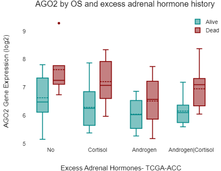
Adrenocortical carcinoma (ACC) is a rare and aggressive malignancy characterized by diagnostic challenges, high recurrence rates, and poor prognosis. This study explored the role of miRNA processing genes in ACC and their potential role as diagnostic and prognostic biomarkers. We analyzed the mRNA expression levels of miRNA machinery components (DROSHA, DGCR8, XPO5, RAN, DICER, TARBP2, and AGO2) utilizing mRNA-Seq data from The Cancer Genome Atlas (TCGA) and The Genotype-Tissue Expression (GTEx) projects. Additionally, protein levels were quantified in tissue samples from the Kolling Institute of Medical Research's tumor bank. Our results demonstrated that among all miRNA processing components, AGO2 exhibited significant overexpression in ACC compared to the normal adrenal cortex and benign adrenal adenoma (P < 0.001). Kaplan-Meier survival analysis indicated that higher AGO2 expression correlated with significantly worse overall survival in ACC patients (HR: 7.07, P < 0.001). Among 32 cancer types in TCGA, the prognostic significance of AGO2 was most prominent in ACC. This study is the first to report AGO2's potential as a diagnostic and prognostic biomarker in ACC, emphasizing its significance in ACC pathogenesis and potential application as a non-invasive liquid biopsy biomarker.

Keywords: AGO2-protein; adrenocortical carcinoma; diagnostic and prognostic biomarkers; miRNA.


Conflict of interest statement

The authors declare that there is no conflict of interest that could be perceived as prejudicing the impartiality of the study reported.

Figures

Figure 1
Figure 1
mRNA expression analysis of genes related to miRNA biogenesis in adrenocortical carcinoma (ACC) and normal adrenal cortex (NAC) tissue samples. The expression levels of miRNA biogenesis genes (AGO2, DROSHA, DGCR8, XPO5, RAN, TARBP2, and DICER1) were compared in ACC and normal adrenal cortex tissue samples using RNA-Seq data from the TCGA and GTEX datasets. Among these genes, AGO2 showed significantly higher expression in ACC samples than in normal samples (P < 0.001), whereas minimal or no expression of AGO2 was detected in normal samples. A full-color version of this figure is available at https://doi.org/10.1530/ERC-24-0061.
Figure 2
Figure 2
Association between miRNA biogenesis gene expression and survival rates in adrenocortical carcinoma (ACC) patients. Gene survival analysis of TCGA RNA-seq data was performed to explore overall survival rates in 79 ACC patients with adrenocortical carcinoma according to high (green) or low (brown) gene expression levels. The analysis revealed a poor prognosis associated with high expression levels of AGO2, DGCR8, XPO5, and RAN, with log-rank P <0.001. TARBP2 showed a weaker prognostic association (log-rank P = 0.014). DROSHA and DICER did not exhibit significant prognostic correlations, with log-rank P = 0.85 and P = 0.57, respectively. Among the genes involved in the miRNA biogenesis pathway, AGO2 emerged as the strongest prognostic indicator in ACC, exhibiting a hazard ratio (HR) of 7.07 and a log-rank test P-value of 2.8e-06.
Figure 3
Figure 3
Kaplan‒Meier gene expression analysis of AGO2-ACC-TCGA. Kaplan‒Meier curves comparing survival between ACC patients with low (< 15.52, blue) and high (≥ 15.52, red) AGO2 expression in the TCGA cohort. The difference in survival was statistically significant (P = 0.0003335, log-rank test statistic = 12.87), indicating a prognostic impact of AGO2 expression on patient outcome.
Figure 4
Figure 4
Protein expression analysis of miRNA biogenesis components in adrenocortical carcinoma (ACC), normal, and benign tissue samples. The protein expression levels of AGO2, DROSHA, DGCR8, XPO5, RAN, TARBP2, and DICER1 in normal, benign, and adrenocortical cancer tissue homogenate samples were measured using ELISA. The results revealed that XPO5, RAN, TARBP2, and DICER1 protein expression was downregulated in cancer samples compared to both normal and benign samples, suggesting a potential role for these proteins in cancer development through post-translational modification. In contrast, AGO2 protein expression was significantly higher in cancer samples than in both normal and benign samples. These findings highlight AGO2 as a strong candidate potential diagnostic biomarker for adrenocortical carcinoma among all the miRNA biogenesis factors analyzed. A full-colour version of this figure is available at https://doi.org/10.1530/ERC-24-0061.
Figure 5
Figure 5
Receiver operating characteristic (ROC) curve for AGO2 protein expression in adrenocortical carcinoma (ACC) patients. The ROC curve illustrates the diagnostic ability of AGO2 protein expression to differentiate between ACC and non-malignant samples. The area under the curve (AUC) was 0.9481 (95% CI: 0.8641 to 1.000), indicating high diagnostic accuracy. A cut-off value of >3.9 for AGO2 protein expression yielded a sensitivity of 88.89% (95% CI: 56.50% to 99.43%) and a specificity of 80.00% (95% CI: 54.81% to 92.95%). The diagonal dashed line represents the line of no discrimination (AUC = 0.5).
Figure 6
Figure 6
Association of AGO2 expression with overall survival (OS) and excess adrenal hormone history in ACC. AGO2 gene expression (log2-transformed) is shown across different hormone production statuses in ACC patients from the TCGA-ACC cohort: No excess hormone production, Cortisol, Androgen, and Androgen|Cortisol. Across all groups, deceased patients (red) have higher AGO2 expression compared to those alive, indicating that elevated AGO2 is associated with poor survival outcomes. Notably, even patients with no excess hormone production show a positive correlation between AGO2 expression and survival, highlighting AGO2 as a potentially better prognostic marker than hormone production status alone.

Similar articles

References

    1. Amini N, Margonis GA, Kim Y, Tran TB, Postlewait LM, Maithel SK, Wang TS, Evans DB, Hatzaras I, Shenoy R, et al. 2016. Curative resection of adrenocortical carcinoma: rates and patterns of postoperative recurrence. Annals of Surgical Oncology 23 126–133. (10.1245/s10434-015-4810-y) - DOI - PMC - PubMed
    1. Assié G, Letouzé E, Fassnacht M, Jouinot A, Luscap W, Barreau O, Omeiri H, Rodriguez S, Perlemoine K, René-Corail F, et al. 2014. Integrated genomic characterization of adrenocortical carcinoma. Nature Genetics 46 607–612. (10.1038/ng.2953) - DOI - PubMed
    1. Bartha Á & Győrffy B. 2021. TNMplot.com: a web tool for the comparison of gene expression in normal, tumor and metastatic tissues. International Journal of Molecular Sciences 22 2622. (10.3390/ijms22052622) - DOI - PMC - PubMed
    1. Bernstein E Caudy AA Hammond SM & Hannon GJ. 2001. Role for a bidentate ribonuclease in the initiation step of RNA interference. Nature 409 363–366. (10.1038/35053110) - DOI - PubMed
    1. Caramuta S Lee L Özata DM Akçakaya P Xie H Höög A Zedenius J Bäckdahl M Larsson C & Lui W-O. 2013. Clinical and functional impact of TARBP2 over-expression in adrenocortical carcinoma. Endocrine-Related Cancer 20 551–564. (10.1530/ERC-13-0098) - DOI - PMC - PubMed

LinkOut - more resources