. 2024 Nov 13;15(11):e0046524.
doi: 10.1128/mbio.00465-24. Epub 2024 Oct 15.

The structural and mechanistic bases for the viral resistance to allosteric HIV-1 integrase inhibitor pirmitegravir

Affiliations

The structural and mechanistic bases for the viral resistance to allosteric HIV-1 integrase inhibitor pirmitegravir

Tung Dinh et al. mBio. .

Erratum in

Abstract

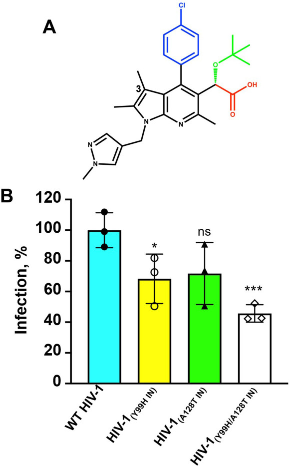
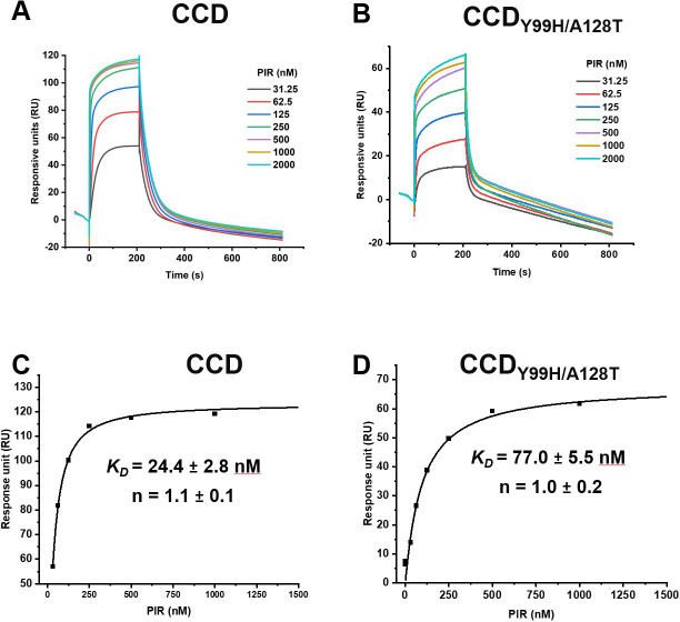
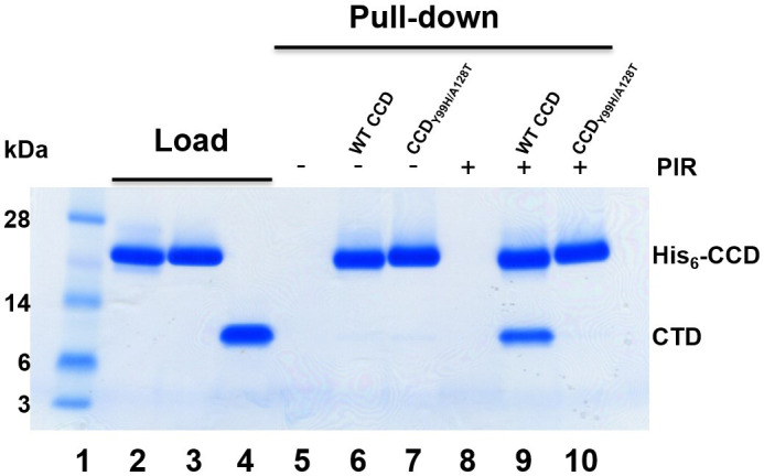
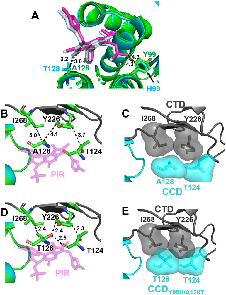
Allosteric HIV-1 integrase (IN) inhibitors (ALLINIs) are investigational antiretroviral agents that potently impair virion maturation by inducing hyper-multimerization of IN and inhibiting its interaction with viral genomic RNA. The pyrrolopyridine-based ALLINI pirmitegravir (PIR) has recently advanced into phase 2a clinical trials. Previous cell culture-based viral breakthrough assays identified the HIV-1(Y99H/A128T IN) variant that confers substantial resistance to this inhibitor. Here, we have elucidated the unexpected mechanism of viral resistance to PIR. Although both Tyr99 and Ala128 are positioned within the inhibitor binding V-shaped cavity at the IN catalytic core domain (CCD) dimer interface, the Y99H/A128T IN mutations did not substantially affect the direct binding of PIR to the CCD dimer or functional oligomerization of full-length IN. Instead, the drug-resistant mutations introduced a steric hindrance at the inhibitor-mediated interface between CCD and C-terminal domain (CTD) and compromised CTD binding to the CCDY99H/A128T + PIR complex. Consequently, full-length INY99H/A128T was substantially less susceptible to the PIR-induced hyper-multimerization than the WT protein, and HIV-1(Y99H/A128T IN) conferred >150-fold resistance to the inhibitor compared with the WT virus. By rationally modifying PIR, we have developed its analog EKC110, which readily induced hyper-multimerization of INY99H/A128T in vitro and was ~14-fold more potent against HIV-1(Y99H/A128T IN) than the parent inhibitor. These findings suggest a path for developing improved PIR chemotypes with a higher barrier to resistance for their potential clinical use.IMPORTANCEAntiretroviral therapies save the lives of millions of people living with HIV (PLWH). However, the evolution of multi-drug-resistant viral phenotypes is a major clinical problem, and there are limited or no treatment options for heavily treatment-experienced PLWH. Allosteric HIV-1 integrase inhibitors (ALLINIs) are a novel class of antiretroviral compounds that work by a unique mechanism of binding to the non-catalytic site on the viral protein and inducing aberrant integrase multimerization. Accordingly, ALLINIs potently inhibit both wild-type HIV-1 and all drug-resistant viral phenotypes that have so far emerged against currently used therapies. Pirmitegravir, a highly potent and safe investigational ALLINI, is currently advancing through clinical trials. Here, we have elucidated the structural and mechanistic bases behind the emergence of HIV-1 integrase mutations in infected cells that confer resistance to pirmitegravir. In turn, our findings allowed us to rationally develop an improved ALLINI with substantially enhanced potency against the pirmitegravir-resistant virus.

Keywords: ALLINI; HIV-1 integrase; antiretroviral drug; pirmitegravir.


Conflict of interest statement

Kyungjin Kim was the chief executive officer of ST Pharm Co. Ltd. No other authors declare a potential conflict of interest.

Figures

Fig 1
Fig 1
(A) The chemical structure of PIR. The separate functional groups are color-coded: carboxylate in red; tert-butoxyl in green; chlorophenyl in blue; and core pyrrolopyridine and methylpyrazole rings in black. The 3-methyl group on the pyrrolopyridine ring is indicated. (B) Infectivity of WT and indicated mutant viruses. The statistical significance was determined between WT and IN mutants by unpaired (two-tailed) t-test. For WT vs Y99H P = 0.0272; WT vs A128T P = 0.0611; WT vs Y99H/A128T P = 0.0008. *, P ≤ 0.05; ***, P ≤ 0.001; ns, not significant (P > 0.05).
Fig 2
Fig 2
DLS analysis of PIR induced aberrant IN multimerization. Five hundred nanomolar PIR was added to 200 nM full-length WT IN (A) or INY99H/A128T, (B) and DLS signals were recorded at indicated times (1–15 min). DMSO controls are shown after incubation of full-length IN proteins for 15 min to indicate that these proteins remained fully soluble in the absence of PIR.
Fig 3
Fig 3
SPR analysis of PIR binding to WT CCD and CCDY99H/A128T. Representative sensorgrams for PIR binding to WT CCD (A) vs CCDY99H/A128T (B). PIR concentrations are indicated. The dissociation constant (KD) and the Hill coefficient (n) for PIR + CCD (C) and PIR + CCDY99H/A128T (D) were determined using the Hill equation.
Fig 4
Fig 4
Affinity pull-down assays to probe PIR-induced CCD-CTD interactions. Lane 1: molecular weight markers; lanes 2–4: loads of His6-CCD (lane 2), His6-CCDY99H/A128T (lane 3), and tag-less CTD (lane 4); lanes 5–7: affinity pull-down using Ni beads of CTD alone (lane 5, control), His6-CCD + CTD (lane 6), His6-CCDY99H/A128T + CTD (lane 7) in the absence of PIR; and lanes 8–10: affinity pull-down using Ni beads of CTD + PIR (lane 8, control), His6-CCD + PIR + CTD (lane 9), His6-CCDY99H/A128T + PIR + CTD (lane 10).
Fig 5
Fig 5
The structural analysis of PIR interactions with WT and drug-resistant proteins. (A) Superimposed crystal structures of WT CCD (green) + PIR (magenta) and CCDY99H/A128T (cyan) + PIR (pale cyan). The closest distances were measured from the 3-methyl group of PIR‘s pyrrolopyridine ring to Cβ of Ala128 and Thr128, as well as from the methyl group on PIR’s tert-butoxy to Tyr99 and His99. (B) The closest distances between indicated CCD and CTD residues are shown in the structure of the WT CTD-CCD + PIR complex. Specifically, the distances from Cα of Ala128 to C4 of Ile268 and C3 of the aromatic ring of Tyr226, as well as from Cβ of Thr124 to C6 of the aromatic ring of Tyr226 are indicated. (C) Van der Waals surface for indicated residues are shown in the structure of WT CTD-CCD + PIR. The inhibitor is not shown for clarity. (D) The closest distances between indicated CCD and CTD residues are shown when the structure of CCDY99H/A128T + PIR is superimposed onto the structure of WT CTD-CCD + PIR. Specifically, the distances from Cγ of Thr128 to C4 of Ile268, from the Thr128 side chain to C3 of the aromatic ring of Tyr226, and from Cβ of Thr124 to C6 of the aromatic ring of Tyr226 are indicated. In addition, the hydrogen bond formed between Thr128 and Thr124 side chains is illustrated. (E) Van der Waals surface for indicated residues reveals steric clashes observed by overlapping, shaded surfaces when the structure of CCDY99H/A128T + PIR superimposed onto the structure of WT CTD-CCD + PIR. The inhibitor is not shown for clarity.
Fig 6
Fig 6
The structural analysis of EKC110 interactions with the CCD and the CTD-CCD. (A) The chemical structure of EKC110. The separate functional groups are color-coded: carboxylate in red; tert-butoxyl in green; chlorophenyl in blue; and core pyrrolopyridine and methylpyrazole rings in black. (B) The crystal structure of the CCD + EKC110 is superimposed onto the CCD + PIR, which reveals a noticeable tilt of the EKC110 pyrrolopyridine core toward A128 compared with PIR. The closest distances from Cβ of Ala128 to C3 of the EKC110’s pyrrolopyridin ring and the 3-methyl substituent of PIR’s pyrrolopyridin ring are shown. In addition, the closest distances between Cγ of Leu102 and the chlorine atoms of PIR and EKC110 are indicated. (C) The crystal structure of the CTD-CCD + EKC110 is superimposed onto the CTD-CCD + PIR, which reveals that Lys266 side chain (gray) forms a salt bridge with the Glu171 side chain (green) in the presence of PIR (magenta), whereas Lys266 side chain (yellow) engages with the pharmacophore carboxylate of EKC110 (blue). (D) The crystal structure of the CTD-CCD + EKC110 is superimposed onto the CTD-CCD + PIR to show repositioning of the CTD in the presence of EKC110 vs PIR. CTDs are shown in yellow and gray in EKC110 + CTD CCD and PIR + CTD CCD structures, respectively. PIR and EKC110 are in magenta and blue. The distances between the Cα atoms for indicated residues (Asp232, Glu246, Asn254, and Asn262) in the EKC110 +CTD CCD vs the PIR + CTD CCD structures are shown.
Fig 7
Fig 7
Structural analysis of EKC110 interactions WT and drug-resistance proteins. (A) Superimposed crystals structures of WT CCD (green) + EKC110 (blue) and CCDY99H/A128T (cyan) + EKC110 (orange). The closest distances from the EKC110’s pyrrolopyridine ring to Cβ of Ala128 and Thr128, as well as from the methyl group on EKC110’s tert-butoxy to Tyr99 and His99 are indicated. (B) The closest distances between indicated CCD and CTD residues in the structure of WT CTD-CCD + EKC110 are shown. Specifically, the distances from Cα of Ala128 to C4 of Ile268 and C3 of the aromatic ring of Tyr226 as well as from Cγ of Thr124 to C6 of the aromatic ring of Tyr226 are indicated. (C) Van der Waals surface for indicated residues are shown in the structure of WT CTD-CCD + PIR. Thr124, which does not encounter any steric hindrance, and the inhibitors are not shown for clarity. (D) The closest distances between indicated CCD and CTD residues are shown when the structure of CCDY99H/A128T + EKC-110 is superimposed onto the structure of WT CTD-CCD + EKC-110. Specifically, the distances from Cγ of Thr128 to C4 of Ile268, from the side chain of Thr128 to C3 of the aromatic ring of Tyr226, and from Cβ of Thr124 to the C6 of the aromatic ring of Tyr226 are indicated. In addition, the hydrogen bond between the side chains of Thr128 and Thr124 is illustrated. (E) Van der Waals surface for indicated residues reveals steric clashes observed by overlapping, shaded surfaces when the structure of CCDY99H/A128T + EKC110 superimposed onto the structure of WT CTD-CCD + EKC110. Thr124, which does not encounter any steric hindrance, and the inhibitors are not shown for clarity.
Fig 8
Fig 8
MD simulation of the CTD interactions with WT CCD vs CCDY99H/A128T in the complex with PIR (A) and EKC110 (B). Displacement of the CTD domain from the PIR + CCDY99H/A128T complex is measured with a center-of-mass displacement of d = 4.85 Å and rotations along the principal axes of inertia of θ = 26.66°, Φ = 23.84°, and Ψ = 11.81°. WT CCD and CCDY99H/A128T are colored cyan and green, respectively. CTDs interacting with WT CCD and CCDY99H/A128T are colored red and blue, respectively.
Fig 9
Fig 9
Interactions of EKC110 with full-length HIV-1 IN. DLS analysis of EKC110 induced aberrant IN multimerization. Five hundred nanomolar EKC110 was added to 200 nM full-length WT IN (A) or INY99H/A128T, (B) and DLS signals were recorded at indicated times (1–15 min). DMSO controls are shown after incubation of full-length IN proteins for 15 min to indicate that these proteins remained fully soluble in the absence of EKC110.

Update of

References

    1. Christ F, Voet A, Marchand A, Nicolet S, Desimmie BA, Marchand D, Bardiot D, Van der Veken NJ, Van Remoortel B, Strelkov SV, De Maeyer M, Chaltin P, Debyser Z. 2010. Rational design of small-molecule inhibitors of the LEDGF/p75-integrase interaction and HIV replication. Nat Chem Biol 6:442–448. doi:10.1038/nchembio.370 - DOI - PubMed
    1. Fader LD, Malenfant E, Parisien M, Carson R, Bilodeau F, Landry S, Pesant M, Brochu C, Morin S, Chabot C, et al. . 2014. Discovery of BI 224436, a noncatalytic site integrase inhibitor (NCINI) of HIV-1. ACS Med Chem Lett 5:422–427. doi:10.1021/ml500002n - DOI - PMC - PubMed
    1. Sharma A, Slaughter A, Jena N, Feng L, Kessl JJ, Fadel HJ, Malani N, Male F, Wu L, Poeschla E, Bushman FD, Fuchs JR, Kvaratskhelia M. 2014. A new class of multimerization selective inhibitors of HIV-1 integrase. PLoS Pathog 10:e1004171. doi:10.1371/journal.ppat.1004171 - DOI - PMC - PubMed
    1. Patel D, Antwi J, Koneru PC, Serrao E, Forli S, Kessl JJ, Feng L, Deng N, Levy RM, Fuchs JR, Olson AJ, Engelman AN, Bauman JD, Kvaratskhelia M, Arnold E. 2016. A new class of allosteric HIV-1 integrase inhibitors identified by crystallographic fragment screening of the catalytic core domain. J Biol Chem 291:23569–23577. doi:10.1074/jbc.M116.753384 - DOI - PMC - PubMed
    1. Maehigashi T, Ahn S, Kim UI, Lindenberger J, Oo A, Koneru PC, Mahboubi B, Engelman AN, Kvaratskhelia M, Kim K, Kim B. 2021. A highly potent and safe pyrrolopyridine-based allosteric HIV-1 integrase inhibitor targeting host LEDGF/p75-integrase interaction site. PLoS Pathog 17:e1009671. doi:10.1371/journal.ppat.1009671 - DOI - PMC - PubMed

Substances

LinkOut - more resources