. 2024 Oct 4;13(19):1651.
doi: 10.3390/cells13191651.

Kisspeptin Alleviates Human Hepatic Fibrogenesis by Inhibiting TGFβ Signaling in Hepatic Stellate Cells

Affiliations

Kisspeptin Alleviates Human Hepatic Fibrogenesis by Inhibiting TGFβ Signaling in Hepatic Stellate Cells

Kavita Prasad et al. Cells. .

Abstract

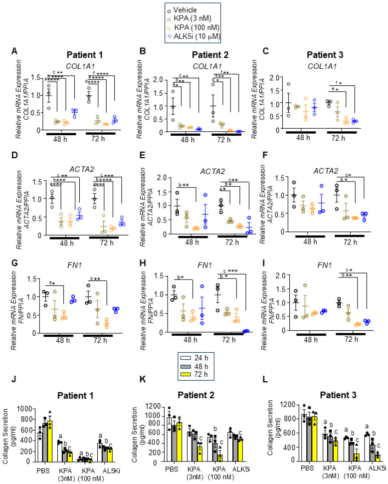
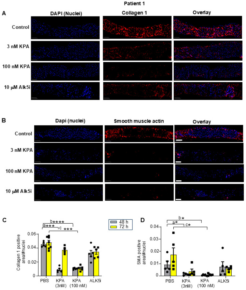
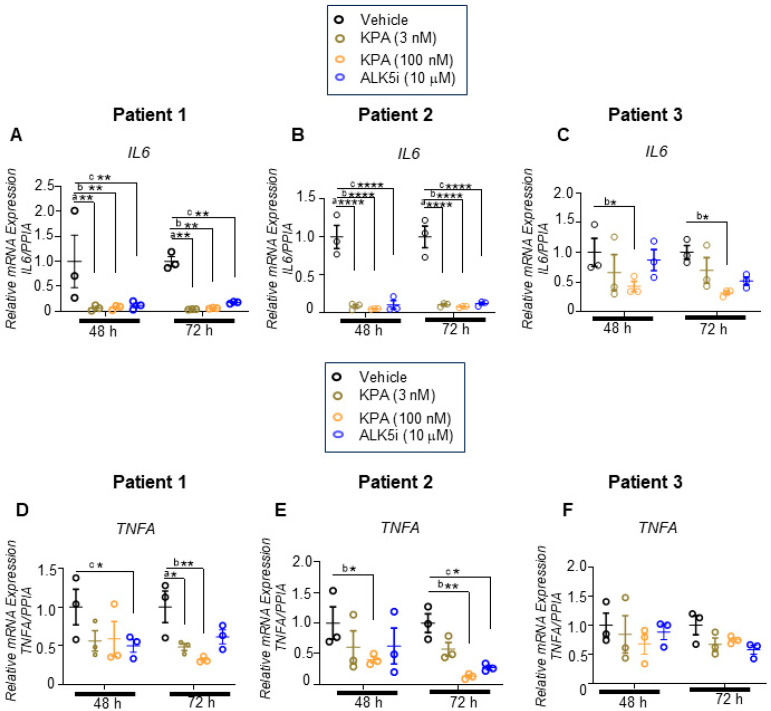
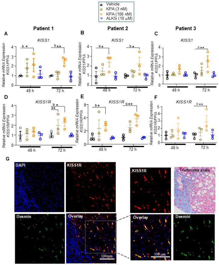
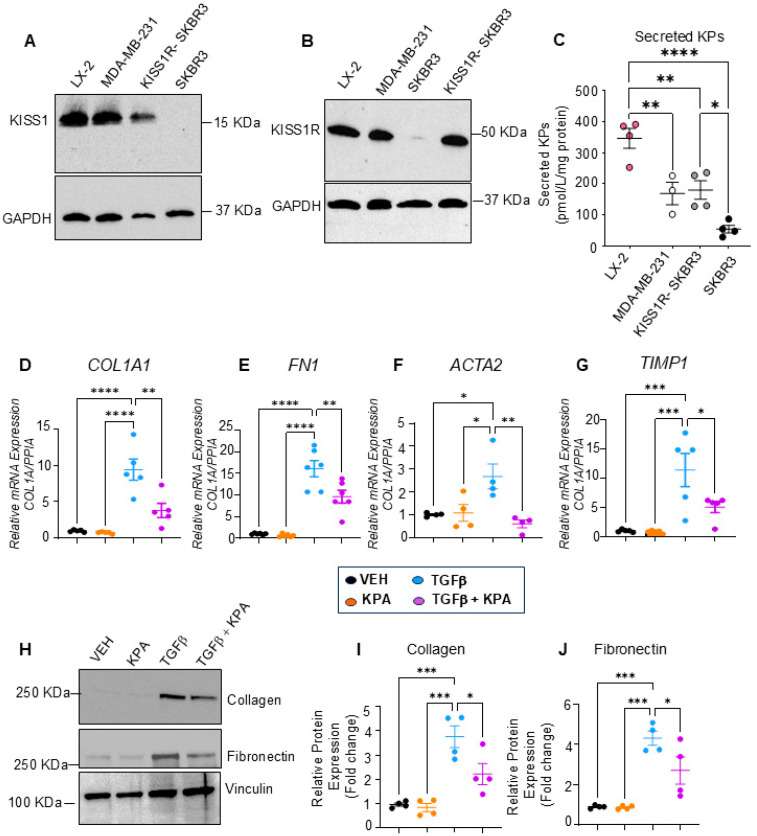
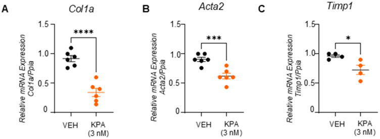
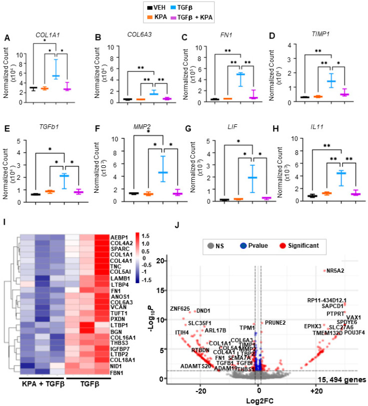
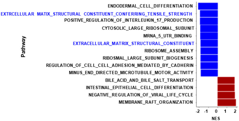
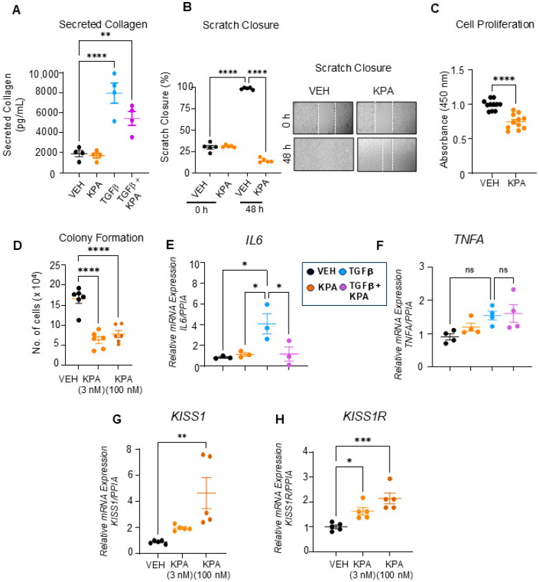
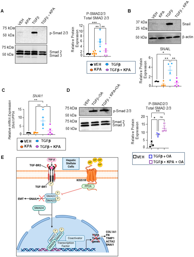
The peptide hormone kisspeptin attenuates liver steatosis, metabolic dysfunction-associated steatohepatitis (MASH), and fibrosis in mouse models by signaling via the kisspeptin 1 receptor (KISS1R). However, whether kisspeptin impacts fibrogenesis in the human liver is not known. We investigated the impact of a potent kisspeptin analog (KPA) on fibrogenesis using human precision-cut liver slices (hPCLS) from fibrotic livers from male patients, in human hepatic stellate cells (HSCs), LX-2, and in primary mouse HSCs. In hPCLS, 48 h and 72 h of KPA (3 nM, 100 nM) treatment decreased collagen secretion and lowered the expression of fibrogenic and inflammatory markers. Immunohistochemical studies revealed that KISS1R is expressed and localized to HSCs in MASH/fibrotic livers. In HSCs, KPA treatment reduced transforming growth factor b (TGFβ)-the induced expression of fibrogenic and inflammatory markers, in addition to decreasing TGFβ-induced collagen secretion, cell migration, proliferation, and colony formation. Mechanistically, KISS1R signaling downregulated TGFβ signaling by decreasing SMAD2/3 phosphorylation via the activation of protein phosphatases, PP2A, which dephosphorylates SMAD 2/3. This study revealed for the first time that kisspeptin reverses human hepatic fibrogenesis, thus identifying it as a new therapeutic target to treat hepatic fibrosis.

Keywords: KISS1R; MASH; MASLD; TGFβ; fibrosis; hepatic stellate cells; kisspeptin.


Conflict of interest statement

The funders had no role in the design of the study; in the collection, analyses, or interpretation of data; in the writing of the manuscript; or in the decision to publish the results. MB is a coinventor on a patent application US20230256051A1 filed by Rutgers University. S.L.F has relationships with the companies listed below; however, these activities are unrelated to the content of this article: Consulting: 89 Bio, Boehringer Ingelheim, Boston Pharmaceuticals, Bristol Myers Squibb, ChemomAb, Foresite Laboratories, Gordian Biotechnology, Glycotest, Glympse Bio, Hepgene, In sitro, Junevity, Korro Bio, Kriya, Laekna, Lerna Therapeutics, Macomics, Mediar, Merck, Morphic Therapeutics, North Sea Therapeutics, Ochre Bio, Overtone Therapeutics, Pfizer Pharmaceuticals, Pliant, Prosciento, RAPT, Sagimet, Satellite Bio, Seal Rock, Scholar Rock, Sunbird Bio, Surrozen, Takeda, Variant Bio. Stock options: Escient, Galectin, Galmed, Genfit, Gordian Biotechnology, Hepgene, Junevity, Lifemax, Metacrine, Morphic Therapeutics, Nimbus, North Sea, Ochre Bio, Therapeutics, Scholar Rock, and Sunbird Bio. Research Activities with Commercial Entities: Abalone Bio (SBIR Grant) and Novo Nordisk. The other authors declare no conflicts of interest that pertain to this work.

Figures

Figure 1
Figure 1
KPA reduces fibrogenic gene expression as well as type 1 collagen secretion in human fibrotic precision-cut liver slices (PCLSs). Human PCLSs generated from patients with fibrotic livers were treated with KPA (3 nM, 100 nM), vehicle (PBS), or TGFβ receptor 1 kinase inhibitor II (ALK5i, 10 mM) for 48 or 72 h. (AI) Changes in fibrogenic gene expression determined by qPCR. a, p < 0.05 vs. PBS (vehicle); b, p < 0.05 vs. PBS (vehicle); and c, p < 0.05 vs. PBS (vehicle) for each time point. (JL) Secreted pro-collagen I alpha 1 protein in culture media measured by ELISA. a, p < 0.05 vs. PBS (vehicle) secretion (24 h); b, p < 0.05 vs. PBS (vehicle) secretion (48 h); and c, p < 0.05 vs. PBS (vehicle) secretion (72 h). For (AI) (data are shown from N = 3 patients), results are expressed as mean +/− S.E.M. * p < 0.05 vs. control. ** p < 0.01 vs. control. *** p < 0.001 vs. control. **** p < 0.0001 vs. control. Two-way ANOVA was used, followed by multiple comparison test.
Figure 2
Figure 2
KPA treatment reduces collagen and smooth muscle actin (SMA) expression in hPCLS. Representative images of human PCLS generated from fibrotic liver biopsy from Patient 1, treated with KPA, vehicle (PBS), or ALK5i for 48 h and immunostained for (A) collagen 1 and (B) smooth muscle actin. Scale bars: 250 mm. (C,D) Quantification of collagen and smooth muscle actin (SMA) immunostaining in hPLCS from Patient 1 treated with KPA (3 nM, 100 nM), vehicle (PBS), or ALK5i (10 mM) for 48 h or 72 h (4 technical replicates). See Figure S2 for additional images from Patients 2 and 3. Results: mean +/− S.E.M. a, p < 0.05 vs. PBS (48 h) for KPA 3 nM; b, p < 0.05 vs. PBS (48 h) for KPA 100 nM; c, p < 0.05 vs. PBS (vehicle, 72 h), * p < 0.05 vs. control. *** p < 0.001 vs. control. **** p < 0.0001 vs. control. Two-way ANOVA was used, followed by multiple comparison test.
Figure 3
Figure 3
KPA reduces inflammatory gene expression in hPCLS. Human PCLSs generated from patients with fibrotic livers were treated with KPA (3 nM, 100 nM), vehicle (PBS), or ALK5i (10 mM) for 48 or 72 h. Changes in gene expression were determined by qPCR for (A,D) Patient 1, (B,E) Patient 2, and (C,F) Patient 3. Data are shown from n = 3 patients. a, p < 0.05 vs. PBS (vehicle); b, p < 0.05 vs. PBS (vehicle); and c, p < 0.05 vs. PBS (vehicle) for each time point. Results are expressed as mean +/− S.E.M. * p < 0.05 vs. control. ** p < 0.01 vs. control. **** p < 0.0001 vs. control. Two-way ANOVA was conducted, followed by multiple comparison test.
Figure 4
Figure 4
KPA treatment upregulates KISS1 and KISS1R expression in hPCLS. Human PCLS generated from patients with fibrotic liver biopsies were treated with KPA (3 nM, 100 nM), vehicle (PBS), or ALK5i (10 mM) for 48 or 72 h. Changes in (AC) KISS1 and KISS1R (DF) gene expression were determined by qPCR in patient livers. Data are shown from n = 3 patients. Results are expressed as mean +/− S.E.M. * p < 0.05 vs. control. ** p < 0.01 vs. control. a, p < 0.05 vs. PBS (vehicle); b, p < 0.05 vs. PBS (vehicle); and c, p < 0.05 vs. PBS (vehicle), for each time point. Two-way ANOVA was conducted, followed by multiple comparison test. (G) Representative confocal images of human hepatic stellate cells in MASH patient biopsies, immunostained for endogenous KISS1R (red) and desmin (green), a marker for stellate cells. Areas of colocalization (yellow) are shown in overlay; magnified images and trichrome staining of liver fibrotic section are shown on the right.
Figure 5
Figure 5
KPA treatment decreases activation of human hepatic stellate LX-2 cells. Representative Western blots showing expression of endogenous (A) KISS1 protein (n = 5 biological replicates) and (B) KISS1R protein expression (n = 4 biological replicates). MDA-MB-231, SKBR3, and KISS1R-SKBR3 were used as reference for expression (n = 4). (C) Secreted kisspeptin protein in culture media measured by ELISA (N-4 biological replicates). (DG) Changes in fibrogenic gene expression in response to KPA (3 nM, 48 h) +/− TGFb (5 ng/mL, 48 h). (n = 4–6 biological replicates) (H) Western blot analysis and (I,J) densitometric analyses of blots, showing changes in fibrogenic protein in response to KPA (3 nM, 72 h) +/− TGFb (5 ng/mL, 72 h). (n = 4 biological replicates) Results are expressed as mean +/− S.E.M. * p < 0.05 vs. control. ** p < 0.01 vs. control. *** p < 0.001 vs. control. **** p < 0.0001 vs. control. One-way ANOVA was conducted, followed by multiple comparison test.
Figure 6
Figure 6
KPA reduces fibrogenic markers in primary mouse HSCs. Gene expression by qPCR analysis showing the effect of KPA (3 nM, 48 h) treatment on (A) Col1a, (B) Acta2, and (C) Timp1. (n = 4–6 biological replicates) Results are expressed as mean +/− S.E.M. Student’s unpaired t-test. * p < 0.05 vs. control. *** p < 0.001 vs. control. **** p < 0.0001 vs. control.
Figure 7
Figure 7
Effect of KPA treatment on gene expression in human hepatic stellate cells by RNA-seq. RNA was extracted from LX-2 cells treated with PBS (vehicle), KPA (3 nM, 48 h), or +/− TGFβ (5 ng/mL, 48 h). Changes in fibrogenic (AF) and inflammatory (G,H) genes were identified by transcriptomic analysis. Data are presented as (I) heatmap and (J) volcano plot of differentially expressed genes (padj < 0.1) in LX-2 cells treated with KPA (3 nM, 48 h) + TGFβ (5 ng/mL, 48 h) compared to TGFβ alone. (n = 3 biological replicates). Results are expressed as mean +/− S.E.M. * p < 0.05 vs. control. ** p < 0.01 vs. control. One-way ANOVA was performed, followed by multiple comparison test.
Figure 8
Figure 8
KPA downregulates molecular pathways related to extracellular matrix production in HSCs. Gene set enrichment analysis of LX-2 cells after 48 h of treatment with KPA (3 nM), TGFβ (5 ng/mL), or TGFβ alone is shown. X-axis represents normalized enrichment scores of gene sets (blue: downregulated pathways; red: upregulated pathways).
Figure 9
Figure 9
KPA treatment decreases collagen secretion, cell migration, and proliferation of LX-2 cells. Quantification of (A) TGFβ-induced collagen secretion following VEH, KPA (3 nM), TGFβ (5 ng/mL), and KPA + TGFβ treatment (n = 4 biological replicates); (B) cell migration following VEH and KPA (3 nM) treatment. KPA treatment decreases (n = 4 biological replicates) (C) LX-2 cell proliferation following VEH and KPA (3 nM) treatment, as assessed using BrdU (n = 11 biological replicates) and (D) soft agar colony formation following VEH, KPA (3 nM, 100 nM) treatment (n = 11 biological replicates) t. Gene expression by qPCR analysis showing the effect of KPA (3 nM, 48 h) treatment on inflammatory markers (E,F) IL6 and TNFA, and (G,H) KISS1 and KISS1R (n = 3–5 biological replicates). Results are expressed as mean +/− S.E.M. Student’s unpaired t-test or one-way ANOVA was performed, followed by multiple comparison test. * p < 0.05 vs. control. ** p < 0.01 vs. control. *** p < 0.001 vs. control. **** p < 0.0001 vs. control.
Figure 10
Figure 10
KPA treatment decreases TGFβ signaling in human HSCs. (A) TGFβ-induced SMAD phosphorylation (n = 5 biological replicates) and (B) SNAIL expression (n = 4 biological replicates) was observed, following KPA (3 nM, 72 h) treatment, as determined by Western blot analysis; densitometric analysis of blots are shown below. (C) TGFβ-induced SNAI1 mRNA expression following KPA (3 nM, 48 h) determined by qPCR (n = 4 biological replicates). (D) TGFβ-induced SMAD 2/3 phosphorylation cells were pretreated with OA (5 nM) for 3 h, following TGFβ1 (5 ng/mL), and/or KPA (3 nM) for 48 as determined by Western blot analysis. OA treatment did not impact cell viability (see Figure S5) (n = 4 biological replicates). (E) Schematic showing proposed signaling pathways by which KISS1R activation with kisspeptin (KP) suppresses hepatic fibrosis in hepatic stellate cells. Results are expressed as mean +/− S.E.M. * p < 0.05 vs. control. ** p < 0.01 vs. control. *** p < 0.001 vs. control. One-way ANOVA was performed, followed by multiple comparison test.

Similar articles

References

    1. Akkiz H., Gieseler R.K., Canbay A. Liver Fibrosis: From Basic Science towards Clinical Progress, Focusing on the Central Role of Hepatic Stellate Cells. Int. J. Mol. Sci. 2024;25:7873. doi: 10.3390/ijms25147873. - DOI - PMC - PubMed
    1. Cogliati B., Yashaswini C.N., Wang S., Sia D., Friedman S.L. Friend or foe? The elusive role of hepatic stellate cells in liver cancer. Nat. Rev. Gastroenterol. Hepatol. 2023;20:647–661. doi: 10.1038/s41575-023-00821-z. - DOI - PMC - PubMed
    1. Llovet J.M., Willoughby C.E., Singal A.G., Greten T.F., Heikenwalder M., El-Serag H.B., Finn R.S., Friedman S.L. Nonalcoholic steatohepatitis-related hepatocellular carcinoma: Pathogenesis and treatment. Nat. Rev. Gastroenterol. Hepatol. 2023;20:487–503. doi: 10.1038/s41575-023-00754-7. - DOI - PubMed
    1. Friedman S.L. Hepatic Fibrosis and Cancer: The Silent Threats of Metabolic Syndrome. Diabetes Metab. J. 2024;48:161–169. doi: 10.4093/dmj.2023.0240. - DOI - PMC - PubMed
    1. Tapper E.B., Parikh N.D. Diagnosis and Management of Cirrhosis and Its Complications: A Review. JAMA. 2023;329:1589–1602. doi: 10.1001/jama.2023.5997. - DOI - PMC - PubMed

Publication types

MeSH terms

LinkOut - more resources