Review
. 2024 Oct 9;13(19):1666.
doi: 10.3390/cells13191666.

Beyond Cancer Cells: How the Tumor Microenvironment Drives Cancer Progression

Affiliations
Review

Beyond Cancer Cells: How the Tumor Microenvironment Drives Cancer Progression

Hussein Sabit et al. Cells. .

Abstract

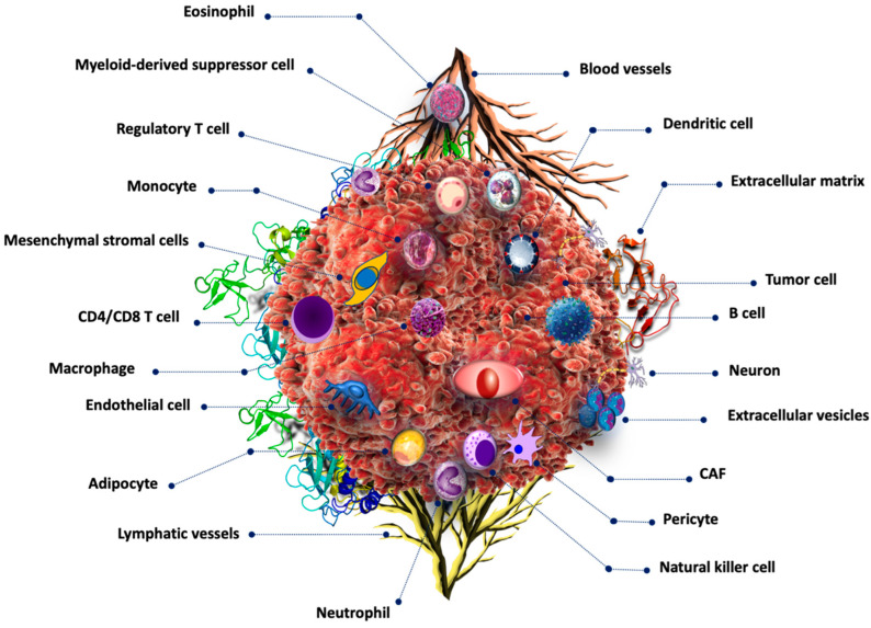
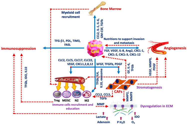
Liver cancer represents a substantial global health challenge, contributing significantly to worldwide morbidity and mortality. It has long been understood that tumors are not composed solely of cancerous cells, but also include a variety of normal cells within their structure. These tumor-associated normal cells encompass vascular endothelial cells, fibroblasts, and various inflammatory cells, including neutrophils, monocytes, macrophages, mast cells, eosinophils, and lymphocytes. Additionally, tumor cells engage in complex interactions with stromal cells and elements of the extracellular matrix (ECM). Initially, the components of what is now known as the tumor microenvironment (TME) were thought to be passive bystanders in the processes of tumor proliferation and local invasion. However, recent research has significantly advanced our understanding of the TME's active role in tumor growth and metastasis. Tumor progression is now known to be driven by an intricate imbalance of positive and negative regulatory signals, primarily influenced by specific growth factors produced by both inflammatory and neoplastic cells. This review article explores the latest developments and future directions in understanding how the TME modulates liver cancer, with the aim of informing the design of novel therapies that target critical components of the TME.

Keywords: ECM; TME; immunotherapy; liver cancer; targeted therapy; tumor microenvironment.


Conflict of interest statement

The authors declare no conflicts of interest.

Figures

Figure 1
Figure 1
Tumor microenvironment components. Cancer cells are surrounded by numerous non-cancerous cells including those related to the immune system such as B cells, T-cells, dendritic cells, monocytes, eosinophils, and basophils, among others. Cancer-associated fibroblasts are also common in the TME.
Figure 2
Figure 2
Tumor cell interaction with the microenvironment. The interplay between cancer cells and The TME is the main factor in cancer progression. Some inducers are released from the tumor cells to affect other components such as the FGF family, VEGF, and IL-8 among others which induce angiogenesis, and other factors including TGF-b1 and FASL induce immunosuppression. Tumor cells also release IL-4 and IL-19 to induce immune cell recruitment and education and release CCL2, IL-1, and IL-6 to activate bone marrow to take part in the production of myeloid cell recruitment. GM-CSF: Granulocyte-macrophage colony-stimulating factor, FGF: Fibroblast growth factor, Ang2: Angiopoietin-2, PDGF: Platelet-derived growth factor, PD-L1: Programmed Cell Death Ligand-1, TIM3: T-cell immunoglobulin domain and mucin domain 3, FASL: Fas ligand, CXCL: chemokines, CAF: cancer-associated fibroblasts, IL: Interleukin, M2: M2 macrophage, N2: Neutrophil, Treg: T-regulatory lymphocyte, MDSC: myeloid-derived suppressor cell, MMP: Matrix metalloproteinase, GF: Growth factor, IDO: Indolamine 2,3 dioxygenase.
Figure 3
Figure 3
Immunological mechanisms regulating tumor growth. (A) Tumor suppressive microenvironment. Tumor cell proliferation is inhibited by activated CD4+, CD8+, NK, M1 macrophages, and neutrophils. (B) Immunosuppressive microenvironment. Tumor cells that secrete factors such as TGFβ1, G-CSF, etc. promote MDSC, Treg cells, and M2 macrophages, which inhibit anti-tumor T-cells and NK cells.

Similar articles

Cited by

References

    1. Zeng D., Wu J., Luo H., Li Y., Xiao J., Peng J., Ye Z., Zhou R., Yu Y., Wang G., et al. Tumor microenvironment evaluation promotes precise checkpoint immunotherapy of advanced gastric cancer. J. Immunother. Cancer. 2021;9:e002467. doi: 10.1136/jitc-2021-002467. - DOI - PMC - PubMed
    1. Massalha H., Bahar Halpern K., Abu-Gazala S., Jana T., Massasa E.E., Moor A.E., Buchauer L., Rozenberg M., Pikarsky E., Amit I., et al. A single cell atlas of the human liver tumor microenvironment. Mol. Syst. Biol. 2020;16:e9682. doi: 10.15252/msb.20209682. - DOI - PMC - PubMed
    1. Muppala S. Significance of the Tumor Microenvironment in Liver Cancer Progression. Crit. Rev. Oncog. 2020;25:1–9. doi: 10.1615/CritRevOncog.2020034987. - DOI - PubMed
    1. Yuen V.W., Wong C.C. Hypoxia-inducible factors and innate immunity in liver cancer. J. Clin. Investig. 2020;130:5052–5062. doi: 10.1172/JCI137553. - DOI - PMC - PubMed
    1. Kaps L., Schuppan D. Targeting Cancer Associated Fibroblasts in Liver Fibrosis and Liver Cancer Using Nanocarriers. Cells. 2020;9:2027. doi: 10.3390/cells9092027. - DOI - PMC - PubMed

LinkOut - more resources