. 2024 Nov 19;98(11):e0042924.
doi: 10.1128/jvi.00429-24. Epub 2024 Oct 15.

Development of a safe and broad-spectrum attenuated PEDV vaccine candidate by S2 subunit replacement

Affiliations

Development of a safe and broad-spectrum attenuated PEDV vaccine candidate by S2 subunit replacement

Ding Zhang et al. J Virol. .

Abstract

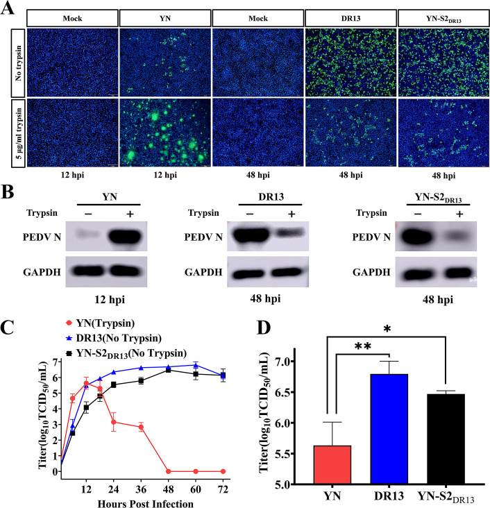
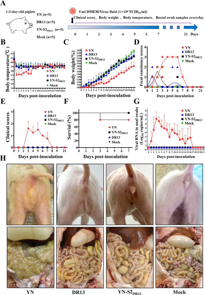
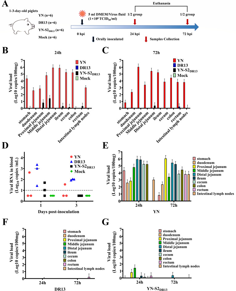
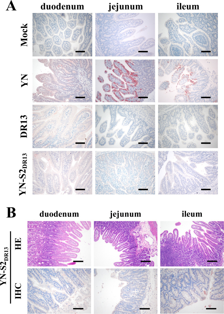
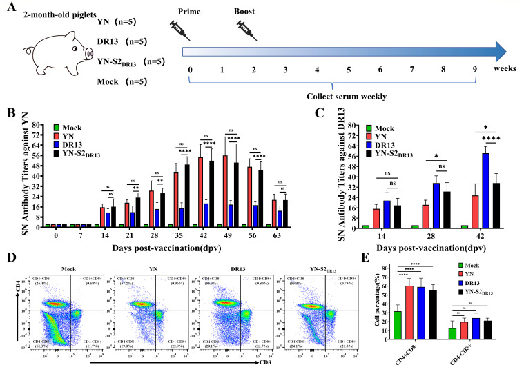
Porcine epidemic diarrhea (PED) has caused serious economic losses to the swine livestock industry. Due to the rapid variation in the PEDV) genome, especially the spike (S) protein, the cross-protection ability of antibodies between different vaccine strains is weakened. Hence, the rapid development of safe, broad-spectrum and highly effective attenuated PEDV vaccine still needs further research. Here, we found that the replacement of the S2 subunit had little effect on S protein immunogenicity. Moreover, the chimeric virus (YN-S2DR13), the S protein of the YN strain was replaced by the DR13 S2 subunit, which lost its trypsin tropism and increased its propagation ability (approximately 1 titer) in Vero cells. Then, the pathogenesis of YN-S2DR13 was evaluated in neonatal piglets. Importantly, quantitative real-time PCR, histopathology, and immunohistochemistry confirmed that the virulence of YN-S2DR13 was significantly reduced compared with that of YN. Immunization with YN-S2DR13 induced neutralizing antibodies against both YN and DR13 in weaned piglets. In vitro passaging data also showed that YN-S2DR13 had good genetic stability. Collectively, these results suggest that YN-S2DR13 has significant advantages as a novel vaccine candidate, including a capacity for viral propagation to high titers with no trypsin requirement and the potential to provide protection against both PEDV G1 and G2 strains infections. Our results also suggests that S2 subunit replacement using reverse genetics can be a rapid strategy for the rational design of live attenuated vaccines for PEDV.

Importance: Emerging highly virulent porcine epidemic diarrhea virus (PEDV) G2 strains has caused substantial economic losses worldwide. Vaccination with a live attenuated vaccine is a promising method to prevent and control PED because it can induce a strong immune response (including T- and B-cell immunity). Previous studies have demonstrated that the S2 subunit of the PEDV spike (S) protein is the determinant of PEDV trypsin independence. Here, we evaluated the pathogenicity, tissue tropism, and immunogenicity of the chimeric virus (YN-S2DR13) via animal experiments. We demonstrated that YN-S2DR13 strain, as a trypsin independent strain, increased intracellular proliferation capacity, significantly reduced virulence, and induced broad-spectrum neutralization protection against PEDV G1 and G2 strains. In vitro passaging data also validated the stability of YN-S2DR13. Our results showed that generating a chimeric PEDV strain that is trypsin-independent by replacing the S2 subunit is a promising approach for designing a live attenuated vaccine for PEDV in the future.

Keywords: chimeric virus; immunogenicity; porcine epidemic diarrhea virus (PEDV); spike protein; virulence.


Conflict of interest statement

The authors declare no conflict of interest.

Figures

Fig 1
Fig 1
Growth characteristic analysis of the PEDV YN-S2DR13 in Vero cells. (A) Immunofluorescent assay of Vero cells infected with different PEDVs at 12 hpi (YN) and 48 hpi (YN-S2DR13 and DR13) with or without trypsin at an multiplicity of infection (MOI) of 0.01. Scale bars, 100 µm. (B) Western blotting analysis of the nucleocapsid protein in the YN (12 hpi), DR13 (48 hpi), and YN-S2DR13 (48 hpi) strains. Glyceraldehyde-3-phosphate dehydrogenase (GAPDH) was used as an internal reference. (C) Vero cells were inoculated with YN, DR13, or YN-S2DR13 in the presence or absence of trypsin at an MOI of 0.01. The supernatant was harvested at 6, 12, 18, 24, 36, 48, 60, and 72 hpi and titrated. Three replicates were performed. Error bars represent the means ± standard deviations. (D) Comparison of the highest titers of the three PEDV strains. Significant differences are indicated as follows: *P < 0.05, **P < 0.01.
Fig 2
Fig 2
Evaluation of the pathogenicity of PEDV YN, DR13, and YN-S2DR13 in piglets. (A) Schematic overview of animal experiments, including infectious doses and types of clinical indicators. (B) Body temperature of piglets. (C) Body weight of piglets. (D) Evaluation of fecal consistency of piglets. (E) Survival rate of piglets. (G) Detection of viral RNA in fecal swabs by RT-qPCR. (H) Representative clinical signs and gross examination of infected piglets.
Fig 3
Fig 3
Experimental design and analysis of gastrointestinal viral loads in infected piglets. (A) Schematic overview of animal experiments, including infectious doses and sample collection schedules. (B and C) Quantification of viral loads in different intestine segments from PEDV-infected piglets at 24 hpi (B) or 72 hpi (C), respectively. (D) Quantification of viral loads in serum from PEDV-infected piglets at 24 hpi and 72 hpi. (E–G) Comparison of viral loads in different gastrointestinal segments at 24 h and 72 h after infection with YN (E), DR13 (F), or YN-S2DR13 (G).
Fig 4
Fig 4
Hematoxylin-eosin staining of the duodenum, jejunum, and ileum of the mock, YN-infected, DR13-infected, and YN-S2DR13-infected piglets. (A) Hematoxylin and eosin-stained tissue sections of duodenum, jejunum, and ileum from YN-infected, DR13-infected, YN-S2DR13-infected piglets, and the mock group (scale bar = 200 µm). Tissues were collected at 3 dpi and processed routinely for slide preparation for HE staining. (B) VH/CD ratios of PEDV-infected piglets euthanized at 3 dpi. Significant differences are indicated as follows: *P < 0.05, **P < 0.01, ***P < 0.001, ****P < 0.0001.
Fig 5
Fig 5
Immunohistochemical staining of the duodenum, jejunum, and ileum of the mock, YN-infected, DR13-infected, and YN-S2DR13-infected piglets. (A) Immunohistochemical staining of the PEDV nucleocapsid (N) in the duodenum, jejunum, and ileum of piglets from YN-infected, DR13-infected, YN-S2DR13-infected piglets, and mock group at 72 hpi. Representative micrographs were taken and are shown above. Scale bar = 100 µm. (B) HE staining and IHC analysis of small intestines of piglets infected with YN-S2DR13 at 24 hpi.
Fig 6
Fig 6
Evaluation of the immunogenicity of PEDV YN, DR13, and YN-S2DR13. (A) Schematic overview of animal experiments, including immune procedures and specimen collection schedule. (B and C) Detection of specific neutralizing antibody levels against strain YN (B) or DR13 (C) in the sera of piglets immunized with YN, DR13, and YN-S2DR13 on different days postvaccination. The significant differences are indicated as follows: ns, not significant; *P < 0.05, **P < 0.01, ***P < 0.001, ****P < 0.0001. (D) Representative flow cytometry data from piglets 28 dpv after immunization with YN, DR13, and YN-S2DR13. (E) Mean percentages of CD3+CD4+CD8- T cells and CD3+CD4-CD8+ T cells in peripheral blood mononuclear cells of pigs immunized with YN, DR13, and YN-S2DR13.
Fig 7
Fig 7
Research and development model of attenuated vaccine for the PEDV G2 strains. (A) Sequence alignment of PEDV G2 spike proteins. Schematic diagram of YN spike protein organization. S1, receptor-binding subunit; S2, membrane fusion subunit. The sequences were aligned using ClustalW2, and the alignment was drawn with ESPript 3.0. Amino acid sequence similarity analysis using DNASTAR software. The analyzed viruses (abbreviations; NCBI accession numbers) were as follows: AJ1102 (JX188454.1), FL2013 (KP765609), GD-1(JX647847), LW/L (JX647847), CH/GDGZ/2012 (KF384500), CH/YNKM-8/2013 (KF761675), JSHA2013 (KR818833), JS-HZ2012 (KC210147), AH2012 (KC210145), CH/HB/ZJK02 (OL870436), XJ-DB2 (KM287429), and MEX/104/2013 (KJ645708). (B) Structures of PEDVs (G2 strains, DR13, and G2-S2DR13) spike proteins. Structural models were predicted using SWISS-MODE based on PEDV S protein structure (PDB ID: 7W6M). G2 strains and DR13 spike proteins are marked yellow and blue, respectively. (C) The model map shows that piglets immunized with the chimeric strain can induce broad-spectrum neutralizing antibodies against both the PEDV G1 and G2 strains.

Similar articles

References

    1. Stevenson GW, Hoang H, Schwartz KJ, Burrough ER, Sun D, Madson D, Cooper VL, Pillatzki A, Gauger P, Schmitt BJ, Koster LG, Killian ML, Yoon KJ. 2013. Emergence of Porcine epidemic diarrhea virus in the United States: clinical signs, lesions, and viral genomic sequences. J Vet Diagn Invest 25:649–654. doi:10.1177/1040638713501675 - DOI - PubMed
    1. Li W, Li H, Liu Y, Pan Y, Deng F, Song Y, Tang X, He Q. 2012. New variants of porcine epidemic diarrhea virus, China, 2011. Emerg Infect Dis 18:1350–1353. doi:10.3201/eid1808.120002 - DOI - PMC - PubMed
    1. Kocherhans R, Bridgen A, Ackermann M, Tobler K. 2001. Completion of the porcine epidemic diarrhoea coronavirus (PEDV) genome sequence. Virus Genes 23:137–144. doi:10.1023/a:1011831902219 - DOI - PMC - PubMed
    1. Lee C. 2015. Porcine epidemic diarrhea virus: an emerging and re-emerging epizootic swine virus. Virol J 12:193. doi:10.1186/s12985-015-0421-2 - DOI - PMC - PubMed
    1. Liu X, Zhang L, Zhang Q, Zhou P, Fang Y, Zhao D, Feng J, Li W, Zhang Y, Wang Y. 2019. Evaluation and comparison of immunogenicity and cross-protective efficacy of two inactivated cell culture-derived GIIa- and GIIb-genotype porcine epidemic diarrhea virus vaccines in suckling piglets. Vet Microbiol 230:278–282. doi:10.1016/j.vetmic.2019.02.018 - DOI - PubMed

MeSH terms

LinkOut - more resources