. 2024 Nov 19;98(11):e0121524.
doi: 10.1128/jvi.01215-24. Epub 2024 Oct 15.

Pan-flavivirus analysis reveals sfRNA-independent, 3' UTR-biased siRNA production from an insect-specific flavivirus

Affiliations

Pan-flavivirus analysis reveals sfRNA-independent, 3' UTR-biased siRNA production from an insect-specific flavivirus

Benoit Besson et al. J Virol. .

Abstract

RNA interference (RNAi) plays an essential role in mosquito antiviral immunity, but it is not known whether viral small interfering RNA (siRNA) profiles differ between mosquito-borne and mosquito-specific viruses. A pan-Orthoflavivirus analysis in Aedes albopictus cells revealed that viral siRNAs were evenly distributed across the viral genome of most representatives of the Flavivirus genus. In contrast, siRNA production was biased toward the 3' untranslated region (UTR) of the genomes of classical insect-specific flaviviruses (cISF), which was most pronounced for Kamiti River virus (KRV), a virus with a unique, 1.2 kb long 3' UTR. KRV-derived siRNAs were produced in high quantities and almost exclusively mapped to the 3' UTR. We mapped the 5' end of KRV subgenomic flavivirus RNAs (sfRNAs), products of the 5'-3' exoribonuclease XRN1/Pacman stalling on secondary RNA structures in the 3' UTR of the viral genome. We found that KRV produces high copy numbers of a long, 1,017 nt sfRNA1 and a short, 421 nt sfRNA2, corresponding to two predicted XRN1-resistant elements. Expression of both sfRNA1 and sfRNA2 was reduced in Pacman-deficient Aedes albopictus cells; however, this did not correlate with a shift in viral siRNA profiles. We suggest that cISFs, particularly KRV, developed a unique mechanism to produce high amounts of siRNAs as a decoy for the antiviral RNAi response in an sfRNA-independent manner.IMPORTANCEThe Flavivirus genus contains diverse mosquito viruses ranging from insect-specific viruses circulating exclusively in mosquito populations to mosquito-borne viruses that cause disease in humans and animals. Studying the mechanisms of virus replication and antiviral immunity in mosquitoes is important to understand arbovirus transmission and may inform the development of disease control strategies. In insects, RNA interference (RNAi) provides broad antiviral activity and constitutes a major immune response against viruses. Comparing diverse members of the Flavivirus genus, we found that all flaviviruses are targeted by RNAi. However, the insect-specific Kamiti River virus was unique in that small interfering RNAs are highly skewed toward its uniquely long 3' untranslated region. These results suggest that mosquito-specific viruses have evolved unique mechanisms for genome replication and immune evasion.

Keywords: flavivirus; insect-specific flavivirus; piRNA; siRNA; small RNAs.


Conflict of interest statement

The authors declare no conflict of interest.

Figures

Fig 1
Fig 1
Comparison of flavivirus-derived small RNAs in U4.4 cells. (A) Maximum likelihood phylogenic tree based on whole genome sequences of the indicated viruses. Branch lengths are proportional to the number of substitutions per site. MBF, mosquito-borne flavivirus; ISF, insect-specific flavivirus. (B) Size profiles of flavivirus-derived small RNAs in reads per million (RPM) from U4.4 cells infected for 72 h at a multiplicity of infection (MOI) of 0.1. Positive-sense RNAs are shown in red and negative-sense RNA in blue. Ratios of viral piRNAs over siRNAs are indicated for each virus. The results are the average of two experiments for all flaviviruses, except for CxFV (n = 1), CFAV [n = 3, of which two in wild-type (WT) U4.4 cells and one in CRISPR CTRL U4.4 cells], and KRV (n = 3). Error bars are the standard deviation between replicates.
Fig 2
Fig 2
KRV vsiRNAs are strongly biased toward the 3′ UTR. (A) Distribution of flavivirus-derived vsiRNAs across the genome of each virus in reads per million (RPM) from U4.4 cells infected at an MOI of 0.1 for 72 h. Start and end of the 3′ UTRs are indicated by dashed vertical lines. (B) Distribution of KRV vsiRNAs on a logarithmic scale with the position of the 3′ UTR indicated. (C) Percentage of vsiRNAs mapping to the 3′ UTR compared to the whole genome sequence for the indicated flaviviruses. The dashed horizontal line indicates the median of 3′ UTR-derived vsiRNA from non-cISFs. (A–C) The results are the average of two experiments for all flaviviruses, except for CxFV (n = 1), CFAV [n = 3, of which two in wild-type (WT) U4.4 cells and one in CRISPR CTRL U4.4 cells] and KRV (n = 3). Error bars are the standard deviation between replicates for each individual nucleotide. Positive-strand RNAs are shown in red and negative-strand RNAs in blue. ** P < 0.01 by two-way analysis of variance (ANOVA) and Fisher’s Least Significant Difference (LSD) test.
Fig 3
Fig 3
KRV has a long and unique 3′ UTR and produces high quantities of sfRNA. (A) Length of the 3′ UTR of all members of the Flavivirus genus with a RefSeq, a complete coding genome, and a 3′ UTR of at least 200 nt. Viruses belong to the clades indicated: cISF, classical insect-specific flaviviruses; dISF, dual-host affiliated insect-specific flaviviruses; MBF, mosquito-borne flaviviruses; NKV, no known vector; TBF, tick-borne flaviviruses. For virus name and accession numbers, see Table S1. (B) Secondary structure prediction of the 3′ UTR of four ISFs. Maximum likelihood phylogenic tree and alignment of the 3′ UTR of listed viruses with conserved regions as described in Ref. (56). Branch lengths are proportional to the number of substitutions per site. Evolutionarily conserved RNA elements are highlighted in color, with structurally homologous elements in the same color. Elements without color represent locally stable RNA structures from single-sequence RNA structure predictions. Exoribonuclease-resistant structures (xrRNA) in KRV, CFAV, and AEFV are shown in blue, including reported pseudoknot interactions (15) with sequence regions downstream of the three-way junction structures. Repeat elements a (Ra) and b (Rb) (19) are depicted in olive and orange, respectively. 3′ stem-loop elements (3′SL) are shown in dark green. The internal 3′SL element of KRV is predicted to adopt a longer closing stem, which lacks evolutionary support in other viruses. The same applies to the extended closing stems of Ra elements in XFV. Percent nucleotide identities of each virus to KRV are indicated for the region between xrRNA2 and the 3′SL. Lengths of the 3′ UTRs are indicated on the right. (C) Northern blot of positive-sense viral RNA in U4.4 cells infected with either NOUV or KRV (in triplicate, #1-3) at an MOI of 0.1 or mock infected for 72 h. Viral RNA was detected using a pool of oligonucleotide probes complementary to the 3′ UTR of KRV, between positions 10361 and 11375. (D) Relative RT-qPCR quantification of KRV RNA in U4.4 cells infected for 72 h at an MOI of 0.1. Data are expressed relative to gRNA copy numbers, and bars indicate means and standard deviation of four replicates. *P < 0.05 and **P < 0.01 by one-way ANOVA and Fisher’s LSD test. Approximative positions of the PCR primers are indicated for each RNA species. (E) Position of 5′ ends of KRV sfRNA1 and sfRNA2 defined by 5′ to 3′ end ligation and sequencing, displayed on the consensus xrRNA structure predicted from SHAPE data for CFAV xrRNA1, which is 90% identical to KRV xrRNA1 and xrRNA2 (15).
Fig 4
Fig 4
cISF-derived siRNA correlates with the sfRNA region of their genome. (A) Percentage of vsiRNAs compared to the whole genome sequence and (B) average density of vsiRNAs per nucleotide in the indicated regions of the genome of CFAV (left) or KRV (right). The size of each region is indicated in italic as a percentage of the genome size. (A and B) The results are the average of three experiments; error bars indicate standard deviations. Positive-strand RNAs are shown in red and negative-strand RNAs in blue. * P < 0.05 and ****P < 0.0001 by two-way ANOVA and Fisher’s LSD test.
Fig 5
Fig 5
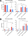
Pacman knockout does not affect vsiRNA profiles. (A) Northern blot of positive-sense [(+) RNA] or negative-sense [(−) RNA] viral RNA in control (CTRL) cells or Pacman-knockout cells (clones g3#3 and g2#13), mock infected (−) or infected with KRV (+) at an MOI of 0.1 for 72 h. Viral RNA was detected using a pool of oligonucleotide probes complementary to the 3′ UTR of KRV, between positions 10361 and 11375. The location of ribosomal RNA (rRNA) based on ethidium bromide staining is indicated. (B and C) Quantification of total siRNAs (B) and sense (+) and antisense (−) vsiRNAs (C) in WT, CRISPR control (CTRL), or Pacman-knockout (KO) U4.4 cell lines infected with ZIKV at an MOI of 0.1 or KRV at an MOI of 10 for 72 h. Viral siRNA levels were normalized to viral gRNA levels. Errors bars represent the standard deviation from two independent cell lines. ns, non-significant. *P < 0.05 by two-way ANOVA and Fisher’s LSD test. (D and E) Distribution of (+) and (−) vsiRNAs across the 3′ end of the genomes of KRV (D) or ZIKV (E) (from nt 9,000 onward) in CTRL and Pacman KO cells. Top panels show (+) vsiRNAs and lower panels show (−) vsiRNAs. The results are the average from two independent cell lines. (F and G) Percentage of vsiRNAs derived from the indicated regions compared to the entire viral genome. The size of each region is indicated in italic as a percentage of the viral genome size. Errors bars represent the standard deviation from two independent cell lines. (H) Distribution of KRV vsiRNAs on a logarithmic scale plotted on the 3′ terminal region of the genome. Read counts in WT/CTRL cells and Pacman-KO cells are superimposed for (+) vsiRNAs (top panel) and (−) vsiRNAs (lower panel) separately. (D and E) Boundaries of 3′ UTRs and sfRNAs are indicated by dashed vertical lines. (D–H) Blue, (+) vsiRNA; red, (−) vsiRNA; darker, WT and CTRL cells; lighter, Pacman-KO cells.

References

    1. Postler TS, Beer M, Blitvich BJ, Bukh J, de Lamballerie X, Drexler JF, Imrie A, Kapoor A, Karganova GG, Lemey P, Lohmann V, Simmonds P, Smith DB, Stapleton JT, Kuhn JH. 2023. Renaming of the genus Flavivirus to Orthoflavivirus and extension of binomial species names within the family Flaviviridae. Arch Virol 168:224. doi:10.1007/s00705-023-05835-1 - DOI - PubMed
    1. Saiyasombat R, Bolling BG, Brault AC, Bartholomay LC, Blitvich BJ. 2011. Evidence of efficient transovarial transmission of Culex flavivirus by Culex pipiens (Diptera: Culicidae). J Med Entomol 48:1031–1038. doi:10.1603/me11043 - DOI - PubMed
    1. Frangeul L, Blanc H, Saleh M-C, Suzuki Y. 2020. Differential small RNA responses against co-infecting insect-specific viruses in Aedes albopictus mosquitoes. Viruses 12:468. doi:10.3390/v12040468 - DOI - PMC - PubMed
    1. Blitvich BJ, Firth AE. 2015. Insect-specific flaviviruses: a systematic review of their discovery, host range, mode of transmission, superinfection exclusion potential and genomic organization. Viruses 7:1927–1959. doi:10.3390/v7041927 - DOI - PMC - PubMed
    1. Moureau G, Cook S, Lemey P, Nougairede A, Forrester NL, Khasnatinov M, Charrel RN, Firth AE, Gould EA, de Lamballerie X. 2015. New insights into flavivirus evolution, taxonomy and biogeographic history, extended by analysis of canonical and alternative coding sequences. PLoS One 10:e0117849. doi:10.1371/journal.pone.0117849 - DOI - PMC - PubMed

LinkOut - more resources