. 2024 Nov 22;9(22):e178381.
doi: 10.1172/jci.insight.178381.

Type 2 innate immunity promotes the development of pulmonary fibrosis in Hermansky-Pudlak syndrome

Affiliations

Type 2 innate immunity promotes the development of pulmonary fibrosis in Hermansky-Pudlak syndrome

Parand Sorkhdini et al. JCI Insight. .

Abstract

Hermansky-Pudlak syndrome (HPS), particularly types 1 and 4, is characterized by progressive pulmonary fibrosis, a major cause of morbidity and mortality. However, the precise mechanisms driving pulmonary fibrosis in HPS are not fully elucidated. Our previous studies suggested that CHI3L1-driven fibroproliferation may be a notable factor in HPS-associated fibrosis. This study aimed to explore the role of CHI3L1-CRTH2 interaction on type 2 innate lymphoid cells (ILC2s) and explored the potential contribution of ILC2-fibroblast crosstalk in the development of pulmonary fibrosis in HPS. We identified ILC2s in lung tissues from patients with idiopathic pulmonary fibrosis and HPS. Using bleomycin-challenged WT and Hps1-/- mice, we observed that ILC2s were recruited and appeared to contribute to fibrosis development in the Hps1-/- mice, with CRTH2 playing a notable role in ILC2 accumulation. We sorted ILC2s, profiled fibrosis-related genes and mediators, and conducted coculture experiments with primary lung ILC2s and fibroblasts. Our findings suggest that ILC2s may directly stimulate the proliferation and differentiation of primary lung fibroblasts partially through amphiregulin-EGFR-dependent mechanisms. Additionally, specific overexpression of CHI3L1 in the ILC2 population using the IL-7Rcre driver, which was associated with increased fibroproliferation, indicates that ILC2-mediated, CRTH2-dependent mechanisms might contribute to optimal CHI3L1-induced fibroproliferative repair in HPS-associated pulmonary fibrosis.

Keywords: Fibrosis; Genetic diseases; Immunology; Innate immunity; Pulmonology.


Conflict of interest statement

Conflict of interest: YZ holds a patent, titled “Methods for the diagnosis and treatment of pulmonary fibrosis in subjects with Hermansky-Pudlak Syndrome.” BRG is a paid full-time employee of AstraZeneca.

Figures

Figure 1
Figure 1. ILC2s are increased in human HPS lungs.
(A) ILC2 staining in human lung tissues. Anti-GATA3, -CD90/Thy1, and a cocktail of 4 antibodies against CD3, CD56, CD20, and CD79α were employed to stain human lung tissues in normal individuals, idiopathic pulmonary fibrosis (IPF) patients, and HPS patients. ILC2s are identified as GATA3+CD90/Thy1+, and antibody cocktail–negative cells (purple cells). The corresponding phase-contrast image shows lung tissue architecture in normal individuals, and pathologic changes in IPF and HPS lungs. (B) Counting of ILC2s (normalized per area) under ×40 magnification using an immunofluorescence microscope. Values are mean ± SEM with a minimum of 4 samples in each group. Comparisons between groups were conducted by 2-way ANOVA with Bonferroni’s post hoc test. ***P ≤ 0.001. Scale bars: 20 μm.
Figure 2
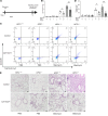
Figure 2. ILC2s are increased in HPS/bleomycin-induced lung fibrosis.
WT and HPS1–/– mice were subjected to i.p. bleomycin administration, primary ILC2s were sorted from mouse lungs, and ILC2 numbers were assessed by flow cytometry. (A) Schematic of the experiment. (B) Gating strategy of CD90-positive, Lineage-negative, T1/ST2-positive, and ICOS-positive ILC2s. (C and D) Representative charts and quantification of increased percentages of pulmonary ILC2s in the lungs of bleomycin-treated HPS1–/– mice. (E) Increased pulmonary ILC2 numbers in the lungs of bleomycin-treated HPS1–/– mice. Values are mean ± SEM with a minimum of 8 mice in each group. Comparisons between groups were conducted by 2-way ANOVA with Bonferroni’s post hoc test. *P ≤ 0.05; **P ≤ 0.01; ***P ≤ 0.001; ****P ≤ 0.0001.
Figure 3
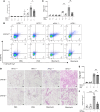
Figure 3. CRTH2 inhibition prevents ILC2 recruitment and fibrosis development in HPS/bleomycin-induced lung fibrosis.
(A) Schematic of the experiment. WT and HPS1–/– mice were subjected to a single dose of intratracheal (i.t.) bleomycin administration. (BE) Mice were treated with CRTH2 inhibitor or its vehicle control. (B) ILC2 number assessed by flow cytometry. (C) Gating strategy of CD90-positive, Lineage-negative, T1/ST2-positive, and ICOS-positive ILC2s. (D) A Sircol assay was used to assess the levels of collagen accumulation in the lung. (E) Representative histopathology of lung sections stained with H&E to depict the degree of fibrosis. Scale bars: 200 μm. *P ≤ 0.05; **P ≤ 0.01 by 2-way ANOVA with Bonferroni’s post hoc test.
Figure 4
Figure 4. ILC2s are recruited via CHI3L1-CRTH2–dependent mechanisms in HPS/bleomycin-induced lung fibrosis.
WT, CRTH2-knockout (CRTH2–/–), HPS1–/–, and HPS1–/– CRTH2–/– double-mutant mice were subjected to intratracheal (i.t.) bleomycin administration. (A) Lung ILC2 number was assessed by flow cytometry. (B) Gating strategy of CD90-positive, Lineage-negative, T1/ST2-positive, and ICOS-positive ILC2s. (C) Sircol assay was used to assess the levels of collagen accumulation in the lung. (D) Representative histopathology of lung sections stained with H&E to depict the degree of fibrosis. (E) HPS1–/– Rag1–/– mice were subjected to a single dose of i.t. bleomycin administration. A Sircol assay was used to assess the levels of collagen accumulation in the lung. (F) An ILC2 recruitment assay was performed in Transwell plates in vitro. Values are mean ± SEM with a minimum of 8 mice in each group. Comparisons between groups were conducted by 2-way ANOVA with Bonferroni’s post hoc test. *P ≤ 0.05; **P ≤ 0.01; ***P ≤ 0.001. Scale bars: 200 μm.
Figure 5
Figure 5. IL-33–mediated ILC2 activation contributes to HPS/bleomycin-induced lung fibrosis.
WT and HPS1–/– mice were subjected to intratracheal (i.t.) bleomycin administration. Mice were treated with IL-33 siRNA (every other day, 3 nmol/mouse) or its scrambled control. (A) Schematic of the experiment. (BD) Numbers of BAL eosinophils, neutrophils, and T cells were not altered by IL-33 siRNA treatment. (E and F) Percentages of lung ILC2s and Th2s were assessed by flow cytometry. (G) Total lung collagen was quantified using a Sircol assay on day 14. (H and I) Gene expression of COL-A1 mRNA and fibronectin mRNA was measured by RT-PCR. (J) Representative histopathology of lung sections stained with H&E to depict the degree of fibrosis. Values are mean ± SEM with 7–9 mice in each group. Comparisons between groups were conducted by 2-way ANOVA with Bonferroni’s post hoc test. *P ≤ 0.05; **P ≤ 0.01; ***P ≤ 0.001; ****P ≤ 0.0001. Scale bars: 100 μm.
Figure 6
Figure 6. ILC2s sorted from bleomycin-challenged HPS1–/– mice express high levels of IL-5, IL-13, amphiregulin (AREG), CRTH2, and CHI3L1.
(AC, G, and I) Gene expression levels for IL-5, IL-13, AREG, and CRTH2. (DF and H) Soluble IL-5, IL-13, AREG, and CHI3L1 produced by ILC2s were measured in the culture supernatants of ILC2s sorted from WT and HPS1–/– mice. Values are mean ± SEM with 7–9 mice in each group. Comparisons between groups were conducted by 2-way ANOVA with Bonferroni’s post hoc test. *P ≤ 0.05; **P ≤ 0.01; ***P ≤ 0.001; ****P ≤ 0.0001.
Figure 7
Figure 7. ILC2s sorted from bleomycin-challenged HPS1–/– mice exhibited a unique profibrotic gene expression profile.
(A) Gene expression heatmap analyzed by Python. There are 46 genes that were differentially expressed. In the upper cluster, upregulated genes are visible. Many of them play a role in a variety of matrix remodeling and collagen production in various lung diseases and cancers. (BD) Three pie charts displaying the molecular function (MF), biological process, and protein class of top genes that were most altered in ILC2s isolated from HPS1–/– mice compared with WT mice upon bleomycin treatment. (E) Reactome analysis highlights the immune system and extracellular matrix organization pathways that were altered in ILC2s isolated from HPS1–/– mice compared with WT mice upon bleomycin treatment.
Figure 8
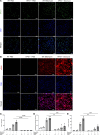
Figure 8. ILC2s stimulate fibroblast proliferation and differentiation in in vitro coculture.
WT and HPS1–/– mice were subjected to i.p. bleomycin administration. Primary ILC2s were sorted from mouse lungs and cocultured with primary fibroblasts. (A) ILC2 and fibroblast costaining in PBS- and bleomycin-treated WT and HPS1–/– mice. BrdU immunostaining was used to identify proliferating cells. (B) α-Smooth muscle actin (α-SMA) immunostaining was used to detect α-SMA expression in primary fibroblasts (original magnification, ×40). (C and D) Quantitative analysis of BrdU-positive cells normalized per area. The cells were incubated with BrdU for 24 hours and incorporated BrdU was detected with the BrdU Cell Proliferation Assay. (E) Quantitative analysis of α-SMA–positive cells normalized per area. Images are representative of 4 independent experiments. Values are mean ± SEM with a minimum of 4 samples in each group. Comparisons between groups were conducted by 2-way ANOVA with Bonferroni’s post hoc test. **P ≤ 0.01; ***P ≤ 0.001; ****P ≤ 0.0001. Scale bars: 100 μm.
Figure 9
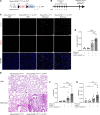
Figure 9. Overexpression of CHI3L1 in ILC2s leads to increased fibrosis development.
(A) Schematic diagram for the generation of Rosa-CHI3L1LSL/LSL IL-7Rcre transgenic mice. Rosa-CHI3L1LSL/LSL and Rosa-CHI3L1LSL/LSL IL-7Rcre mice were subjected to i.p. bleomycin administration. (B) Schematic of the experiment. (C) CHI3L1 and GATA3 staining in mouse lung tissues. Anti-CHI3L1 and -GATA3 antibodies were employed to stain lung tissues from Rosa-CHI3L1LSL/LSL and Rosa-CHI3L1LSL/LSL IL-7Rcre mice. CHI3L1 is identified as red. GATA3 is identified as green and employed as a positive identifier of ILC2s. (D) Total bronchoalveolar lavage (BAL) collagen was quantified using a Sircol assay. (E) Representative histopathology of lung sections stained with H&E to depict the degree of fibrosis in Rosa-CHI3L1LSL/LSL and Rosa-CHI3L1LSL/LSL IL-7Rcre mice that received bleomycin or vehicle (PBS). (F and G) Gene expression of COL-A1 mRNA and fibronectin mRNA was measured by RT-PCR. Values are mean ± SEM with 7–9 mice in each group. Comparisons between groups were conducted by 2-way ANOVA with Bonferroni’s post hoc test. *P ≤ 0.05; **P ≤ 0.01; ***P ≤ 0.001; ****P ≤ 0.0001. Scale bars: 100 μm.

References

    1. Gahl W, Huizing M. Hermansky-Pudlak syndrome. In: Adam MP, et al, eds. GeneReviews. University of Washington; 2012.
    1. Schinella RA, et al. Hermansky-Pudlak syndrome with granulomatous colitis. Ann Intern Med. 1980;92(1):20–23. doi: 10.7326/0003-4819-92-1-20. - DOI - PubMed
    1. Anderson PD, et al. Hermansky-Pudlak syndrome type 4 (HPS-4): clinical and molecular characteristics. Hum Genet. 2003;113(1):10–17. doi: 10.1007/s00439-003-0933-5. - DOI - PubMed
    1. Gahl WA, et al. Genetic defects and clinical characteristics of patients with a form of oculocutaneous albinism (Hermansky-Pudlak syndrome) N Engl J Med. 1998;338(18):1258–1264. doi: 10.1056/NEJM199804303381803. - DOI - PubMed
    1. Hermansky F, Pudlak P. Albinism associated with hemorrhagic diathesis and unusual pigmented reticular cells in the bone marrow: report of two cases with histochemical studies. Blood. 1959;14(2):162–169. doi: 10.1182/blood-2016-01-696435. - DOI - PubMed

MeSH terms